INTRODUCTION
Involutional entropion is a condition in the eld-erly population in which the eyelid margin is turned in against the globe with the eyelid margin and touching the cornea. It is a common lower eyelid
malposition encountered by most ophthalmologists, including ophthalmic practitioners or trainees who do not specialise in oculoplastic surgery, in outpa-tient clinics. When left untreated, this condition can cause ocular discomfort, several corneal disorders, and ocular inflammations that may be associated with visual disturbance. Surgical treatment should be considered if there are persistent symptoms and changes to the ocular surface.
The underlying pathogenesis of involutional en-tropion is thought to be due to 4 factors : 1. hori-zontal eyelid laxity, 2. vertical eyelid laxity with
ORIGINAL
Efficacy of the Quickert procedure for involutional
en-tropion : the first case series in Asia
Tatsuro Miyamoto
1), Hiroshi Eguchi
1), Takashi Katome
1), Toshihiko Nagasawa
1),
Yoshinori Mitamura
1), Geoffrey Crawford, FRANZCO, FRACS
2) 1)Department of Ophthalmology, Institute of Health Biosciences, the University of Tokushima Graduate School, Tokushima, Japan ; and 2)
Centre for Ophthalmology and Visual Science, University of Western Australia and the Lions Eye Institute, Western Australia, Australia
Abstract : Purpose : To report the efficacy of the Quickert procedure in the first case se-ries of involutional entropion in an elderly Asian population, and to introduce the tech-nique to Asian ophthalmologists including general ophthalmologisits and ophthalmic trainees. Methods : We conducted a retrospective review of 13 consecutive patients un-derwent the Quickert procedure for involutional entropion by occasional eyelid surgeons at Tokushima University Hospital or Mino Tanaka Hospital from September 2003 to April 2010. Demographic data, including gender, age, history of previous eyelid surgery, systemic disease, recurrence of entropion, postoperative complications, and symptoms were analyzed. Results : There were 5 male (38.5%%) and 8 female (61.5%%) subjects with a mean age of 77.8 years. Three patients underwent previous surgery for entropion were included. Entropion was rectified in all patients by a single Quickert procedure, and no recurrence was observed for a maximum of 89 months after the surgery. Although notching of the eyelid margin and mild symblepharon were observed in one patient, no symptoms associated with these complications were reported. Conclusion : The Quickert procedure can be one of the surgical procedures of choice for involutional entropion and should be common surgical approach for occasional eyelid surgeons in Asia as well as in western countries. J. Med. Invest. 59 : 136-142, February, 2012
Keywords : the Quickert procedure, involutional entropion, occasional eyelid surgeon
Received for publication November 14, 2011 ; accepted Decem-ber 19, 2011.
Address correspondence and reprint requests to Hiroshi Eguchi, M.D., Ph.D., Department of Ophthalmology, Institute of Health Biosciences, the University of Tokushima Graduate School, 3 18 15, Kuramoto cho, Tokushima 770 8503, Japan and Fax : +088 -631 - 4848.
attenuation or disinsertion of the lower lid retractors, 3. overriding of the preseptal orbicularis oculi mus-cle, and 4. relative enophthalmos (1-4). Horizontal eyelid laxity is probably the main underlying fac-tor (5), and enophthalmos has been disproved as a cause (6). Therefore, various surgical procedures that address one, two, or all of these factors have been described, and it is widely accepted that surgi-cally addressing the causative factors is necessary to successfully correct entropion (5, 7). In terms of pre-operative examinations, horizontal eyelid laxity is es-timated by the pinch test and medial canthal tendon (MCT) laxity grading by the lateral distraction test (8). However, a mild degree of horizontal eyelid lax-ity (clinically “undetected”) predisposes to the fail-ure of surgical correction (5) ; therefore, most gen-eral ophthalmologists or ophthalmic trainees find it difficult to precisely evaluate horizontal eyelid laxity and even oculoplastic specialists can overlook this condition.
Quickert described a horizontal eyelid shorten-ing procedure that was combined with the modi-fied Wies entropion repair method as the Quickert procedure in 1972 (9). Since it can address all the aforementioned factors causing involutional en-tropion and the surgical technique is relatively easy, it has become entrenched as one of the popular pro-cedures for the treatment of involutional entropion in Western countries (4, 5, 9-11). However, to our knowledge, there are no journal articles describing the use of this technique in Asian population. If the procedure is reasonable for pathophysiologically
correcting involutional entropion, easy to perform even for occasional eyelid surgeons or ophthalmic trainees and gives good results, it should be consid-ered for practice by Asian ophthalmologists. The aim of this article is to introduce the Quickert pro-cedure to Asian ophthalmologists, including general ophthalmologists and ophthalmic trainees, by report-ing the first case series of involutional entropion in an elderly Asian population.
PATIENTS AND METHODS
Demographic data (Table 1)We retrospectively examined 13 consecutive eld-erly patients (13 lower eyelids) with involutional entropion who underwent the Quickert proce-dure between September 2003 and April 2010 at Tokushima University Hospital or Mino Tanaka Hospital. The mean age was 77.8 years (range, 69-86 years) at the time of surgery ; 5 (38.5%) were male and 8 (61.5%) were female. Of these, 3 eye-lids had previously undergone surgery to correct entropion. One patient (case 5) underwent the Jones procedure at Tokushima University Hospital a year earlier and another patient (case 12) underwent probable tarsal stripping at some practitioner of plastic surgeon. The rest (case 3) had unknown eyelid surgery three times at other hospitals by three different occasional eyelid surgeons. There were no patients with systemic connective tissue disease.
Table 1 Demographic data of the 13 cases
Case Age Gender Eye Cornea Surgery date Follow - up(mo) Others 1 78 F OD SPK 2003/09/08 89 None 2 86 M OD SPK 2005/05/15 81 Anemia 3 86 F OD SPK 2006/11/24 51 Three surgeries * 4 81 F OD Erosion 2007/06/29 44 PKP 5 77 F OD SPK 2007/11/02 17 Jones procedure (2006/02/21**) 6 81 F OD SPK 2008/05/24 33 Blepharoptosis
7 76 F OD SPK 2008/09/09 29 Optic atrophy, Iritis 8 79 M OD SPK 2008/10/10 28 DSAEK, PKP, Glaucoma
9 76 M OS SPK 2008/11/25 27 Hepatocirrhosis, Thrombocytopenia 10 73 F OD SPK 2009/04/10 22 Cataract surgery, Buckling surgery 11 75 F OD SPK 2009/12/05 14 Conjunctivochalasis
12 75 M OS Erosion 2010/04/23 10 Parkinson disease, Tarsal stripping 13 69 M OD SPK 2010/04/27 10 Nephrosis
* : Past history of three unknown surgeries for involutional entropion ** : Date of previous operation at Tokushima University Hospital
M : Male ; F : Female ; SPK : Superficial punctate keratitis ; PKP : Penetrating keratoplasty ; DSAEK : Descemet’s stripping automated endothelial keratoplasty ; mo : month.
Surgical technique
Moxifloxacin hydrochloride 0.5% (Vegamox!!,
Alcon) or 0.3% gatifloxacin hydrate (Gatiflo!!, Senju)
ophthalmic solutions were applied to the conjunc-tival sac 3 times daily for 3 days prior to surgery. After the application of 1 drop of 0.4% oxybupro-caine hydrochloride (Benoxil!!, Santen), the lower
eyelid was anaesthetised with approximately 2 ml of 1% lidocaine hydrochloride and a 1 : 100,000 di-lution of epinephrine (1% Xylocaine!! Injection,
AstraZeneca) subcutaneously and subconjunctively. All surgeries were performed by occasional eyelid surgeons (HE or TM), not oculoplastic specialists, according to the “Quickert procedure” (4, 9, 10).
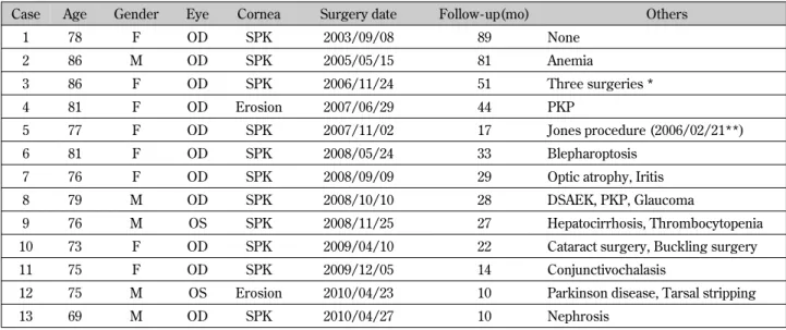
A full-thickness vertical 5-mm incision was in-itially made perpendicular to the eyelid margin, start-ing at 5 mm from the lateral canthus, approximately one-quarter of the eyelid length. A second full-thickness eyelid incision was made horizontally from the inferior aspect of the initial incision 5 mm below the lid margin and below the tarsal plate to the inferior punctum. A third incision was, made laterally from the inferior aspect of the initial inci-sion to the lateral canthus (Fig. 1A). The medial flap
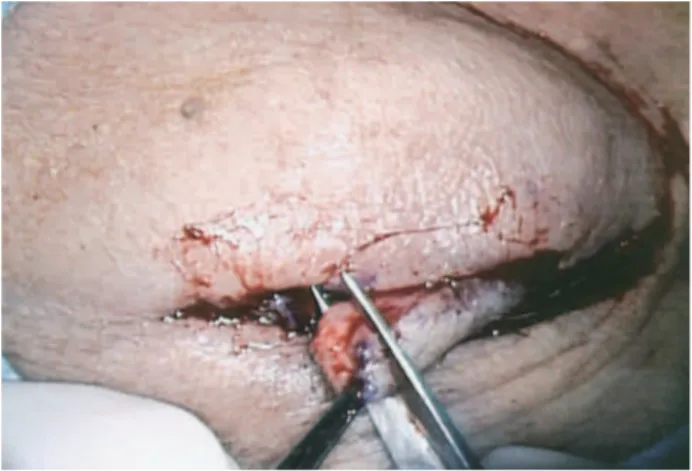
of the eyelid was then folded over the lateral flap and tightened to give moderate tension. The fourth in-cision was made at the point marked on the medial flap vertical to the eyelid margin and carried to the inferior tarsal border where it joined the second in-cision (Fig. 1B). Thus, the block shape of the over-lapping full-thickness eyelid was excised. The tar-sal plate was approximated 2 absorbable sutures (8-0 Vicryl, Ethicon) (Fig. 1C), and the margin of
the eyelid flap was approximated by passing one non-absorbable 6-0 Nylon suture (Nescosuture!!,
Alfresa Pharma) through the grey line. The skin margin of the approximated flap was closed using interrupted 6-0 nylon sutures. Three double armed 4-0 absorbable sutures (4-0 PDS or 4-0 Vicryl, Ethicon) were placed through the lower lid retrac-tor layer (Fig. 1D). They were passed under the orbicularis oculi muscle anterior to the tarsal plate in the upper wound edge to emerge in the skin 2 mm inferior to the lash line. The sutures for lower lid retractor plication were placed at two points in the medial flap and at one point in the lateral flap (Fig. 1E). The double arms of the suture were placed 2-3 mm apart and tied firmly without bol-sters. Finally, the skin of the second and third inci-sions was closed with interrupted 6-0 nylon sutures (Fig. 1F).
Figure 1A :Incision marked
Full - thickness eyelid incision was made. The numbers indicate the order of the incision
Figure 1B :Overlapping eyelid incision
Overlapping full - thickness eyelid incision was excised to correct the horizontal eyelid laxiety.
Figure 1C :Close the lid margin
The two 8 - 0 absorbable sutures were placed through the edge of tarsal plate close to the lid margin to avoid eyelid notching.
Postoperative care
The topical antimicrobials stated above and fluorometholone (0.1% Flumetholon!!, Santen)
oph-thalmic solutions were applied 3 times daily for 2
weeks, and betamethasone sodium phosphate-fra-diomycin sulphate (Rinderon!!-A, Shionogi)
oint-ment was instilled in the suture sites twice daily until suture removal. The skin sutures were re-moved at 7 days postoperation. The first postopera-tive review in the outpatient clinic was at 4-5 days after surgery. All patients were examined again for the recurrence of entropion, corneal disorders, ocu-lar symptoms, and complications at 1, 3, 6, and 12 months postoperation as scheduled visits and once a year while they could return.
RESULTS
The follow-up period ranged from 10 to 89 months (mean, 35 months). No patient was lost to a sched-uled follow-up, except one patient who died within 8 months of the surgery. Entropion was corrected in all patients by a single Quickert procedure (Figs. 2A, B), and there was no recurrence in any of the
Figure 1D :The sutures through the lower lid retractor
Absorbable sutures were placed through the lower lid retractor layer.
Figure 1E :Three double armed sutures
Three double armed sutures were passed under the orbicularis anterior to the tarsal plate to emerge 2 mm inferior to the lash line.
Figure 1F :Close the skins
The second and third incisions were closed with 6 - 0 nylon su-tures.
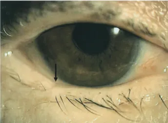
Figure 2A :Preoperative photograph
Marked entropion and cilia touching the cornea are observed.
Figure 2B :Postoperative photograph
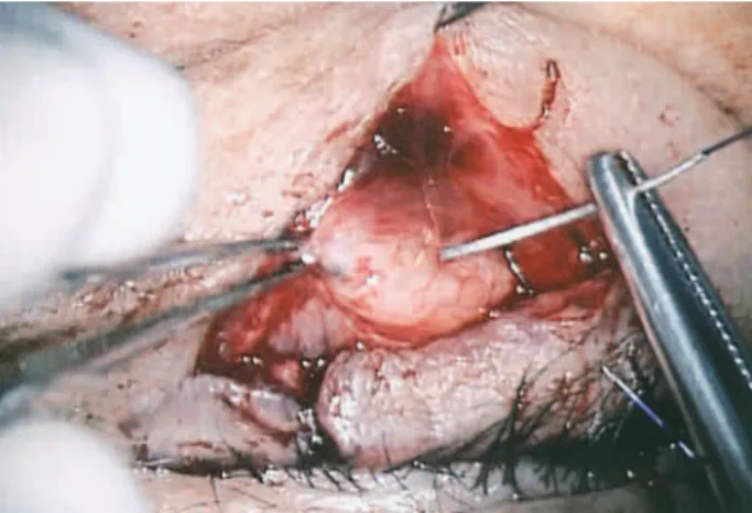
patients. Corneal disorders and ocular symptoms were decreased or had disappeared in all patients at each visit. Although mild notching of the eyelid margin and negligible symblepharon were observed in one patient (Figs. 3A, B), no symptoms associ-ated with these complications were reported.
DISCUSSION
Involutional entropion is a more common eyelid malposition in elderly Asian populations than in non-Asians, and 11.4% of the total ophthalmic plastic surgery operations completed on Asians are for en-tropion repair (12) It is possible that the actual num-ber of the elderly population with involutional en-tropion who visit ophthalmic practitioners and the
number of patients who require surgical treatment are higher than this reported value. In other words, it remains possible that a large number of Asian ophthalmologists, including ophthalmic practitioners and trainees who do not specialise in ophthalmic plastic surgery, are required to perform some sur-gical treatments for involutional entropion.
Although numerous procedures have been re-ported that address one, two, or all the causes of involutional entropion, there is no perfect method and all the technique are associated with advantages and disadvantages. Moreover, the major etiological cause is still a matter of debate. Danks et al. specu-lated that horizontal laxity is probably the main un-derlying factor and stated that lid shortening should be performed in all primary surgeries and reopera-tions.5In terms of horizontal laxity, it is generally
rectified by the tarsal strip procedure (13) or full-thickness eyelid resection (1, 4, 5). The former pro-cedure preserves the almond-shaped configuration of the lateral canthus while avoiding phimosis or eye-lid notching (14) ; however, ophthalmic practitioners or trainees may be hesitant to dissect the lateral can-thus deep enough to reach the periosteum of the orbital rim because most of them provide routine medical care mainly for the eyeball, or at the most, for the eyelid. The latter procedure can be easily performed by most ophthalmologists using common scissors, but it is more invasive than the former procedure and is associated with an increased prob-ability of notching the eyelid margin.
Meanwhile, Jones et al. reported that vertical laxity of the eyelid is the most important cause of involu-tional entropion (15), and this opinion was supported by Kakizaki et al. who advocates the shortening of the lower lid retractor, addressing only the vertical laxity, as the first operational procedure for involu-tional entropion (16). Kakizaki et al. also stated that the looseness of the orbicularis oculi muscle is not the main cause of involutional entropion on the basis of the findings of perioperative magnetic resonance imaging of the lower lid (17). However, in Jones pro-cedure, a meticulous knowledge of the lower eyelid anatomy is required to precisely isolate and tight-ened the lower lid retractor, and it may be a diffi-cult procedure for the occasional eyelid surgeons to perform. Barnes et al. suggested that an ideal op-eration should be effective, with minimum discom-fort, rapid recovery and be simple enough for a trainee to perform (18). Rougraff et al. analyzed the published recurrence rates and suggested that the correction of at least 2 of 3 involutional changes
Figure 3A :Complications of the eyelid margin
Minimal notching of the eyelid margin is observed.
Figure 3B :Complications of the conjunctiva palpebrae
conferred a higher long-term success rate (19) ; therefore, we believe that rectifying as many causes of involutional entropion as possible is preferable. For general ophthalmologists and trainees, particu-larly when they do not have confidence in preopera-tive examination of horizontal and/or vertical laxity of the eyelid, it is certain that they choose a proce-dure that can address all the possible causes of involutional entropion.
We prefer the Quickert procedure because it can address horizontal eyelid laxity by full-thickness eyelid shortening and vertical eyelid laxity by tuck-ing the lower lid retractor between the orbicularis oculi muscle and the tarsal plate and can prevent the preseptal orbicularis oculi muscle overriding by creating fibrosis with the sutures. In other words, the procedure can address all the known causes of involutional entropion at single surgery. Besides, the surgical technique can be performed by most ophthalmic practitioners and trainees because it is not always necessary to have meticulous knowledge of the lower lid lamellae. Full-thickness eyelid re-section enables us to easily master the anatomy of the cross-section of the lower lid. Every ophthal-mologist may understand the conjunctiva as a glossy mucosa and the orbicularis oculi muscle as a bloody striped tissue. The lower lid retractor can then be identified at a glance as it is whitish in appearance and is located between the orbicularis oculi muscle and the conjunctiva. When correcting involutional entropion, tucking the full-thickness layers of the lower lid retractor is important because the lower lid retractor consists of a double layer and the pos-terior layer represents the main tractional compo-nent of the lower lid (19). When only the anterior layer is addressed using the Jones procedure or if the lower lid retractors are not adequate isolated, it is possible for entropion to recur. In this case series, we treated a case of recurrent involutional entropion that was initially treated by an occasional eyelid surgeon using the Jones procedure. Over-looked horizontal eyelid laxity or insufficient tuck-ing of the lower lid retractor without a clear under-standing of the double layer may be the cause of recurrence. All of the recurred cases can be treated by a single Quickert procedure performed by an occasional eyelid surgeon (HE) without any support from oculoplastic specialists. We believe that a cross-section of the eyelid, that enables us to understand the full-thickness of the lower lid retractor even after a previous Jones procedure has obscured the layer, contributes to the excellent results for this
technique.
Although the success rate of Quickert procedure is reported to be the highest in several procedures, (5, 20), we have to take into account that patients inclusion of surgery for involutional entropion may elder patients who do not have high rates of life expectancy. In this cases series, the success rate is 100% and all cases are followed for at least 10 months, except for a case who died 8 months after the surgery, and the follow-up duration ranges to 89 months at maximum. Thus, this case series proves Quickert procdure have a good success rate for the long-term result. Mild notching of the eyelid margin and negligible symblepharon at the first vertical full-thickness incision occurred in only one patient with respect to postoperative complica-tions in this case series. This patient had under-gone three previous unknown surgeries by three different occasional eyelid surgeons. The symble-pharon was possibly caused by a severe postopera-tive inflammation of the eyelid tissue due to the lack of normal anatomy. None of the patients experi-enced symblepharon after a primary Quickert pro-cedure. At present, no complaints regarding these complications have been reported.
In conclusion, the Quickert procedure is easy for most ophthalmologists to perform, and it is the logical approach for rectifying involutional entropion. It can also be applied to recurrent cases in which the normal anatomy of the eyelid has already been lost after unknown surgery. The recurrence rate is very low in the long-term outcome. The Quickert procedure should be considered as one of the sur-gical treatments of choice for involutional entropion in the elderly Asian population.
CONFLICT OF INTEREST
The authors declared no conflict of interest.
REFERENCES
1. Dalgleish R, Smith JLS : Mechanics and histol-ogy of senile entropion. Br J Ophthalmol 50 : 79-91, 1966
2. Dryden R, Leibsohn J, Wobig J : Senile en-tropion pathogenesis and treatment. Arch Oph-thalmol 96 : 1883-1885, 1978
3. Jones LT : The anatomy of the lower eyelid and its relation to the cause and cure of entropion.
Am J Ophthalmol 1 : 29-36, 1960
4. Collin JRO, Rathbun JE : Involutional entropion. A review with evaluation of procedure. Arch Ophthalmol 96 : 1058-1064, 1978
5. Danks JJ, Rose GE : Involutional lower lid entropion. To shorten or not to shorten? Oph-thalmology 105 : 2065-2067, 1998
6. Kersten RC, Hammer BJ, Kulwin DR : The role of enophthalmos in involutional entropion. Ophthal Plast Reconstr Surg 13 : 195-198, 1997 7. O’Sullivan EP, Howe LJ, Barnes E, Barnes J, Olver JM : Factors affecting the success rate of Quickert and Wies procedures for lower lid entropion. Orbit 17 : 59-60, 1999
8. Oliver JM, Sathia J, Wright M : Lower eyelid medial canthal tendon laxity grading : an in-terobserver study of normal subjects. Ophthal-mology 108 : 2321-2325, 2001
9. Quickert MH : The eyelids. Malposition of the lid. In : Sorby A, editor. Modern Ophthalmol-ogy, 2nded. London : Butterworth, v. 4A, sec II,
1972, pp.937-954.
10. Marcovits AS : Variations on the theme of in-volutional entropion and the Quickert repair. Ann Ophthalmol 12 : 1028-1032, 1980
11. Tyers AG, Collin JRO : Colour atlas of ophthal-mic plastic surgery, 3rd ed. London :
Butter-worth, vi. sec A, 2008, pp. 98-100.
12. Carter SR, Chang J, Aguilar GL, Rathbun JE, Seiff R : Involutional entropion and ectropion of
the Asian lower eyelid. Ophthal Plast Reconstr Surg 16 : 45-49, 2000
13. Anderson RL, Gordy DD : The tarsal strip pro-cedure. Arch Ophthalmol 97 : 2192-2196, 1979 14. Rougraff PM, Tse DT, Johnson TE, Feuer W : Involutional entropion repair with fornix sutures and lateral tarsal strip procedure. Ophthal Plast Reconstr Surg 17 : 281-287, 2000
15. Jones LT, Reeth MJ, Tsujimura JK : Senile en-tropion. Am J Ophthalmol 55 : 463-469, 1963 16. Kakizaki H, Zako M, Mito H, Kato Y, Iwaki M :
Modified operation to correctly detect and fix the lower eyelid retractor in involutional en-tropion. Jpn J Ophthalmol 49 : 330-332, 2005 17. Kakizaki H, Zako M, Mito H, Katori N, Iwaki
M : Magnetic resonance imaging of pre and postoperative lower eyelid states in involutional entropion. Jpn J Ophthalmol 48 : 363-367, 2004 18. Barnes JA, Bunce C, Oliver JM : Simple effec-tive surgery for involutional entropion suitable for the general ophthalmologist. Ophthalmol-ogy 113 : 92-96, 2006
19. Kakizaki H, Zhao J, Nakano T, Asamoto K, Zako M, Iwaki M, Miyaishi O : The lower eye-lid retractor consists of definite double layer. Ophthalmology 113 : 2346-2350, 2006
20. Oliver JM, Barnes JA : Effective small-incision surgery for involutional lower eyelid entropion. Ophthalmology 107 : 1982-1988, 2000