Two cases of eosinophilic pustular folliculitis associated with
pregnancy
Dear Editor,
Although the pathogenesis of eosinophilic pustular folliculitis (EPF), a clinical entity proposed by Ofuji et al.,1 remains
unclear, it has been speculated that T-helper type 2 (Th2) immune responses are important.2It is reported that success-ful pregnancy is a Th2-related phenomenon.3 To our
knowl-Correspondence: Yoshio Urano, M.D., Ph.D., Division of Dermatology, Tokushima Red Cross Hospital, 103 Irinokuchi, Komatsushima City, Tokushima 773-8502, Japan. Email: dermniss@tokushima-med.jrc.or.jp
*Present address: Department of Dermatology, Institute of Biomedical Sciences, Tokushima University Graduate School, Tokushima, Japan.
Letters to the Editor
218
Ó2015 The Authors. The Journal of Dermatology published by Wiley Publishing Asia Pty Ltd
on behalf of Japanese Dermatological Association
This is an open access article under the terms of the Creative Commons Attribution NonCommercial NoDerivs
License, which permits use and distribution in any medium, provided the original work is properly cited, the use is
non commercial and no modifications or adaptations are made.
edge, six cases of EPF associated with pregnancy have been reported in the published work. We report two additional cases of EPF associated with pregnancy.
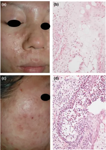
Serological tests for HIV were negative in both cases. In case 1, a 30-year-old Japanese woman in the 10th week of her first pregnancy presented with a 5-week history of pruritic eruptions. Topical steroid was ineffective. Her medical history was unremarkable except atopic dermatitis. Physical examina-tion revealed many pustules on coalescent, erythematous pla-ques on the edematous face (Fig. 1a). A number of papules and pustules were disseminated on the trunk, arms and legs without tendency to coalesce. Blood tests revealed a white blood cell count of 17 090/lL (19.3% eosinophils) and a total immunoglobulin E level of 561 IU/mL. Histological examination showed many eosinophils in the outer root sheath and seba-ceous gland (Fig. 1b). Oral prednisolone was effective.
Pred-nisolone was discontinued immediately after the spontaneous abortion due to intrauterine infection in the 16th week of preg-nancy. Eruptions recurred in approximately 2 weeks. Oral indo-methacin was ineffective. Dapsone therapy was effective in combination with prednisolone but was stopped because she desired to get pregnant, although prednisolone therapy was continued. There was little recurrence of skin lesions until her second pregnancy. She suffered from itchy papules mainly on the hands and feet during the first and second pregnancy tri-mesters. She gave birth to a healthy boy at full term. Pred-nisolone was tapered and discontinued after delivery. No relapse was observed for 2 years.
In case 2, a 26-year-old Japanese woman in the 15th week of her first pregnancy presented with a 3-day history of pruritic eruptions. Her medical history was unremarkable except atopic dermatitis. Physical examination revealed erythematous pla-ques with pustules and erosions on the edematous face, ears and neck (Fig. 1c), and papules on the dorsal surfaces of the hands. Blood tests revealed a white blood cell count of 14 030/lL (11.3% eosinophils). Histological findings were simi-lar to those of case 1 (Fig. 1d). Topical steroid was effective. She gave birth to a healthy boy at full term. No recurrence was observed for 5 years.
Out of eight cases of EPF associated with pregnancy, including our two cases, four suffered from the disease after the start of pregnancy. The others got it prior to pregnancy and experienced exacerbation of EPF during pregnancy. In one case, the exacerbation was associated with each of three pregnancies.4 In another case, EPF developed prior to the
onset of pregnancy, deteriorated in pregnancy and resolved following delivery.5In one of ours (case 1), EPF developed after
the start of the first pregnancy and relapsed during the second pregnancy. Although further information is needed, these may indicate that pregnancy and EPF are correlated through Th2-type immune responses.2,3
CONFLICT OF INTEREST:
None declared.Yoshihiro MATSUDATE,
1,* Yuki MIYAOKA,
2Yoshio URANO
1 1Division of Dermatology, Tokushima Red Cross Hospital, Komatsushima, and2Miyaoka Dermatology Clinic, Tokushima, Japan doi: 10.1111/1346-8138.13179
REFERENCES
1 Ofuji S, Ogino A, Horio T, Oseko T, Uehara M. Eosinophilic pustular folliculitis. Acta Derm Venereol 1970; 50: 195 203.
2 Nervi SJ, Schwartz RA, Dmochowski M. Eosinophilic pustular folliculi tis: a 40 year retrospect. J Am Acad Dermatol 2006; 55: 285 289. 3 Marzi M, Vigano A, Trabattoni D et al. Characterization of type 1 and
type 2 cytokine production profile in physiologic and pathologic human pregnancy. Clin Exp Immunol 1996; 106: 127 133.
4 Kus S, Candan I, Ince U, Uygur T. Eosinophilic pustular folliculitis (Ofuji’s disease) exacerbated with pregnancies. J Eur Acad Dermatol Venereol 2006; 20: 1347 1348.
5 Mabuchi T, Matsuyama T, Ozawa A. Case of eosinophilic pustular folliculitis associated with pregnancy. J Dermatol 2011; 38: 1 2.
(a)
(c)
(b)
(d)
Figure 1. Clinical and histological features of (a,b) case 1 and (c,d) case 2. (a) There were coalescent, erythematous plaques with pustules on the edematous face. (b) There were eosinophils, neutrophils and mononuclear cells in the sebaceous gland (hematoxylin eosin [HE], original magnification9200). (c) There were erythematous plaques with pustules and erosions on the edematous face. (d) There was a vesicle containing eosinophils, neutrophils, mononuclear cells and degenerated epithelial cells in the outer root sheath and sebaceous gland. An inflammatory cell infiltration composed of eosinophils and mononuclear cells was observed around the hair follicle (HE,9200).