REVIEW
Nailfold capillaroscopy : a comprehensive review on common
findings and clinical usefulness in non-rheumatic disease
Natalia Mansueto, Cinzia Rotondo, Addolorata Corrado, and Francesco Paolo Cantatore Department of Medical and Surgical Sciences - Rheumatology Unit, University of Foggia, Italy
Abstract : Nailfold video-capillaroscopy (NVC) is a useful diagnostic tool, used to early detect abnormalities in micro-circulation, providing a qualitative description of microvascular anomalies in Raynaud’s phenomenon. NVC role in the diagnosis of Systemic Sclerosis is well known. In other rheumatic conditions such as connective tissue diseases, vasculitis, and arthritis, the NVC anomalies are often included in a scleroderma like pattern. The use of NVC in non-rheumatic diseases (NRD), with remarkable microvascular damage, as diabetes, is not stan-dardized yet, although several research studies are carrying on. The aim of this article is to provide a resume of published results in order to lay the groundwork for the employment of NVC both in the diagnosis and follow up of microvascular complication in NRD. Furthermore, we mention NVC findings in pathologies without well rec-ognize microvascular damages in their pathogenesis : micro-vessels abnormalities may suggest a different point of view. J. Med. Invest. 68 : 6-14, February, 2021
Keywords : nailfold videocapillaroscopy, microcirculation, Raynaud’s phenomenon, diabetes, glaucoma
INTRODUCTION
Nailfold video-capillaroscopy (NVC) is a non-invasive diag-nostic test used to study microvascular abnormalities, predomi-nantly, in many rheumatological disorders, as connective tissue diseases (CTD). In particular, in systemic sclerosis (SSc), NVC allows detection of pathognomonic microvascular alterations and their activity phases, clustered in specific scleroderma patterns (early, active, and late) (1). The importance of NVC in SSc is evidenced by the inclusion of NVC scleroderma patterns among items in the new 2013 classification criteria for SSc. In addition, NVC is now considered a key finding in the very early stage of disease, in the clinical assessment and in the treatment management (2). In other CTD, as systemic lupus erythemato-sus, inflammatory myositis, undifferentiated connective tissue diseases and mixed connective tissue disease, although non-spe-cific, the NVC patterns can provide a valid support for the diag-nosis (3). This useful tool reproduces in vivo amplified images of skin microcirculation. NVC is a simple and reproducible test that can be assessed both in adults and in children (4). Moreover, NVC is used in diagnosis and follow-up of Raynaud’s phenome-non (RP), an intense vasospasm of the small arteries, charac-terized by three phases, as ischemia, cyanosis and reperfusion (5). There are two different types of RP : primary RP (PRP), that does not underlie a pathology, and secondary RP (SRP) that is considered an early manifestation of a CTD, e.g. SSc. In patients with RP, NVC and other non-invasive methods, as laser techniques, may supplement clinical examination and provides a more accurately differential diagnosis that guide the correct treatment both in PRP than in SRP (6, 7). Furthermore, NVC
may be used to assess the effectiveness of pharmacological thera-pies (vasoactive drug and / or immunosuppressive treatments) in RP patients (8-11). The utility of NVC in non-rheumatic disease (NRD) is not well studied. Various diseases cause microvascular damage, that can be detected by using capillaroscopy. NVC allows to evaluate the measurements of individual capillaries (length, shape, and diameter of each capillary loop, the number of capillaries) and the dynamic parameters (blood flow velocity, using a software program) (12). Even if abnormal NVC findings are observed in up to 10% of healthy subjects, it has been ob-served that in SSc patients NVC features mirror microvascular changes, which play crucial roles in disease pathophysiology, and correlate with SSc duration and peculiar autoantibody profiles (13). SSc microvascular changes are due to endothelial cell dysfunction leading to their transition in to active myofibro-blast, overproduction of vasoconstrictors (as endothelin 1), and decrease of serum levels vasodilators (as nitric oxide) (14). The sustained impairment of the microvascular tone causes opening of the endothelial junctions with the increase of micro-vessels permeability and progressive microvascular leak, inducing mi-crohemorrhages and local edema in SSc patients (15). Microhem-orrhages represent the consequence of capillaries loops damage, appearing frequently in the early stage of microvascular disease, and representing the ‘bridge’ between the presence of giant capil-laries and desertification (16) (Fig.1).
Ectasia and giant capillaries, representing the early stage of peripheral microangiopathy and the initial response to aberrant perfusion, should be considered as the “red flags” for CTD not yet detected (17) (Fig. 2).
Tissue hypoxia, due to vessels abnormalities and poor blood
The Journal of Medical Investigation Vol. 68 2021
List of abbreviation
AA = alopecia aerata, CTD = connective tissue disease, DM = di-abetes mellitus, DR = diabetic retinopathy, HC = healthy control, HHT = hereditary hemorrhagic telangiectasia, HTG = high tension glaucoma, ILD = interstitial lung disease, IOP = intraocular pressure, KD = Kindler syndrome, NRD = non rheumatic disease, NTG = normal tension glaucoma, NVC = nailfold video-capillaroscopy, PAH = pulmo-nary arterial hypertension, POAG = primary open angle glaucoma, RA = rheumatoid arthritis, SCD = sickle cell disease, SH = systemic hypertension, SSc = systemic sclerosis, XFS = exfoliating syndrome
Received for publication March 24, 2020 ; accepted December 7, 2020. Address correspondence and reprint requests to Natalia Mansueto, MD, Department of Medical and Surgical Sciences - Rheumatology Unit, University of Foggia, Italy, Ospedali Riuniti, Foggia viale Luigi Pinto 1, 71122, Italy.
flow, is a potent booster of vascular endothelial grow factor (VEGF), which has been shown to induce the formation of cha-otic vessels, called neo-angiogenesis, and evidenced at NVC as meandering loop or bushy and branched capillaries organized in clusters (18). Tissue’s chronic hypoxia led to irreversible mi-croangiopathy, which is characterized at NVC by the presence of avascular areas, hallmarks of long-standing SSc disease and predictors of poor prognosis (19). Although some findings, such as tortuosity and elongated loops, are not included in “sclero-derma pattern”, it is interesting to find out why they appear in normal and pathological conditions (Fig. 3).
Tortuosity is not considered as a pathological feature, even if individual’s capillaries have a tendency to became tortuous and dilated with age (20). Elongated loops are found in different pa-thologies, and seem to be more common in patients with rheuma-toid arthritis (RA), not overlap with CTD (21). The reason why this finding were often found in RA is still unclear, even if it can be considered as an aspect of inter-individual variability (22). In this review we examine the most particular NVC findings in NRD, characterized by the involvement of microcirculation in their pathogenesis.
METHOD
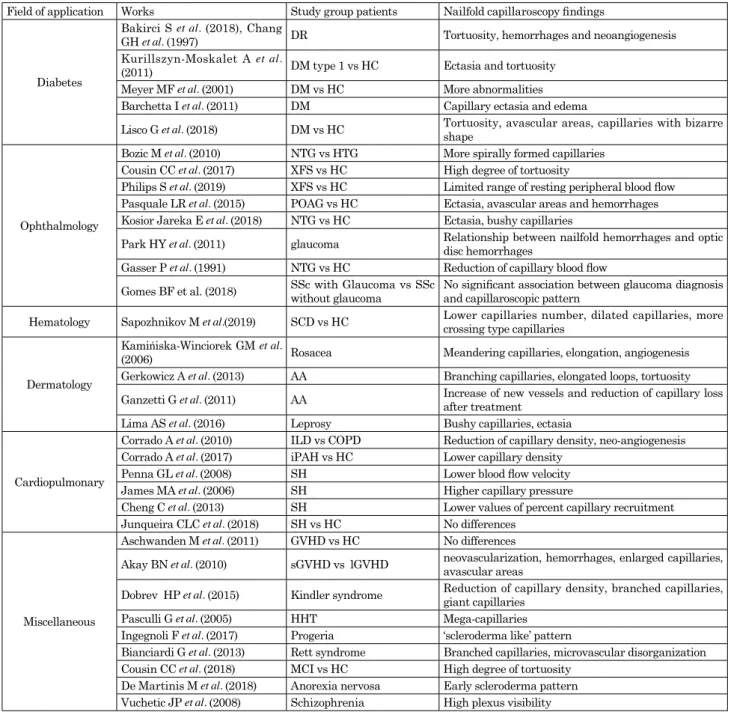
An extensive research was performed using PUBMED and GOOGLE SCHOLAR databases to identify studies. All studied published between 1990 and 2019 were included. The following “Key words” were used : “nailfold videocapillaroscopy” ; “micro-circulation” ; “microvascular damage”. At this stage we consid-ered the pathologies mentioned in more than one paper, and we directed literature research adding the most common pa-thologies to the keywords previous mentioned. We also included in our research the studies regarding rare pathologies, even if we found one single paper. For each key word we added : “and diabetes”, “and glaucoma”, “and sickle cell disease”, “and derma-tology disease”, “and interstitial lung disease”, “and pulmonary arterial hypertension”, “and hereditary disorders”, “and Alzhei-mer disease”, “and psychiatric diseases”. The research included studies covering all the NRD in which nailfold capillaroscopy was performed. Manual searching of references from potentially relevant articles was undertaken in order to include additional studies. Conference abstracts and texts not in English were ex-cluded. In this revision were identified 41 papers that respected the inclusion and exclusion criteria selected. These were divided into different groups belonging to a specific medical branch or pathology, as diabetes, glaucoma, sickle cell disease, dermatol-ogy, cardiopulmonary, miscellaneous. In table 1 are described NVC abnormal findings in NRD in study and in SSc. Table 2 summarized all NVC findings in the key papers included in this review, relating to NVC fields of application.
NAILFOLD CAPILLAROSCOPY IN DIABETES
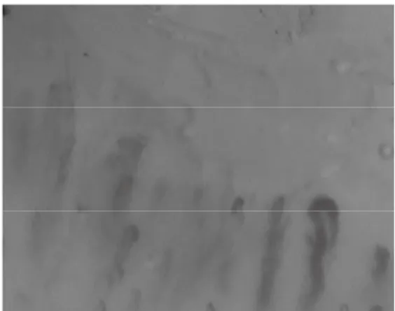
Diabetes mellitus (DM) is a chronic endocrinopathy, defined as hyperglycemic condition, complicated by microvascular struc-tural changes, as diabetic retinopathy (DR), nephropathy and neuropathy. These are worldwide recognized as the leading caus-es of mortality and morbidity. DR is the most common microvas-cular complication of DM, and it is considered the most frequent cause of preventable blindness. Endothelial dysfunction plays an important role in the development of microvascular damage in the DM earliest stage. Many diagnostic tools are used to assess microvascular damage, but the NVC is not widely used for this purpose. Bakirci S et al. performed NVC in 64 patients with DM type 2 to investigate whether the use of NVC could predict the outcome of DR as microvascular complication. No findings reached statistical significance, when patients with and without retinopathy were compared in term of NVC parameters, even if the rate of patients with tortuous capillaries, bleeding area, dilated and giant capillaries, and neo-angiogenesis was higher in the DR-positive group (23). Kuryliszyn-Moskal A et al. found that patients with type 1 DM, compared with healthy subjects, showed higher number of dilated and tortuous capillaries, and increased capillary density. Furthermore, patients with poor metabolic control had more capillaroscopic abnormalities (24). Figure 1. Nailbed hemorrhages (150x magnification ; HORUSvideodermatoscope)
Figure 2. Enlarged capillary (150x magnification ; HORUS videodermatoscope)
Figure 3. Tortuous capillaries (150x magnification ; HORUS videodermatoscope)
Table 1. Main nailfold capillaroscopic findings in most common diseases.
SSc diabetes glaucoma dermatology SCD ILD i-PAH AD RS
Tortuosity V V V V - - - V -Elongation - - - V - - - - -Ectasia V V V - V - V - V Hemorrhages V V V - - - -Avascular area V V V - V V - - -Angiogenesis V V - V - V - - V Giant capillaries V V - - -
-AD : Alzheimer disease ; ILD : interstitial lung disease ; i-PAH : idiopatic pulmonary hypertension ; RS : Rett syndrome ; SCD : sickle cell disease ; SSc : Systemic sclerosis ; V : yes.
Table 2. Common findings in nailfold-videocapillaroscopy in different fields of application.
Field of application Works Study group patients Nailfold capillaroscopy findings
Diabetes
Bakirci S et al. (2018), Chang
GH et al. (1997) DR Tortuosity, hemorrhages and neoangiogenesis Kurillszyn-Moskalet A et al.
(2011) DM type 1 vs HC Ectasia and tortuosity Meyer MF et al. (2001) DM vs HC More abnormalities Barchetta I et al. (2011) DM Capillary ectasia and edema
Lisco G et al. (2018) DM vs HC Tortuosity, avascular areas, capillaries with bizarre shape
Ophthalmology
Bozic M et al. (2010) NTG vs HTG More spirally formed capillaries Cousin CC et al. (2017) XFS vs HC High degree of tortuosity
Philips S et al. (2019) XFS vs HC Limited range of resting peripheral blood flow Pasquale LR et al. (2015) POAG vs HC Ectasia, avascular areas and hemorrhages Kosior Jareka E et al. (2018) NTG vs HC Ectasia, bushy capillaries
Park HY et al. (2011) glaucoma Relationship between nailfold hemorrhages and optic disc hemorrhages Gasser P et al. (1991) NTG vs HC Reduction of capillary blood flow
Gomes BF et al. (2018) SSc with Glaucoma vs SSc without glaucoma No significant association between glaucoma diagnosis and capillaroscopic pattern Hematology Sapozhnikov M et al.(2019) SCD vs HC Lower capillaries number, dilated capillaries, more crossing type capillaries
Dermatology
Kamińiska-Winciorek GM et al.
(2006) Rosacea Meandering capillaries, elongation, angiogenesis Gerkowicz A et al. (2013) AA Branching capillaries, elongated loops, tortuosity Ganzetti G et al. (2011) AA Increase of new vessels and reduction of capillary loss after treatment Lima AS et al. (2016) Leprosy Bushy capillaries, ectasia
Cardiopulmonary
Corrado A et al. (2010) ILD vs COPD Reduction of capillary density, neo-angiogenesis Corrado A et al. (2017) iPAH vs HC Lower capillary density
Penna GL et al. (2008) SH Lower blood flow velocity James MA et al. (2006) SH Higher capillary pressure
Cheng C et al. (2013) SH Lower values of percent capillary recruitment Junqueira CLC et al. (2018) SH vs HC No differences
Miscellaneous
Aschwanden M et al. (2011) GVHD vs HC No differences
Akay BN et al. (2010) sGVHD vs lGVHD neovascularization, hemorrhages, enlarged capillaries, avascular areas Dobrev HP et al. (2015) Kindler syndrome Reduction of capillary density, branched capillaries, giant capillaries Pasculli G et al. (2005) HHT Mega-capillaries
Ingegnoli F et al. (2017) Progeria ‘scleroderma like’ pattern
Bianciardi G et al. (2013) Rett syndrome Branched capillaries, microvascular disorganization Cousin CC et al. (2018) MCI vs HC High degree of tortuosity
De Martinis M et al. (2018) Anorexia nervosa Early scleroderma pattern Vuchetic JP et al. (2008) Schizophrenia High plexus visibility
AA : alopecia areata ; COPD : chronic obstructive pulmonary disease ; DM : diabetes mellitus ; DR : diabetic retinopathy, GHDV : graft versus host disease ; lGVHD : lichenoid graft versus host disease ; sGHDV : sclerodermoid graft versus host disease ; HC : healthy con-trol ; HHT : hereditary hemorrhagic telangiectasia ; SH : systemic hypertension ; ILD : interstitial lung disease ; iPAH : idiopathic pul-monary arterial hypertension ; MCI : mild cognitive impairment ; NVC : nailfold videocapillaroscopy ; SCD : sickle cell disease.
Meyer MF et al. put in relationship capillaroscopic changes (as increased diameter, tortuosity, reduction in capillary number) in patients with DM (25). Barchetta et al. correlated microangio-pathic lesions in the retinal vessels and changes in NVC finding, in patients with type 1 DM and those with type 2 DM, a healthy control group (HC) was included. They reported that patients with DM had capillary ectasia and nailfold edema compared to HC (26). In another study, the relation between microangiopathy, evaluated by ophthalmoscopy, and the presence of bushy capil-laries and avascular areas at NVC was observed in DM patients (27). Chang CH et al. reported a correlation between DR and capillary abnormalities as tortuous capillaries, bushy capillaries and capillary enlargement ; moreover these capillary changes were more common in patients with severe DM (28). Lisco G et al., in their cross sectional and single-center study, examined by means of the NVC the prevalence of capillaroscopic patterns in both type 1 and type 2 DM ; in addiction they assessed the relationship between NVC abnormalities, level of glycemic con-trol, and the presence of DM complication or DM comorbidities. In this study, NVC alterations, including tortuosity, ectasias, avascular areas, and capillaries with bizarre shapes were found to be more prevalent in DM patients than in HC. No additional differences in NVC abnormalities (such as bleeding, visibility of venous subpapillary plexus, and rarefaction areas) were found between diabetics and HC. Composite NVC patterns, defined as the presence of at least two DM nailfold alterations, were observed in both groups of patients with microvascular complica-tion (such as retinopathy), and those with DM comorbidity (such as hypertension, dyslipidemia, and carotid atherosclerosis). All these findings suggest the existence of a “diabetic capillaropa-thy”, as described in several studies (26, 29, 30), suggesting a possible use of NVC to detect and predict DM related microvas-cular complications (31).
NAILFOLD CAPILLAROSCOPY IN GLAUCOMA
Glaucoma is a severe ocular disease that causes progressive visual field loss due to the damage of ganglion retinal cells. The most common type of glaucoma is the primary open angle glau-coma (POAG), categorized in high-tension glauglau-coma (HTG) and normal-tension glaucoma (NTG) (32). Bozic M et al., conducted a pilot study on the utility of NVC in NTG patients and HTG patients. Ophthalmologic examination and NVC were performed on 30 NTG patients and 30 HTG patients. NVC parameters evaluated in the study were capillary row density, capillary diameter, number of spiralized capillaries, permeability of the loops, and loop resistance. The number of spiraled capillaries was more intensively found in NTG patients. According to this finding, the authors hypothesized a possible role of microvascu-lar damage in patients with glaucoma, especially in cases where intraocular pression was not directly involved in nerve damage (33). Exfoliating syndrome (XFS) consists of production and deposition of fibrillar granular materials that may produce ele-vate intraocular pressure (IOP), optic nerve damage, and glau-coma. Cousin CC et al. studied nailfold capillaries morphology in XFS, founding a higher degree of tortuosity in XFS patients than in HC, although the clinical consequences in ocular micro-vasculature is unclear (34).Philips S et al., in their cross-sectional and clinic-based study, performed NVC in 111 participants, 30 with NTG, 30 with HTG, 30 with XFS and 21 HC. The aim of their study was to examine peripheral resting blood flow in patients with glauco-ma, observing plasma gap blood movement in capillaries. They found a lower and limited range of resting peripheral blood flow in patients with XFS compared to HC. This limited range may
represent stiffened vasculature or impaired maximal flow in peripheral vasculature of XFS patients (35). Pasquale LR et al., in their multicentric study, enrolled 199 patients with POAG and 124 HC. The NVC was performed on the fourth and the fifth finger of each subject’s non dominant hand. The presence of hemorrhages, dilated capillary loops, and avascular areas were evaluated by NVC ; data on demographic factors, family history of glaucoma, systemic disease and related were collected. They found that all capillary abnormalities, as dilated capillary loops, avascular zones, and hemorrhages were associated with POAG, but the number of this abnormalities was not associated with the severity of the disease. Just a positive correlation between the hemorrhages number and visual field loss severity was observed. These data support the hypothesis of extra-ocular cap-illary involvements in POAG patients (36). Kosior-Jarecka E et al. focused their attention on NTG patients. NVC was performed in 80 NTG patients and in 2 HC groups (58 young healthy and 50 age-matched volunteers). They observed that abnormal NVC pattern was more common in NTG than in young controls. Mi-cro-bleedings were the most common findings in NTG patients. Dilated capillary loops and branching capillaries were more fre-quent in NTG patients than in age-matched HC. They also found an association between NVC abnormalities and disc hemorrhag-es and paracentral scotoma in glaucoma (37). Previously, Park HY et al. suggested that nailfold hemorrhages could be a marker of optic disc hemorrhages in patients with glaucoma. Further-more, disc hemorrhages were also associated with nailfold capillaries loss, even if the association was stronger with nailfold hemorrhages. No more differences were found in NTG patients and POAG (38). Gasser P et al. evaluated blood flow velocity, a dynamic parameter, in 30 NTG patients, 30 HTG patients and 30 HC. Capillary blood flow velocity was significantly reduced in NTG patients. After cold provocation, 25 of 30 NTG patients, had a measurable blood standstill of more than 12 second, while only three of 30 HC had a measurable blood standstill. No morphological parameters showed significantly differences between the studied groups (39). These findings suggest the role of microvascular abnormalities in NTG patients, that could play an independent role in the pathogenesis of the disease. An interesting study, conducted by Gomes BF et al., investigated the possible association between glaucoma and NVC pattern in 30 SSc patients. Up to 23% of SSc patients had glaucoma. No dif-ferences in NVC patterns were found between patients with and without glaucoma. Furthermore, no differences regarding intra-ocular pressure were found in patients with different severity of NVC patterns. The presence of glaucoma did not interfere with NVC pattern. The high prevalence of glaucoma in SSc patients, may suggest a possible role of systemic vascular damage in the pathogenesis of the disease (40).
NAILFOLD CAPILLAROSCOPIC FINDING IN
HEMA-TOLOGICAL DISEASE : SICKLE CELL DISEASE
Sickle cell disease (SCD) is a disorder characterized by repet-itive vaso-occlusive crises causing microvascular obstruction, tissue ischemia and pain, with consequent chronic multi-organ ischemic sequelae. Sapozhnikov M et al. performed NVC in 71 SCD patients and 70 age matched HC. Capillary number was lower and the final capillary score (measure of capillary dropout inversely related to capillary density) was higher in the SCD group compared to HC. SCD group had a higher percentage of crossing type capillaries. On multivariate linear analyses, final capillary score was independently associated with SCD after ad-justing for age, body mass index, and gender. Furthermore, SCD was associated with more dilated capillaries and lower capillary
density, but the similar number of hemorrhages. These changes appear unrelated to disease severity, frequency of sickle crises, and number of transfusions (41).
NAILFOLD CAPILLAROSCOPY IN
DERMATOLO-GY : ROSACEA, ALOPECIA AERATA, AND LEPROSY
Rosacea is an inflammatory dermatosis with a reported prev-alence of at least 10% in Caucasian adults. It affects facial skin causing facial erythema and / or papulopustular skin lesions. Kamińiska-Winciorek GM et al. examined microcirculation by NVC in 16 female patients with rosacea. In this study, NVC ab-normalities were found in all patients, in particular, meandering capillaries, elongations, and an increased number of capillaries were described. In the rosacea group, abnormalities of the color of the visual area were found in 14 cases (88%) with indistinct field of vision in 3 cases (19%). This result may suggest a possible use of NVC as diagnostic tool for the study of microvasculature architecture in rosacea (42).Gerkowicz A et al. performed NVC in patients with alopecia areata (AA), a T-cell mediate autoimmune disease that leads to partial or total hair loss of scalp and body hair. Other AA clinical features include nail changes. Tortuous loops were the most common findings in patients with AA. In these patients, single tortuous loop was more frequent than multiple tortuous loops. Branching capillaries and features of neovascularization were frequently observed. None of the patients had an extremely elongated loop. These results confirmed the presence of two types of NVC images in patients with AA : in the first type, there were no abnormalities ; in the second type there were anomalies such as branching capillaries, dilated loops, and tortuosity. No differences among patients with different type of AA were found. Nail changes were not related with NVC findings. NVC abnormalities in these patients suggest a possible role of skin microcirculation dysfunction in patients with AA (43). Ganzetti G et al. performed NVC before and after dyphenilcycloprope-none treatment in AA patient. At 24 weeks, it was observed an increase of new vessels and a significant hair regrowth in the areas with more marked angiogenesis. A possible role of nailfold capillaroscopy in the follow up of alopecia before and after treat-ment it was suggested (44).
A study about NVC alterations in patients with Hansen’s bacilli was carried out in order to investigate abnormalities in patients with leprosy and their relationship with clinical parameters. In this research, 60% of patients had some NVC abnormalities, such as micro-hemorrhages, dilated, bushy and corkscrew capillaries, although these changes were not specific for the disease manifestations. However, a large number of evi-dences marked the role of the interaction of Hansen’s bacilli with endothelium in pathogenesis of the disease (45).
NAILFOLD CAPILLAROSCOPIC FINDINGS IN
CAR-DIOPULMONARY DISEASE
The role of NVC in the identification of SSc patients with an higher risk of the organ involvements, is well known and studied (19, 46, 47). Cardiopulmonary diseases, such as interstitial lung disease (ILD) and pulmonary arterial hypertension (PAH) are heterogeneous pathological conditions, frequently associated with SSc, although the idiopathic form is more severe and life-treating. NVC is a simple and non-invasive tool that allows the early detection of local microvascular changes in CTD ; so, it was used to evaluate the possible presence of different NVC patterns among patients with cardiopulmonary diseases, both
idiopathic form and CTD related form. Different studies state the comparison between capillaroscopic findings in SSc patients with and without ILD (48, 49), suggesting a possible role of NVC in the early detection of lung involvement in CTD (50).
Patients with ILD often present lower capillary density and higher quantity of bushed / ramified capillaries (51, 52), while the presence of giant capillaries may be associated with reduced capacity of the lung for carbon monoxide (53). These data may support the hypothesis of a prognostic role for NVC, and a ILD physiopathology mechanism based on vascular damage (52). Corrado A et al. compared NVC findings in patients with idio-pathic ILD and patients with SSc related disease (SSc-ILD). In this study, NVC was performed on 23 SSc-ILD, 20 patients with idiopathic ILD and 22 patients with chronic obstructive pulmonary disease to evidence the abnormalities among the groups. For each patient were evaluated : density of capillaries, capillary width, capillary length, presence of microhemorrhages and angiogenetic aspect, such as branched capillaries and avas-cular areas. The main findings of the study were the detection of minor capillaroscopic alterations in patients with idiopathic ILD compared to SSc-ILD patients. The density of capillaries was significant reduced, and neo-angiogenetic aspects were more diffused in idiopathic ILD patients than in patients with chronic obstructive pulmonary disease, matched for age, arterial oxygen saturation, and diffusing capacity of the lung for carbon monox-ide (DLCO) values. These findings confirm the role of vascular damage in the ILD pathogenesis and put the accent on the role of NVC as a useful tool to diagnose CTD in patients that have lung’s involvement as the onset manifestation (54).
As regards the patients with systemic hypertension (SH), most of the changes in peripheral resistance affect the microvascular network, and the functional characteristics of arterioles. Micro-vascular rarefaction has also been described in the early stages of SH. These findings had led to hypothesize that the microvas-cular network is not only one of the putative factors responsible for increased pressure but is also a key target of SH (55). To evaluate if capillary rarefaction persists despite treatment with angiotensin converting enzyme inhibitors, thiazide diuretics and / or beta-blockers, Penna GL et al. performed NVC in 28 well-controlled essential SH patients and 19 normotensive sub-jects, evaluating the functional capillary densities at baseline, during post-occlusive hyperemia and after venous congestion. SH patients showed lower mean functional capillary density at baseline, during post-occlusive reactive hyperemia, and during venous congestion responses. Mean capillary diameters were not different in SH group, but their red blood cell velocity at baseline was significantly lower. Regardless of the type of therapy used, SH patients showed microvascular abnormalities that reflect the increased of vascular resistance, a clinical feature of SH (56). It has not been fully clarified the difference between a common microcirculation aging process and micro-vessels abnormalities induced by SH in the elderly. James MA et al. studied capillary pressure, density, and skin microvascular function in elderly subjects (aged > 60 years) with untreated hypertension, elder-ly normotensive subjects, and young normotensive subjects. Capillary pressure was higher in both elderly groups compared with young normotensives, but capillary density did not differ among the three groups (57). Even Cheng C et al. evaluated the role of microvascular abnormalities in the pathogenesis of SH, validating the NVC, comparing its findings with forearm blood flow, a well-established measure of vascular function. In their study, typical values for capillary counts (capillaries / mm2) have been 55-80 for baseline, 65-90 for post-ischemic, and 90-105 for venous occlusion. Values for percent capillary recruitment, a new parameter introduced by Cheng C. et al. in order to en-able direct comparison of the total number of actively perfused
(functional) capillaries between individuals, were lower among hypertensive than normotensive patients (58). Junqueira CLC et al. evaluated microcirculation by NVC in patients with resistant, or mild-to-moderate hypertension compared to normotensive patients. Functional capillary density (the number of capillaries with flowing red blood cells by unit tissue area) and capillary diameters showed no significant differences among the three groups (59).
PAH is a group of diseases characterized by elevated pul-monary arterial resistance leading to right heart failure. Both idiopathic form and secondary form (usually related to CTD and SSc) are described. Corrado A et al. in a recent study, com-pared capillaroscopic abnormalities in patients with idiopathic PAH and those with PAH secondary to SSc (SSc-PAH). In this study the presence of NVC anomalies were found in 38% of id-iopathic PAH subjects, in particular, the capillary density was significantly lower in patients with idiopathic PAH than in HC. Furthermore, idiopathic PAH patients had a higher capillary width than HC. A more severe microangiopathy, characterized by higher rate of active / late scleroderma pattern, lower capillary density, and neo-angiogenesis, was found in SSc-PAH patients compared to SSc patients without PAH. These findings confirm that PAH patients (both idiopathic and SSc related forms) had more aggressive micro-vessels involvements and support the hypothesis of systemic microvascular involvement in idiopathic PAH which can be detected by NVC, although it is not clear if the systemic microcirculation anomalies are causal or consequential to PAH disease (60).
NAILFOLD CAPILLAROSCOPY IN MISCELLANEOUS
DISEASES : FROM HEREDITARY DISORDERS TO
ALZHEIMER DISEASE
As useful and feasible diagnostic tool, NVC was performed in different diseases, in order to reach new knowledges in the pathogenesis, clinical involvements and treatment efficacy.
Aschwanden M et al. performed NVC in 11 patients with sclerodermoid graft-versus-host disease. All these patients received peripheral blood stem cells to treat their hematologic diseases. The role of vascular pathology in chronic graft versus host disease is less well understood. In this study nine patients showed a good visibility of the capillaries, with a uniform pat-tern. Rare hemorrhages were seen in three patients and just a patient showed enlarged capillaries (61). These results countered with those of Akay’s study about NVC in patients with graft versus host disease. In the latter study severe capillaries abnor-malities were found, probably due to the presence of RP in 17% of the patients enrolled (62).
Kindler syndrome (KS) is a type of hereditary epidermolysis bullosa characterized by skin fragility, photosensitivity and progressive poikilodermatous changes. Dobrev HP et al. reported the case of a 54-year old woman with KS. Pronounced alter-ations in finger capillaries, such as reduction in capillary density, neo-angiogenesis, branching capillaries, enlarged and giant capillaries was observed at NVC. These changes may be due to a mechanism that compensate the loss of capillaries caused by chronic finger trauma (63).
In hereditary hemorrhagic telangiectasia (HHT), a domi-nantly inherited disorder, cutaneous telangiectasias have been studied by skin biopsy and recently by NVC. Pasculli G et al. per-formed NVC in 88 patients with HHT : pseudo-mega-capillaries and mega-capillaries were found in six patients (7%) ; the other 82 patients, as well as HC, had normal NVC patterns (64).
Ingegnoli F et al. showed a case of scleroderma like pattern in adult progeria (Werner syndrome). This patient was referred
to a Rheumatology Division for the suspicion of SSc because he suffered from large and deep ulcers on both legs. NVC showed a scleroderma like pattern and the genetic tests diagnosed Werner Syndrome. This clinical case demonstrates the possibility of NVC anomalies, such as dilated capillaries and tortuosity, in patient with a rare genetic disorder, suggesting microvascular damage as key point in the pathogenesis of the disease. To the best of our knowledge, this is the first report describing the NVC abnormalities in Werner syndrome (65).
Bianciardi et al. performed NVC in patients with Rett Syn-drome, a neurobiological post-natal disease that represents the second common cause of mental retardation. Its non-neurological phenotype is characterized by cold and blue hands and feet : in these subjects NVC showed branched capillaries, dilated capil-laries, and microarchitecture disorganization. These findings indicate the presence of previously unrecognized microvascular abnormalities in Rett syndrome (66).
Cerebrovascular disease (CVD) is highly associated with Alzheimer’s disease (AD), but its role is not entirely understood. Cousin CC et al. studied NVC abnormalities in patients with AD and mild cognitive impairment in comparison to patients with normal cognition. In 56% of AD patients NVC showed an in-crease of capillary tortuosity, suggesting a role of microvascular changes in the pathogenesis of AD (67).
Few studies explored the role of microvascular changes in patients with psychiatric diseases such as schizophrenia and anorexia nervosa. RP has been described in anorexia nervosa with evidence of three different NVC patterns : normal, non-spe-cific and early scleroderma pattern. This results underlined a correlation between RP in patients with anorexia nervosa and early scleroderma pattern at NVC, suggesting that patients with RP and anorexia have NVC pattern similar to patients suffering from CTD (68).
Vuchetich JP et al. performed NVC in patients with schizo-phrenia evaluating the nail plexus visibility. This study revealed that patients with a higher plexus visibility had significantly more negative symptoms and poorer social functioning (69).
SUMMARY
Endothelial dysfunction plays an important role in the patho-genesis of microvascular complications in several diseases, such as diabetes mellitus, pulmonary disease (ILD and PAH), hema-tological diseases and CTD.
NVC is a safe and useful investigational tool that could assess an early detection of endothelial dysfunctions, allowing a quali-tative description of the microvascular abnormalities. The most common diagnostic use of NVC is in the differentiation between primary and secondary RP. Nowadays, well recognized and standardized patterns (early, active, and late scleroderma pat-terns) are established in SSc patients (2). Although understud-ied, NVC abnormal findings, without specific pattern, could be related to clinical parameters in various pathologies. In diabetes mellitus, NVC could be used to early detect organ involvements and microvascular complications, to improve and potentiate the treatment management.
In glaucoma, sickle cell disease, Werner syndrome, and Rett syndrome, NVC seems to discover new etiopathogenetic path-ways. In particular, in the Werner syndrome an association between scleroderma like pattern and the presence of deep ulcers are described, suggesting a severe microangiopathy (50).
In idiopathic ILD, idiopathic PAH, and SH the improvement of NVC findings could be useful to assess treatment efficacy.
Even though the NVC finding of tortuosity, elongation, ecta-sia, and hemorrhages could be common, but not standardized,
their presence in association with systemic disease could suggest an early endothelial dysfunction worthy of depth clinical assess-ment and therapeutic enhanceassess-ment.
At the time, we need more studies to open new scenarios of NVC technique’s application in NRD.
FUNDING
The study had no financial support.
COMPETING INTEREST
NoneDISCLOSURE STATEMENT
N. Mansueto had no disclosures C Rotondo had no disclosures A Corrado had no disclosures FP Cantatore had no disclosuresREFERENCES
1. Cutolo M, Paolino S, Smith V : Nailfold capillaroscopy in rheumatology : ready for the daily use but with care in ter-minology. Clin Rheumatol 38 (9) : 2293-2297, 2019
2. Ingegnoli F, Smith V, Sulli A, Cutolo M : Capillaroscopy in Routine Diagnostics : Potentials and Limitations. Curr Rheumatol Rev 14(1) : 5-11, 2018
3. Cantatore FP, Corrado A, Covelli M, Lapadula G : Mor-phologic study of the microcirculation in connective tissue diseases. Ann Ital Med Int 15(4) : 273-81, 2000
4. Ocampo-Garza SS, Villarreal-Alarcón MA, Villarreal-Treviño AV, Ocampo-Candiani J : Capillaroscopy : A Valuable
Diag-nostic Tool. Actas Dermosifiliogr 110(5) : 347-352, 2019 5. Hughes M, Herrick AL : Raynaud’s Phenomenon. Best
Pract Res Clin Rheumatol 30(1) : 112-132, 2016
6. Grattagliano V, Iannone F, Praino E, De Zio A, Riccardi MT, Carrozzo N, Maggi P, Lapadula G : Digital laser dop-pler flowmetry may discriminate “limited” from “diffuse” systemic sclerosis. Microvasc Res 80(2) : 221-6, 2010 7. Ruaro B, Sulli A, Smith V, Pizzorni C, Paolino S, Alessandri
E, Cutolo M : Microvascular damage evaluation in systemic sclerosis : the role of nailfold videocapillaroscopy and laser techniques. Reumatismo 69(4) : 147-155, 2017
8. Ruaro B, Smith V, Sulli A, Pizzorni C, Tardito S, Patanè M, Paolino S, Cutolo M : Innovations in the Assessment of Primary and Secondary Raynaud’s Phenomenon. Front Pharmacol April 16, 2019
9. Caramaschi P, Volpe A, Pieropan S, Tinazzi I, Mahamid H, Bambara LM, Biasi D : Cyclophosphamide treatment improves microvessel damage in systemic sclerosis. Clin Rheumatol 28(4) : 391-5, 2009
10. Guiducci S, Bellando Randone S, Bruni C, Carnesecchi G, Maresta A, Iannone F, Lapadula G, Matucci Cerinic M : Bosentan fosters microvascular de-remodelling in sys-temic sclerosis. Clin Rheumatol 31(12) : 1723-5, 2012 11. Cutolo M, Zampogna G, Vremis L, Smith V, Pizzorni C,
Sulli A : Longterm effects of endothelin receptor antag-onism on microvascular damage evaluated by nailfold capillaroscopic analysis in systemic sclerosis. J Rheumatol 40(1) : 40-5, 2013
12. Grassi W, De Angelis R : Capillaroscopy : questions and answers. Clin Rheumatol 26(12) : 2009, 2007
13. Sulli A, Ruaro B, Smith V, Pizzorni C, Zampogna G, Gallo M, Cutolo M : Progression of nailfold microvascular damage and antinuclear antibody pattern in systemic sclerosis. J Rheumatol 40 : 634-9, 2013
14. Cutolo M, Soldano S, Smith V : Pathophysiology of systemic sclerosis : current understanding and new insights. Expert Rev Clin Immunol 15 : 753-64, 2019
15. Bruni C, Frech T, Manetti M, Rossi FW, Furst DE, De Paulis A, Rivellese F, Guiducci S, Matucci-Cerinic M, Bellando-Randone S : Vascular Leaking, a Pivotal and Early Pathogenetic Event in Systemic Sclerosis : Should the door be closed? Front Immunol 9 : 2045-8, 2018
16. Cutolo M, Sulli A, Secchi ME, Olivieri M, Pizzorni C : The Contribution of Capillaroscopy to the Differential Diagnosis of Connective Autoimmune Diseases. Best Pract Res Clin Rheumatol 21(6) : 1093-108, 2007
17. Cortes S, Cutolo M : Capillaroscopic patterns in rheumatic diseases. Acta Reumatologica Portuguesa 32 : 29-36, 2007 18. Beyer C, Schett G, Gay S, Distler O, Distler JH : Hypoxia
in the pathogenesis of systemic sclerosis. Arthritis Res Ther 11 : 220, 2009
19. Pizzorni C, Sulli A, Paolino S, Ruaro B, Smith V, Trombetta AC, Cutolo M : Progression of Organ Involvement in Sys-temic Sclerosis Patients with Persistent “Late” Nailfold Capillaroscopic Pattern of Microangiopathy : A Prospective Study. J Rheumatol 44(12) : 1941-1942, 2017
20. Jones BF, Oral M, Morris CW, Ring EFJ : A proposed taxon-omy for nailfold capillaries based on their morphology. IEEE Transactions on Medical Imaging 20(4) : 333-341, 2001 21. Lambova SN, Muller Ladnen U : Capillaroscopic Pattern in
Inflammatory Arthritis. Microvasc Res 83(3) : 318-22, 2012 22. Cutolo M, Sulli A, Smith V : How to perform and interpret capillaroscopy. Best Pract Res Clin Rheumatol 27(2) : 237-248, 2013
23. Bakirci S, Celik E, Acikgoz SB, Erturk Z, Tocoglu AG, Imga NN, Kaya M, Tamer A : The evaluation of nailfold vid-eocapillaroscopy findings in patients with type 2 diabetes with and without diabetic retinopathy. North Clin Istanb 6(2) : 146-150, 2018
24. Kuryliszyn-Moskal A, Dubicki A, Zarzycki W, Zonnenberg A, Górska M : Microvascular abnormalities in capillarosco-py correlate with higher serum IL-18 and sE-selectin levels in patients with type 1 diabetes complicated by microangi-opathy. Folia Histochem Cytobiol 49(1) : 104-10, 2011 25. Meyer MF, Pfohl M, Schatz H : Assessment of diabetic
al-terations of microcirculation by means of capillaroscopy and laser-Doppler anemometry. Med Klin (Munich) 96(2) : 71-7, 2001
26. Barchetta I, Riccieri V, Vasile M, Stefanantoni K, Comberiati P, Taverniti L, Cavallo MG : High prevalence of capillary abnormalities in patients with diabetes and association with retinopathy. Diabet Med 28(9) : 1039-44, 2011
27. Maldonado G, Guerrero R, Paredes C, Ríos C : Nailfold cap-illaroscopy in diabetes mellitus. Microvasc Res 112 : 41-46, 2017
28. Chang CH, Tsai RK, Wu WC, Kuo SL, Yu HS : Use of dynamic capillaroscopy for studying cutaneous microcir-culation in patients with diabetes mellitus. Microvasc Res 53(2) : 121-7, 1997
29. Cicco G, Cicco S : Hemorheological aspects in the microvas-culature of several pathologies. Adv Exp Med Biol 599 : 7-15, 2007
30. Uyar S, Balkarli A, Erol MK, Yesil B, Tokuç A, Durmaz D, Görar S, Çekin AH : Assessment of the Relationship
between Diabetic Retinopathy and Nailfold Capillaries in Type 2 Diabetics with a Noninvasive Method : Nailfold Vid-eocapillaroscopy. J Diabetes Res 2016 : 7592402, 2016 31. Lisco G, Cicco G, Cignarelli A, Garruti G, Laviola L,
Giorgino F : Computerized Video-Capillaroscopy Alteration Related to Diabetes Mellitus and Its Complications. Adv Exp Med Biol 1072 : 363-368, 2018
32. Friedman DS, Wolfs RC, O’Colmain BJ, Klein BE, Taylor HR, West S, Leske MC, Mitchell P, Congdon N, Kempen J : Eye Diseases Prevalence Research. Prevalence of open-angle glaucoma among adults in the United States. Arch Ophthal-mol 122(4) : 532-8, 2004
33. Božić M, Senćanić PH, Spahić G, Kontić D, Marković V, Marjanović I, Stojkovic M, Dorđević-Jocić J : Is nail fold cap-illaroscopy useful in normotensive and primary open angle glaucoma? A pilot study. Curr Eye Res 35(12) : 1099-104, 2010
34. Cousins CC, Kang JH, Bovee C, Wang J, Greenstein SH, Turalba A, Shen LQ, Brauner S, Boumenna T, Blum S, Levkovitch-Verbin H, Ritch R, Wiggs JL, Knepper PA, Pasquale LR : Nailfold capillary morphology in exfoliation syndrome. Eye (Lond) 31(5): 698-707, 2017
35. Philip S, Najafi A, Tantraworasin A, Pasquale LR, Ritch R : Nailfold Capillaroscopy of Resting Peripheral Blood Flow in Exfoliation Glaucoma and Primary Open-Angle Glaucoma. JAMA Ophthalmol 137(6) : 618-625, 2019 36. Pasquale LR, Hanyuda A, Ren A, Giovingo M, Greenstein
SH, Cousins C, Patrianakos T, Tanna AP, Wanderling C, Norkett W, Wiggs JL, Green K, Kang JH, Knepper PA : Nailfold Capillary Abnormalities in Primary Open-An-gle Glaucoma : A Multisite Study. Invest Ophthalmol Vis Sci 56(12) : 7021-7028, 2015
37. Kosior-Jarecka E, Bartosińska J, Łukasik U, Wróbel-Dudzińska D, Krasowska D, Chodorowska G, Żarnowski T : Results of Nailfold Capillaroscopy in Patients with Normal-Tension Glaucoma. Curr Eye Res 43(6) : 747-753, 2018
38. Park HY, Park SH, Oh YS, Park CK : Nail bed hemor-rhage : a clinical marker of optic disc hemorhemor-rhage in patients with glaucoma. Arch Ophthalmol 129(10) : 1299-304, 2011 39. Gasser P, Flammer J : Blood-cell velocity in the nailfold
capillaries of patients with normal-tension and high-tension glaucoma. Am J Ophthalmol 111(5) : 585-8, 1991
40. Gomes BF, Souza R, Valadão T, Kara-Junior N, Moraes HV, Santhiago MR : Is there an association between glaucoma and capillaroscopy in patients with systemic sclerosis? Int Ophthalmol 38(1) : 251-256, 2018
41. Sapozhnikov M, Rehman M, Johnson C, Daich J, Salciccioli L, Gillette P, Lazar JM : Characterization of microvascular disease in patients with sickle cell disease using nailfold capillaroscopy. Microvasc Res 125 : 103877, 2019
42. Kamińiska-Winciorek GM, Brzezińska-Wcisło LA : Assess-ment of microcirculatory system with conventional capil-laroscopy in patients with rosacea--preliminary study. Wiad Lek 59(9-10) : 618-22, 2006
43. Gerkowicz A, Krasowska D, Pietrzak A, Michalak-Stoma A, Bartosińska J, Juszkiewicz-Borowiec M, Chodorowska G : Videocapillaroscopic alterations in alopecia areata. Biomed Res Int 2013 : 160203, 2013
44. Ganzetti G, Campanati A, Simonetti O, Cataldi I, Giuliodori K, Offidani AM : Videocapillaroscopic pattern of alopecia areata before and after diphenylciclopropenone treatment. Int J Immunopathol Pharmacol 24(4) : 1087-91, 2011 45. Lima AS, Pizzol VI, Fritsch S, Fonseca GP, Mulinari-Brenner
FA, Muller CS, Ottoboni VC : Nailfold capillaroscopy in leprosy. An Bras Dermatol 91(5) : 686-687, 2016
46. Markusse IM, Meijs J, de Boer B, Bakker JA, Schippers
HPC, Schouffoer AA, Ajmone Marsan N, Kroft LJM, Ninaber MK, Huizinga TWJ, de Vries-Bouwstra JK : Predicting cardiopulmonary involvement in patients with systemic sclerosis : complementary value of nailfold videocapillaroscopy patterns and disease-specific autoantibodies. Rheumatology (Oxford) 56(7) : 1081-1088, 2017
47. Cutolo M, Damjanov N, Ruaro B, Zekovic A, Smith V : Im-aging of connective tissue diseases : Beyond visceral organ imaging? Best Pract Res Clin Rheumatol 30(4) : 670-687, 2016
48. Smith V, Riccieri V, Pizzorni C, Decuman S, Deschepper E, Bonroy C, Sulli A, Piette Y, De Keyser F, Cutolo M : Nailfold capillaroscopy for prediction of novel futuresevere organ involvement in systemic sclerosis. J Rheumatol 40 : 2023-8, 2013
49. Wu W, Jordan S, Becker MO, Dobrota R, Maurer B, Fretheim H, Ye S, Siegert E, Allanore Y, Hoffmann-Vold AM, Distler O : Prediction of progression of interstitial lung disease in patients with systemic sclerosis : the SPAR model. Ann Rheum Dis 77 : 1326-32, 2018
50. Smith V, Distler O, Cutolo M : Might Nailfold Capillarosco-py Be a “Proxy” for Lung Involvement in Connective Tissue Diseases? J Rheumatol 46(9) : 1061-1063, 2019
51. Castellví I, Simeón-Aznar CP, Sarmiento M, Fortuna A, Mayos M, Geli C, Diaz-Torné C, Moya P, De Llobet JM, Casademont J : Association between nailfold capillaroscopy findings and pulmonary function tests in patients with sys-temic sclerosis. J Rheumatol 42(2) : 222-227, 2015
52. Caetano J, Paula FS, Amaral M, Oliveira S, Alves JD : Nailfold Videocapillaroscopy Changes Are Associated With the Presence and Severity of Systemic Sclerosis-Related Interstitial Lung Disease. J Clin Rheumatol 25(3) : e12-e15, 2019
53. Guillen-del-Castillo A, Simeòn-Aznar CP, Callejas-Moraga EL, Tolosa-Vilella C, Alonso-Vila S, Fonollosa-Pla V : Quan-titative Videocapillaroscopy Correlates With Functional Respiratory Parameters : A Clue for Vasculopathy as a Pathogenic Mechanism for Lung Injury in Systemic Sclero-sis. Arthritis Res Ther 20(1) : 281, 2018
54. Corrado A, Carpagnano GE, Gaudio A, Foschino-Barbaro MP, Cantatore FP : Nailfold capillaroscopic findings in sys-temic sclerosis related lung fibrosis and in idiopathic lung fibrosis. Joint Bone Spine 77(6) : 570-4, 2010
55. Vicaut E : Microcirculation and arterial hypertension. Drugs 59 : 1-10, 1999
56. Penna GL, Garbero Rde F, Neves MF, Oigman W, Bot-tino DA, Bouskela E : Treatment of essential hyperten-sion does not normalize capillary rarefaction. Clinics (Sao Paulo) 63(5) : 613-8, 2008
57. James MA, Tullett J, Hemsley AG, Shore AC : Effects of aging and hypertension on the microcirculation. Hypertension 47(5) : 968-74, 2006
58. Cheng C, Daskalakis C, Falkner B : Non-invasive assess-ment of microvascular and endothelial function. J Vis Exp 29(71) : e50008, 2013
59. Junqueira CLC, Magalhães MEC, Brandão AA, Ferreira E, Cyrino FZGA, Maranhão PA, Souza MDGC, Bottino DA, Bouskela E : Microcirculation and biomarkers in pa-tients with resistant or mild-to-moderate hypertension: a cross-sectional study. Hypertens Res 41(7) : 515-523, 2018 60. Corrado A, Correale M, Mansueto N, Monaco I, Carriero A,
Mele A, Colia R, Di Biase M, Cantatore FP : Nailfold capil-laroscopic changes in patients with idiopathic pulmonary arterial hypertension and systemic sclerosis-related pulmo-nary arterial hypertension. Microvasc Res 114 : 46-51, 2017 61. Aschwanden M, Halter JP, Walker UA, Staub D, Tichelli
A, Daikeler T, Jaeger KA, Tyndall A : Nail fold capillaros-copy differs widely between systemic sclerosis and chronic graft vs host disease of the skin. Rheumatology (Oxford) 50(6) : 1168-9, 2011
62. Akay BN, Sanli H, Topcuoglu P, Arat M, Akyol A : Nailfold capillary abnormalities are prevalent in sclerodermoid graft-versus-host disease and readily detected with derma-toscopy. Br J Dermatol 162(5) : 1076-82, 2010
63. Dobrev HP, Vutova NI : Nailfold capillaroscopic changes in Kindler syndrome. Intractable Rare Dis Res 4(4) : 214-6, 2015
64. Pasculli G, Quaranta D, Lenato GM, Suppressa P, Lastella P, Guanti G, Sabbà C : Capillaroscopy of the dorsal skin of the hands in hereditary hemorrhagic telangiectasia. QJM 98(10) : 757-63, 2005
65. Ingegnoli F, Crotti C : Nailfold scleroderma-like capillary abnormalities in Werner syndrome (adult progeria). Vasc Med 22(3) : 246-247, 2017
66. Bianciardi G, Acampa M, Lamberti I, Sartini S, Servi M, Biagi F, Bocchi V, Hayek J, Pastorelli M : Microvascular abnormalities in Rett syndrome. Clin Hemorheol Microcirc 54(1) : 109-13, 2013
67. Cousins CC, Alosco ML, Cousins HC, Chua A, Steinberg EG, Chapman KR, Bing-Canar H, Tripodis Y, Knepper PA, Stern RA, Pasquale LR : Nailfold Capillary Morphol-ogy in Alzheimer’s Disease Dementia. J Alzheimers Dis 66(2) : 601-611, 2018
68. De Martinis M, Sirufo MM, Ginaldi L : Raynaud’s phe-nomenon and nailfold capillaroscopic findings in anorexia nervosa. Curr Med Res Opin 34(3) : 547-550, 2018
69. Vuchetich JP, Liska JL, Dionisio DP, Stanwyck JJ, McGuire KA, Sponheim SR : Elevated nailfold plexus visibility ag-gregates in families and is associated with a specific neg-ative symptom pattern in schizophrenia. Psychiatry Res 160(1) : 30-7, 2008