Title Chronic ischemic proctitis: case report and review
Author(s) Alternative
Kishikawa, H; Nishida, J; Hirano, E; Nakano, M; Arakawa, K; Morishita, T; Kawashima, J; Koide, O; Tanaka, Y; Ishii, H
Journal Gastrointestinal endoscopy, 60(2): 304-308 URL http://hdl.handle.net/10130/1574
Chronic ischemic proctitis: case report and review
Hiroshi Kishikawa, M.D. 1) Jiro Nishida, M.D. 1) Erika Hirano, M.D. 1) Masaru Nakano, M.D.1) Koki Arakawa, M.D. 2) Tetsuo Morishita, M.D.2) Junko Kawashima, M.D.3) Osamu Koide, M.D.4) Yoichi Tanaka, M.D. 4) and Hiromasa Ishii, M.D. 5)
1)
Departments of Gastroenterology, 2)Internal Medicine, 3)Dermatology and 4)Clinical Laboratory, Tokyo Dental College Ichikawa General Hospital, Chiba, Japan
5)
Department of Internal Medicine, School of Medicine, Keio University, Tokyo, Japan KEY WORDS: chronic ischemic proctitis, venous occlusion, and rectal bleeding
Running title: Chronic ischemic proctitis
Address for correspondence and reprint requests: Hiroshi Kishikawa, M.D.
Department of Gastroenterology, Ichikawa General Hospital, Tokyo Dental College
5-11-13 Sugano Ichikawa
Chiba 272-8513, Japan
Phone, 47-322-0151 Fax, 47-325-4456 E-mai; ---
The rectum is spared in most patients with spontaneous ischemic colitis because of its abundant collateral blood supply, and thus ischemic proctitis is a rare clinical entity. In most reported cases, it has been of acute onset after aortoiliac surgery, radiotherapy, or other vascular intervention. This report describes the endoscopic diagnosis and the treatment of recurrent rectal hemorrhage in a patient with spontaneous chronic ischemic proctitis. Chronic ischemic proctitis should be included in the differential diagnosis of lower-GI bleeding, especially for elderly bed-ridden patients with atherosclerotic disease.
CASE REPORT
A 64-year-old woman was hospitalized because of severe anal pain and fever. There was a 2-week history of diarrhea and fecal incontinence. She had been undergoing hemodialysis for 5 years because of chronic renal failure secondary to diabetic nephropathy. There was no history of abdominal pain, nausea, radiation therapy, excessive straining during
defecation, or hematochezia. On examination, the patient appeared well nourished and had cushingoid features. Body temperature was 39.5℃. The perianal skin was swollen and tender. Laboratory test results included the following; Hb level, 6.5g/dL, (normal: 11.5-15 g/dL); leukocyte count, 15000 /μL; (3500-8500/μL); serum creatinine concentration, 8.5
mg/dL (0.47-0.79 mg/dL); blood urea nitrogen, 55 mg/dL (8-23 mg/dL); and C-reactive protein, 25 mg/dL (<0.5 mg/dL). A perianal abscess was diagnosed, and incision and drainage was performed, followed by administration of antibiotic therapy
dark-red stool. Thickening of the rectal wall with stranding of the pararectal fat (Fig. 1) was noted on CT. Sigmoidoscopy revealed a large circular ulcer in the distal rectum and protuberant vessels in a small mucosal defect adjacent to the circular rectal ulcer (Fig. 2A). Hemostasis was achieved by endoscopic hemoclip application, and there was no further bleeding for 1 month. Stool cultures grew no pathogens. Elective colonoscopy performed 2 weeks later revealed a large, circular rectal ulcer surrounded by slightly injected mucosa. Multiple ulcer scars and scattered round ulcers were observed continuously from the rectum to the sigmoid colon (Fig. 2B), but the mucosa proximal to the sigmoid colon was intact. A therapeutic trial of salazosulfapyridine (3 g per day) for 2 weeks was of no benefit. All oral intake was stopped and hyperalimentation was initiated. The hematochezia had decreased, but loose stools and fecal incontinence persisted without improvement. At 1 month after the start of hyperalimentaion, the patient complained of severe genital pain and noted passage of liquid stools via her vagina, suggesting the development of a rectovaginal fistula. Colonoscopy at 2 months after starting hyperalimentation revealed healing of the rectal ulcer, but there were several longitudinal ulcers in the ascending colon, with interspersed normal mucosa (Fig. 2C). The rectovaginal fistula was not visualized endoscopically. Biopsy specimens from an ulcer scar in the sigmoid colon revealed mucosal crypt atrophy and fibrosis of the lamina propia. There was no evidence of malignancy or mucosal prolapse syndrome. Surgical repair of the rectovaginal fistula by transverse colostomy and sphincteroplasty was scheduled, but after 3 months of
disseminated intravascular coagulopathy, and she died 1 month later.
At autopsy, the colonic mucosa appeared pale, and there were scattered circular ulcers from the rectum to the sigmoid colon. Longitudinal or patchy ulcers were observed throughout the ascending and descending colon and into the terminal ileum.
Histopathologically, there was focal loss of epithelial cells in the upper half of the sigmoid mucosa, a classic finding for ischemia. There also were histopathologic features of chronic ischemic change, including mucosal atrophy and fibrosis of the lamina propia (Fig. 3A). Thrombosis of the mesenteric veins was present from the rectosigmoid colon to the ascending colon, but no significant arterial lesions were identified (Fig. 3B). The clinical and pathological findings were those of chronic ischemic proctitis.
DISCUSSION
The splenic flexure and the descending colon, a watershed area in terms of blood supply
between the territories of the superior and inferior mesenteric arteries, are the most common sites of non-occlusive ischemic colitis.1 Conversely, because the rectum is supplied by an extensive arterial network formed by branches of the inferior mesenteric, internal iliac, and internal pudendal arteries, ischemic proctosigmoiditis and ischemic proctitis account for only about 5% of all cases of ischemic disease of the large bowel. In most reports, the onset of ischemic proctitis has been acute and secondary to aortoiliac surgery, radiotherapy, or vascular intervention.2, 3 The present case is a rare example of spontaneous, chronic ischemic proctitis with the classic histopathologic findings of superficial epithelial necrosis, and mucosal fibrosis.
Bharucha et al. 2 reported 10 cases of acute or chronic ischemic proctosigmoiditis. They defined chronic ischemic proctosigmoiditis as ischemic lesions limited to the rectosigmoid colon (within 30 cm of the dentate line), identified either endoscopically or surgically, that produce symptoms of at least 4 weeks duration.However, because it can be difficult to measure the exact length of the involved bowel because of stretching during colonoscopy, and because the duration of symptoms does not always correlate with episodes of ischemia, the term “chronic ischemic proctitis” is used by us for ischemic lesions that include the rectum (irrespective of the proximal extent) when there is histopathologic evidence of chronic ischemia. In the case presented, the typical histopathologic findings of chronic ischemia in the rectosigmoid colon, and the ischemic lesion detected during colonoscopy, made the diagnosis of chronic ischemic proctitis.
Devroede et al. 4 reported a series of 36 cases of chronic rectal ischemia. In that series, the diagnosis was based on histopathologic findings of mucosal atrophy and mucosal fibrosis, the proctoscopic findings, and a clinical history of fecal incontinence or anal pain. This diagnosis of chronicity seems justified, and the endoscopic finding of scattered whitish areas in the rectum was similar to the findings in our patient. However, because Devroede et al. 4 did not rule out the possibility of antecedent infection, the diagnosis of ischemic proctitis cannot be considered definitive, even though the clinical details of some of their cases were almost identical to those described here.
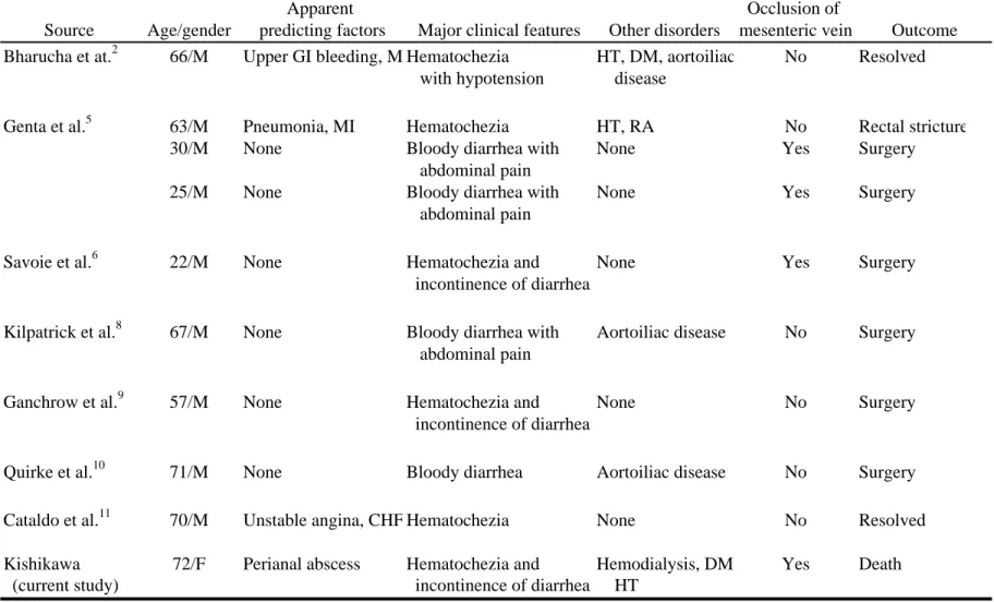
Reported cases of spontaneous chronic ischemic proctitis that meets our criteria for diagnosis are listed in Table 1.2, 5, 6, 8-11 All patients had one or more of the following
symptoms: diarrhea, fecal incontinence, abdominal pain, rectal pain, and bloody diarrhea. Idiopathic fecal incontinence and bloody diarrhea were the most common symptoms. The endoscopic findings of chronic ischemic proctitis are not well described. Atrophic mucosa with scattered white scars reflected repeated episodes of ischemia and healing. Chronic ischemic proctitis should be suspected whenever ulcer formation or erosion is observed endoscopically in the context of atrophic mucosa and multiple whitish scars in the rectosigmoid colon with mucosal crypt atrophy or fibrosis evident in biopsy specimen. CT findings in ischemic colitis are well documented, and although non-specific, they often suggested the diagnosis in the proper clinical setting.12 The CT findings in ischemic proctitis also are nonspecific. Wiesner et al. 12 suggested that ischemic proctitis should be considered when mural thickening confined to the rectum and sigmoid colon is associated with perirectal fat stranding, especially in elderly patients with known cardiovascular risk factors.
A review of published information suggests that the patients with ischemic proctitis fell into two groups: (1) those with atherosclerotic disease who have been hospitalized for some other disorder, and (2) those with mesenteric venous occlusion.
Six of the 10 reported patients with ischemic proctitis were bedridden, elderly, and had been hospitalized for another illness, such as myocardial infarction, pneumonia, or heart failure.2, 8-11 The reason why bedridden patients with atherosclerotic disease are at high risk for ischemic proctitis is poorly understood. Bharucha et al. 2 noted that elderly patients who are hospitalized are prone to acute ischemic proctosigmoiditis as complication.
Vasoconstriction may be a key mechanism in the pathogenesis of rectal ischemia. Thoren et al. 13 observed that reflex vasoconstriction induced by mild central hypovolemia reduces jejunal mucosal perfusion in supine resting humans. They used negative pressure in the lower body to elicit controlled reductions in central blood volume in healthy volunteers and suggested that this unexplained reflex might contribute to the intestinal ischemia that often occurs in hypovolemic, critically ill patients.
Ischemic colitis also can be induced by drugs. Cocaine is the only reagent that induces focal rectal ischemia, and this effect of the drug demonstrates the importance of
vasoconstriction in the pathogenesis of ischemic proctitis. Niazi et al. 14 described rectal involvement in 5 of 7 patients addicted to cocaine who developed hemorrhagic colitis, an uncommon feature of ischemic colitis. Cocaine is a potent vasoconstrictor that induces GI ischemia by constricting the mesenteric vasculature and by increasing platelet aggregation. Because the mesenteric vasculature is densely innervated by sympathetic postganglionic vasoconstrictor fibers, it may be especially susceptible to sustained cocaine-induced vasoconstriction.
Four of the 10 reported cases of ischemic proctitis, including the present case, have been associated with mesenteric venous occlusion.5-7, 9 In the case presented, thrombosis of the mesenteric veins of both the ascending and the sigmoid colon was observed at autopsy. Mesenteric venous thrombosis is responsible for the majority of cases of venous occlusion; inflammatory and autoimmune disease with venous involvement are other rare causes.15 Myointimal hyperplasia of the mesenteric veins has been suggested as an etiology for
mesenteric venous occlusion. 5-7, 9, 15 Unlike common types of ischemic colitis, idiopathic myointimal hyperplasia of the mesenteric veins may occur in young patients without atherosclerotic cardiovascular disease.5, 6,9 It is surprising that several postmortem studies have found no evidence of bowel infarction in about 50% of patients with mesenteric venous thrombosis. In some cases, extensive collateral circulation in the
arcades may have prevented bowel infarction, even when the superior mesenteric vein was completely obstructed. Thus, mesenteric venous occlusion alone is insufficient to cause bowel infarction, and other factors, including hypotension, mucosal hypoperfusion because of vasoconstriction, and atherosclerotic arterial narrowing, are contributory. In our patient, vasoconstriction because of repeated hypotensive episodes during hemodialysis and thrombotic occlusion of the mesenteric veins are likely cause of the chronic ischemic proctitis that progressed into the more proximal colon.
Treatment of ischemic proctitis, acute or chronic, depends on the level of ischemia.3 Superficial mucosal ischemia should be treated conservatively, with close monitoring for signs of sepsis or perforation. Surgery is required for necrosis of the bowel wall. Our review of published data found that surgery was performed in 6 of the 10 patients with chronic ischemic proctitis. Nelson et al. 3 performed surgical resection in 4 of 6 cases of acute ischemic proctitis, an extremely high proportion for a common type of colonic ischemia. The reason why so many patients with ischemic proctitis require surgery is unknown, given the extensive collateral blood supply of the rectosigmoid colon. For our patient, the outcome may have been better if resection had been considered at an earlier
stage.
In summary, a rare case of massive rectal bleeding because of chronic ischemic proctitis is described. Chronic ischemic proctitis should be included in the differential diagnosis of hematochezia, especially in elderly bedridden patients with atherosclerotic disease. Characteristic endoscopic abnormalities, radiologic findings, and the clinical features are important clues to the diagnosis. The histopathologic findings suggest that mesenteric venous occlusion may be an important pathogenetic mechanism. Further studies of the diagnosis, the treatment, and the pathogenesis of chronic ischemic proctitis are needed to characterize this rare clinical entity.
REFERENCES
1) Gandhi SK, Hanson MM, Vernava AM, Kaminski DL, Longo WE. Ischemic colitis. Dis Colon Rectum 1996;39:88-100
2) Bharucha AE, Tremaine WJ, Johnson CD, Batts KP. Ischemic proctosigmoiditis. Am J Gastroenterol 1996;91:2305-9
3) Nelson RL, Briley S, Schuler JJ, Abcarian H. Acute ischemic proctitis. Report of six cases. Dis Colon Rectum 1992;35:375-80
4) Devroede G, Vobecky S, Masse S, Arhan P, Leger C, Duguay C, et al. Ischemic fecal incontinence and rectal angina. Gastroenterology 1982;83:970-80
5) Genta RM, Haggitt RC. Idiopathic myointimal hyperplasia of mesenteric veins. Gastroenterology 1991;101:533-9
hyperplasia of mesenteric veins: report of a case. Dis Colon Rectum 1999;42:1093-6 7) Flaherty MJ, Lie JT, Haggitt RC. Mesenteric inflammatory veno-occlusive disease. A seldom recognized cause of intestinal ischemia. Am J Surg Pathol 1994;18:779-84 8) Kilpatrick ZM, Farman J, Yesner R, Spiro HM. Ischemic proctitis. JAMA 1968;205:64-70
9) Ganchrow MI, Clark JF, Ferguson JA. Ischemic proctitis with obliterative vascular change: report of a case. Dis Colon Rectum 1970;13:470-4
10) Quirke P, Campbell I, Talbot IC. Ischaemic proctitis and adventitial fibromuscular dysplasia of the superior rectal artery. Br J Surg 1984;71:33-8
11) Cataldo PA, Zarka M. Formalin instillation for ischemic proctitis with unrelenting hemorrhage: report of a case. Dis Colon Rectum 2000;43:261-3
12) Wiesner W, Mortele KJ, Glickman JN, Ji H, Khurana B, Ros PR. CT findings in isolated ischemic proctosigmoiditis. Eur Radiol 2002;12:1762-7
13) Thoren A, Ricksten SE, Lundin S, Gazelius B, Elam M. Baroreceptor-mediated reduction of jejunal mucosal perfusion, evaluated with endoluminal laser Doppler flowmetry in conscious humans. J Auton Nerv Syst 1998; 68(3):157-63
14) Niazi M, Kondru A, Levy J, Bloom AA. Spectrum of ischemic colitis in cocaine users. Dis Dis Sci 1997;42:1537-41
15) Grendell JH, Ockner RK. Mesenteric venous thrombosis. Gastroenterology 1982;82:358-72
FIGURE LEGENDS
Figure 1. Unenhanced CT image showing circumferential thickening of rectal wall
(arrowheads) stranding of pararectal fat, and normal-appearing sigmoid colon.
Figure 2. A, Retroverted colonoscopic view of distal rectum showing irregular,
circumferential ulcer (arrow) adjacent to dentate line and a Dieulafoy-like protruding vessel (arrowhead). B, Colonosopic view of sigmoid colon, showing multiple scars (arrowheads), edematous mucosa, and a round ulcer with a slightly injected margin (arrow). C, Colonoscopic view of longitudinal ulcers (arrow) against background of normal-appearing mucosa in ascending colon.
Figure 3. A, Photomicrograph of autopsy specimen from sigmoid colon, showing mucosal
crypt atrophy, fibrosis of lamina propia, intact musclaris mucosa, and distorted regenerated crypt (arrow) (H & E orig. mag. ×50), findings consistent with a diagnosis of chronic ischemia. B, Photomicrograph of section from autopsy specimen of sigmoid colon, showing accurately thrombosed mesenteric vein (arrow). Artery (arrowhead) to the right of the vein appears normal. There is marked fibrous thickening in submucosa. (H & E, orig. mag. ×50).
Table 1. Previous reports of chronic ischemic proctitis
Apparent Occlusion of
Source Age/gender predicting factors Major clinical features Other disorders mesenteric vein Outcome
Bharucha et at.2 66/M Upper GI bleeding, M Hematochezia HT, DM, aortoiliac No Resolved
with hypotension disease
Genta et al.5 63/M Pneumonia, MI Hematochezia HT, RA No Rectal stricture
30/M None Bloody diarrhea with None Yes Surgery
abdominal pain
25/M None Bloody diarrhea with None Yes Surgery
abdominal pain
Savoie et al.6 22/M None Hematochezia and None Yes Surgery
incontinence of diarrhea
Kilpatrick et al.8 67/M None Bloody diarrhea with Aortoiliac disease No Surgery
abdominal pain
Ganchrow et al.9 57/M None Hematochezia and None No Surgery
incontinence of diarrhea
Quirke et al.10 71/M None Bloody diarrhea Aortoiliac disease No Surgery
Cataldo et al.11 70/M Unstable angina, CHF Hematochezia None No Resolved
Kishikawa 72/F Perianal abscess Hematochezia and Hemodialysis, DM Yes Death
(current study) incontinence of diarrhea HT