IRUCAA@TDC : Need for greater consensus on protection of patient anonymity and rights in facial photographs: a survey of international and domestic oral surgery journals.
7
0
0
全文
(2) Bull Tokyo Dent Coll (2012) 53(4): 207–212. Short Communication. Need for Greater Consensus on Protection of Patient Anonymity and Rights in Facial Photographs: A Survey of International and Domestic Oral Surgery Journals Masuro Shintani and Jeremy Williams Oral Health Science Center, Tokyo Dental College, 1-2-2 Masago, Mihama-ku, Chiba 261-8502, Japan Received 29 June, 2012/Accepted for publication 5 September, 2012. Abstract Although the International Committee of Medical Journal Editors has announced that masking the eye area in clinical photographs is inadequate for protection of patient anonymity, such examples can frequently be found in the field of oral surgery, indicating a large gap between the ideal and reality. In this study, two internationally and one domestically distributed journal published between 2009 and 2011 were analyzed. All articles containing clinical photographs of a patient’s facial area were extracted and assessed based on 3 criteria: 1) extent of facial area visible, 2) necessity of showing eye area, and 3) presence or absence and form of eye masking. Showing the eye area was judged necessary in a total of 69.7% and 72.4% of photographs in the international journals, but in only 34.4% in the domestic journal. No eye masking was observed in 46.0% of photographs in one international journal and in only 4.7% in the domestic journal. Inappropriate masking occurred in 57.8% in the domestic journal. These results indicate that usage of eye masking reflects the editorial policy of a journal, influencing both author and reader consciousness. Although there may be problems in adhering to privacy regulations in a clinical setting, more needs to be done to ensure patient privacy in both journals and an educational setting. Key words:. Confidentiality — Photography — Case reports — Informed consent — Oral surgery journals. Introduction In dentistry, and particularly in oral surgery, the use of clinical photographs which include the face of the patient for case reports and educational purposes is more common than in other medical fields. The eye, especially, is a rich source of individual characteristics which can be used in personal identification.. Therefore, the established method of pro tecting the patient’s identity has been to mask the area around the eyes. However, an earlier study suggested that eye masking was completely insufficient for protection of patient anonymity if the patient was visibly familiar to the viewer12). Moreover, in the 1995 edition of the Uniform Requirements for Manuscripts Submitted to. 207.
(3) 208. Shintani M et al.. Biomedical Journals, the International Com mittee of Medical Journal Editors (ICMJE) also noted that masking the eye area was inadequate to protect patient anonymity5). Nevertheless, a large number of clinical pho tographs still rely on this method. The purpose of the present study was to investigate how guidelines on the protection of patient anonymity are actually implemented and how effective such methods of protection are in facial photographs by comparing their use in 3 oral surgery journals.. Materials and Methods Articles from 3 oral surgery journals ( Journal of Oral and Maxillofacial Surgery, JOMS; International Journal of Oral and Maxillofacial Surgery, Int-JOMS; Japanese Journal of Oral and Maxillofacial Surgery, Jp-JOMS) published over a 3-year period between 2009 and 2011 which included all or part of the patient’s face in photographs were evaluated. Assessment of the photographs was carried out by a Japanese dentist between September 2011 and February 2012 based on 3 criteria: 1) extent of facial area visible, 2) necessity of showing eye area, and 3) presence or absence and form of eye masking. Rating for each criterion was performed as follows. Criterion 1 (extent of facial area visible): F, entire face shown; PE, part of face, including eye area, shown; or P, part of face, not including eye area, shown. Criterion 2 (necessity of showing eye area), paying careful attention to the description and contents of each case: Y, showing eye area was unavoid able in elucidating the case; or N, showing eye area was avoidable. Criterion 3 (presence or absence and form of eye masking): N, no eye masking; M0, sufficient area was masked (for the purposes of this study, “sufficient” should be taken to indicate that the eyelid, optic fissure, and entire orbital region extending to the superciliary space were concealed); M1, insufficient area was masked (as in relation to “sufficient” as explained above); or M2, shape. or size of eye masking was inappropriate or perfunctory.. Results In all 3 journals, over 60% of clinical photographs showed the entire face (Table 1). When combined with those which showed only part of the face, 76.7% of photographs in JOMS included the eye area, while 89.1 and 77.1% did so in Int-JOMS and Jp-JOMS, respectively. The fact that the highest per centage among the three journals was seen in Int-JOMS may reflect the wider scope of that journal, which has a readership that includes not only oral surgeons, but also otolaryngologists and plastic surgeons. The percentages in JOMS and Jp-JOMS are very close. However, this is misleading, as the reasons for showing the eye area were different in each case. Treatment may involve more than just the oral region in oral and maxillofacial surgery, encompassing areas associated with symptoms unique to facial nerve paralysis, craniofacial developmental defects or diseases which extend to the entire cranial area. Accordingly, the validity of including the eye area was 69.7% in articles in JOMS and 72.4% in Int-JOMS, but only 34.4% in Jp-JOMS. Masked eye areas were observed in 53.9% of articles in JOMS, 76.5% in Int-JOMS and 95.3% in Jp-JOMS, showing marked differ ences between the 3 journals. Of those clinical photographs which featured masked eye areas, 38.9% in JOMS, 58.2% in Int-JOMS and 21.9% in Jp-JOMS were rated M1; namely, an insufficient area was masked. Furthermore, in Jp-JOMS, 57.8% of clinical photograph masking was inappropriate or perfunctory.. Discussion 1. Journal regulations In the current analysis, 46.0% of facial clinical photographs in JOMS had no eye masking. This figure should be interpreted.
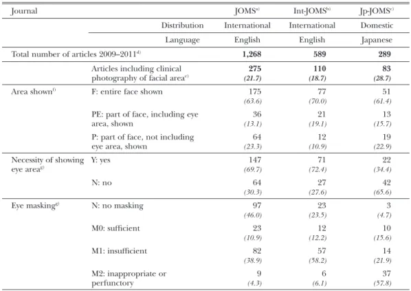
(4) 209. Photographs in Oral Surgery Journals. Table 1 Clinical photographs showing facial area in 3 oral surgery journals Journal. JOMSa). Int-JOMSb). Jp-JOMSc). Distribution. International. International. Domestic. Language. English. English. Japanese. 1,268. 589. 289. 275. 110. 83. (21.7). (18.7). (28.7). Total number of articles 2009–2011. d). Articles including clinical photography of facial areae) Area shownf). F: entire face shown. 77. 51. (70.0). (61.4). PE: part of face, including eye area, shown. 36. 21. 13. (13.1). (19.1). (15.7). P: part of face, not including eye area, shown. 64. 12. 19. (23.3). (10.9). (22.9). Necessity of showing Y: yes eye areag) N: no Eye maskingg). 175 (63.6). N: no masking M0: sufficient M1: insufficient M2: inappropriate or perfunctory. 147. 71. 22. (69.7). (72.4). (34.4). 64. 27. 42. (30.3). (27.6). (65.6). 97. 23. 3. (46.0). (23.5). (4.7). 23. 12. 10. (10.9). (12.2). (15.6). 82. 57. 14. (38.9). (58.2). (21.9). 9. 6. 37. (4.3). (6.1). (57.8). Journal of Oral and Maxillofacial Surgery International Journal of Oral and Maxillofacial Surgery c) Japanese Journal of Oral and Maxillofacial Surgery d) Reviews, editorials, correspondence and presentation abstracts omitted e) Total numbers of articles (%) f) All facial photography (%) g) All photography including eye area (F+PE) (%) a). b). in the light of the journal’s regulations, which stipulate that a signed patient release must be obtained on submission for use of photographs in which the subject is identifi able9). It is clear that most authors submitting to JOMS strictly adhere to journal regulations. However, an overly-strict enforcement of patient consent policies may cause various problems in a clinical setting10) and a subse quent reduction in publications. As indicated in the clinical photographs in JOMS, the publication of facial pictures with appropriate eye masking in journal articles requires discretion. The regulations of Int-JOMS require that if identifiable features are not essential to the. presentation of a case, those areas should be cropped. In cases where patient consent has not been obtained and the figure contains recognizable features, retouching with eye masking or other rendering methods is requested6). The guidelines of Jp-JOMS specify that the eye area should be masked in all clinical pho tographs7), and in this study all but 3 articles included clinical photographs encompassing either the whole face or part of the face and masking of the eyes in all cases. However, although the submission guidelines for JpJOMS state that the journal complies with the Declaration of Helsinki regarding the preservation of subjects’ and patients’ rights8),.
(5) 210. Shintani M et al.. there is no mention of the need to obtain informed consent. These contradictions may negate the very preservation of individual anonymity that such guidelines are intended to preserve. This suggests that many authors and editors of Jp-JOMS hope that simply mask ing the patient’s eyes will render informed consent unnecessary, reflecting an unwelcome neglect of obligations. 2. Eye masking area If the unique features around the eyes are not concealed, individuals can still be easily identified. The superciliary space, in particu lar, is unique to each individual. Therefore, not only the eyelid and the optic fissure, but also the entire orbital region extending to the superciliary space must be concealed3,4). In all 3 journals in the current investigation, how ever, placing a black rectangle over the eye area alone was generally considered sufficient to protect patient anonymity. Therefore, in many cases, with or without masking, the sub ject of the photograph may have been easily recognizable to long-term acquaintances12). Undoubtedly, however, the preservation of anonymity is very much in the interest of acquaintances of the patient, too. In Jp-JOMS, concealment was inappro priate or perfunctory in 57.8% of clinical photographs (M2). When combined with those in which an insufficient area was concealed (M1), the overall percentage was 79.7%. Although adhering to editorial guidelines, in some cases, the authors were unable to conceal the patient’s eyes sufficiently to pro tect privacy due to the symptoms involved. Moreover, use of unorthodox forms of eye masking such as placing a line across the eyes or covering the iris with a small dot may have undermined patient dignity. 3. Quality and respect for subject of clinical photographs In JOMS, many photographs in which the patient’s eyes were not masked appear to have been taken by a professional clinical photographer of considerable skill, judging by the particularly high standard of work. On. the other hand, some photographs were of particularly low quality. If the patient is selfconscious of facial damage or some deformity, they may be reluctant to give consent for publication. In either case, the skills of a trained professional clinical photographer would probably be of benefit to both clinician and patient alike. In a busy clinical setting, however, patient needs must come first, with speed of treatment a priority. This may explain why the use of digital images is on the increase and why high-quality clinical photographs are becoming harder to find. However, awareness of the need for better clinical photographs would result in records which were more beneficial for both patients and clinicians alike1). The dissemination of such high-quality photographs is, of course, essential from the point of view of medical education and clinical training. A survey on patient attitudes toward medical photography reported that 84% of patients would consent to a photograph of their body being used for the purposes of medical education, 71% for publication in a medical journal, and 70% for publication in a book2). This suggests that patients would be more cooperative in allowing such images to be published than might be supposed. 4. Awareness gaps Worldwide, the actual implementation of ICMJE recommendations leaves much to be desired. There is still a significant level of ignorance on the subject, and it may not even be possible to obtain informed consent for clinical photographs in some cases11). One problem is that there is no common ground in terms of an international perspective or standard, and there is a tendency to follow independently established norms within spe cific geographical areas, especially where a journal has a limited domestic distribution such as Jp-JOMS, for example. On the other hand, the article including facial photographs in JOMS and Int-JOMS in this study had been submitted from 40 and 29 countries, respectively. National or regional circumstances vary, and no fixed correlation was found regarding the presence or extent.
(6) 211. Photographs in Oral Surgery Journals. of eye masking or whether this was effective in protecting patient anonymity. Even in articles submitted within the same country and conforming to a preferred journal style, clinical photographs showing a disregard for the patient’s rights can still be observed, and there remain awareness gaps among authors. Due to varying editorial standards, therefore, the optimal direction for journal guidelines remains to be elucidated, a fact often reflected in the results. 5. Breaking away from corrupt practice As demonstrated in the present study, any abuse of patient privacy or dignity in the pub lication of clinical photographs in medical journals can be verified. Furthermore, many medical journals strictly adhere to the high standard of demands on the protection of patient rights, which acts as an indirect deterrent for misuse of clinical photographs in other journals. On the other hand, determining whether such standards are adhered to is more dif ficult in a closed setting such as an oral presentation or medical lecture, for example. Clinical photographs used in an educational setting are mostly taken at institutional clin ics. In many cases, the patient is asked for permission to use such photographs for educational purposes or academic reports. Usually, the patient will give consent. How ever, the utilization of the photographs is entrusted to educator’s conscience, and there is no surveillance or monitoring method to prevent abuse or misuse of such photographs. Moreover, the number of clinical photographs being used is vast, particularly in a classroom setting. Consideration of patient dignity and the quality of such images under such circum stances may leave much to be desired. If the protection of patient anonymity in such images is poor, there repeated use, especially for pedagogical purposes, may result in medical students failing to learn how such material should be presented and the same mistakes being repeated ad infinitum. Therefore, there is a need to rethink the importance of pre senting correct examples of such photography. in an educational setting. The same principle applies to publication: if standards are high, this will lead to the production of further material of equally high standard. The present results suggest that protection of patient privacy must be consis tent and uniformly respected in journals with an international or domestic distribution in non-English-speaking countries.. Postscript This article is based on the investigation performed on three journals in the field of oral surgery only, and will not have any influ ence on the editorial policy of The Bulletin of Tokyo Dental College.. References 1) Berle I (2008) Clinical photography and patient rights: the need for orthopraxy. J Med Ethics 34:89–92. 2) Cheung A, Al-Ausi M, Hathorn I, Hyam J, Jaye P (2005) Patients’ attitudes toward medical photography in the emergency department. Emerg Med J 22:609. 3) Clover AJP, Fitzpatrick E, Healy C (2010) Analysis of methods of providing anonymity in facial photographs; a randomised controlled study. Ir Med J 103:243–245. 4) Engelstad ME, McClellan M, Jacko JA, Melton GB (2011) Deidentification of facial images using composites. J Oral Maxillofac Surg 69: 3026–3031. 5) International Committee of Medical Journal Editors. Uniform Requirements for Manu scripts Submitted to Biomedical Journals: Ethical Considerations in the Conduct and Reporting of Research: Privacy and Confiden tiality. http://www.icmje.org/ethical_5privacy. html (accessed 14 August 2012) 6) International Journal of Oral and Maxillofa cial Surgery — Guide for authors. http://www. elsevier.com/wps/find/journaldescription.cws_ home/623046/authorinstructions (accessed 14 August 2012) 7) Japanese Journal of Oral and Maxillofacial Surgery — Guideline for protection of patients’ privacy and research ethics. http://www. jsoms.or.jp/wp-content/uploads/2011/12/.
(7) 212. Shintani M et al.. privacy20090428.pdf (in Japanese, accessed 14 August 2012) 8) Japanese Journal of Oral and Maxillofacial Surgery — Author info. http://www.jsoms.or.jp/ wp-content/uploads/2011/12/tokokitei20111221. pdf (in Japanese, accessed 14 August 2012) 9) Journal of Oral and Maxillofacial Surgery — Author info. http://www.joms.org/authorinfo (accessed 14 August 2012) 10) Laskin DM (1998) Patient consent for publi cation. J Oral Maxillofac Surg 56:413. 11) MacIntosh T (2006) Ethical considerations for clinical photography in the global south.. Dev World Bioeth 6:81–88. 12) Slue WE Jr. (1989) Unmasking the lone ranger. N Engl J Med 321:550–551. Reprint requests to: Dr. Masuro Shintani Oral Health Science Center, Tokyo Dental College, 1-2-2 Masago, Mihama-ku, Chiba 261-8502, Japan Tel: +81-43-270-3842 E-mail: shintani@tdc.ac.jp.
(8)
図
関連したドキュメント
Laplacian on circle packing fractals invariant with respect to certain Kleinian groups (i.e., discrete groups of M¨ obius transformations on the Riemann sphere C b = C ∪ {∞}),
A NOTE ON SUMS OF POWERS WHICH HAVE A FIXED NUMBER OF PRIME FACTORS.. RAFAEL JAKIMCZUK D EPARTMENT OF
She reviews the status of a number of interrelated problems on diameters of graphs, including: (i) degree/diameter problem, (ii) order/degree problem, (iii) given n, D, D 0 ,
A lemma of considerable generality is proved from which one can obtain inequali- ties of Popoviciu’s type involving norms in a Banach space and Gram determinants.. Key words
It is suggested by our method that most of the quadratic algebras for all St¨ ackel equivalence classes of 3D second order quantum superintegrable systems on conformally flat
Moreover, it is important to note that the spinodal decomposition and the subsequent coarsening process are not only accelerated by temperature (as, in general, diffusion always is)
Next, we prove bounds for the dimensions of p-adic MLV-spaces in Section 3, assuming results in Section 4, and make a conjecture about a special element in the motivic Galois group
de la CAL, Using stochastic processes for studying Bernstein-type operators, Proceedings of the Second International Conference in Functional Analysis and Approximation The-
