IRUCAA@TDC : Fluid extravasation of the articular capsule as a complication of temporomandibular joint pumping and perfusion
7
0
0
全文
(2) Bull. Tokyo dent. Coll., Vol. 43, No. 4, pp. 237⬃242, November, 2002. 237. Clinical Report. FLUID EXTRAVASATION OF THE ARTICULAR CAPSULE AS A COMPLICATION OF TEMPOROMANDIBULAR JOINT PUMPING AND PERFUSION KENICHI SASAKI, RYUICHIROU WATAHIKI, HIDETOSHI TAMURA, MOTOI OGURA and MASAYUKI SHIBUYA Department of Oral and Maxillofacial Surgery, Kameda General Hospital, 929 Higashi-cho, Kamogawa, Chiba 296-8602, Japan. Received 6 March, 2002/Accepted for Publication 7 August, 2002. Abstract This report is a retrospective study of fluid extravasation as a complication of temporomandibular joint pumping and perfusion. Contrast-enhanced 3D-CT of the upper joint compartment was performed for presurgical diagnosis before temporomandibular joint arthroscopic surgery in our hospital from 1996 to 2000. From these cases, 43 joints and 38 patients were selected because they had not improved under conservative treatment during the previous six months. Fluid extravasation of the articular capsule was recognized in 9 joints (20.9%) in 9 patients, 3 males and 6 females. Two of the nine patients had undergone arthroscopic observation before surgery. This test had revealed only thin articular capsule, not a perforation, in any of these cases. The data indicate only extremely tiny perforations or infiltration leakage due to the fluid pressure in the upper joint compartment during pumping or perfusion. Oral and maxillofacial surgeons should be aware of this complication. Key words:. TMD — Fluid leakage of the capsule— Fluid extravasation of the capsule— Contrast-enhanced 3D-CT. INTRODUCTION Recently, arthroscopic surgery1,2,4,5,8,9) and arthrocentesis3) of the temporomandibular joint (TMJ) have been widely used, and many surgeons have reported their utility. But, some surgeons have reported complications of arthroscopic surgery, including lateral pharyngeal edema1,2,9).. We have previously reported our results with contrast-enhanced 3D-CT of the upper joint compartment6–8), and we have sometimes recognized the extravasation of the contrasting medium from the TMJ capsules. We report the rate of extravasation, the specifics of the regions, and the causes of this phenomenon.. 237.
(3) 238. K. SASAKI et al.. Fig. 1 Normal appearance of contrast-enhanced 3D-CT of the upper joint compartment. PATIENTS AND METHODS 1. Patients The contrast-enhanced 3D-CT of upper joint compartment was used to examine 43 joints in 38 patients between December of 1996 and April of 2000. The ages of the patients ranged from 16 to 79 years. All of them presented with closed lock and had not shown any improvement during the previous six or more months under conservative treatment. 2. Methods For 3D-CT scanning, spiral CT data acquisition was employed with a slice width of 1 mm and a reconstruction width of 0.5 mm, 120 kVp Tube voltage, 150 mA Tube current using an X force SH Helical CT (Toshiba Medical Co.). CT values were set in the range of 2,000 Hounsfield Units (HU) to 4,095 HU and gained 3D image using X vigor 3D image processor (Toshiba Medical Co.). Onishi’s method4) was adopted for contrast medium injection. The contrast medium was injected into the upper joint compartment of TMJ using a 23 gauge needle. An X-ray television system was used for confirming the technique. 3D reconstruction of the CT data was performed using X force SH (Toshiba Medical Co., Japan). Fig. 2 Three dimensional view of the same case shown in Fig. 1. and X Vigor (Toshiba Medical Co., Japan). The contrast medium was Optiray® 350 (Mallinckrodt Medical Co., USA). First, the contrast medium was infused until slight pressure was felt without pumping. Then, the contrasted image was obtained, permitting diagnosis of intra-articular status (Figs. 1, 2). We investigated the extravasation of the articular capsule. In the retrospective study, arthroscopic observations has been made during arthroscopic surgery in two cases, according to the criteria for the surgery..
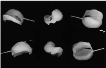
(4) 239. FLUID EXTRAVASATION OF TMJ ARTICULAR CAPSULE. Table 1 Extravasation cases and regions Patient number. Gender. Age in years. Affected TMJ. Region of extravasation. 1* 2* 3 4 5 6 7 8 9. Female Female Female Female Male Female Male Male Female. 55 25 57 26 29 46 20 45 60. Left Left Left Left Right Right Left Left Left. Anteromedial Anteromedial Lateral Medial Anteromedial Anteromedial Lateral Anteromedial Anteromedial. * Together with arthroscopic observation. Fig. 3 Extravasation of contrasted medium at the outside of the anteromedial capsule of the upper joint space on the transverse CT.. RESULTS The numbers of patients and joints in whom we recognized extravasation of the articular capsule were 9 patients and 9 joints, 3 males and 6 females (Table 1). The rate of extravasation was 20.9%. Next, we investigated the location of the extravasation and found that it most frequently occurred in the anteromedial region (6 cases), the lateral (2 cases), and the medial (1 case) (Figs. 3–10). Most of these cases of extravasation were recognized gradually during a 15 minute period. Arthroscopic observation during arthroscopic surgery was applied to 2 of our 9 cases. We found that the anteromedial regions of the articular capsules were extremely thin in. Fig. 4 A same case as Fig. 3; contrast-enhanced 3D-CT was made.. Anterior. Extravasation. Adhesion Medial. Fig. 5 Superior view of the upper joint space. Extravasation was recognized at the anteromedial region of the upper joint as the dark area. Wide adhesion was indicated by the lack of contrast medium in the intermediate and posterior portions of the upper joint space..
(5) 240. K. SASAKI et al.. Fig. 7 A same case as Fig. 6; contrast-enhanced 3D-CT was made.. Anterior Fig. 6 Extravasation of contrasted medium at the medial region of the upper joint on the transverse CT.. the upper joint compartments of both cases, but no perforation of the capsule could be detected (Figs. 11, 12). The fluid extravasation was absorbed over time without any treatment or influences on the function of temporomandibular joint. No lateral pharyngeal edema was experienced in this study.. DISCUSSION Arthroscopic surgery1,2,4,5,8,9) or arthrocentesis3) are often used to treat internal derangement of TMJ. One complication of arthroscopic surgery is lateral pharyngeal edema1,2,9). Green and Van Sickel1) and White9) have reported that this kind of side effect occurs in 0.45–2% of arthroscopic surgeries. This is a low but still significant value, because a vast extravasation can cause the severe complication of obstructive airway. However, the questions of how and where it will occur is not addressed in any current articles. Kakudo et al. has suggested that the causes of leakage during perfusion are the embolization of outflow needle and too much pumping pressure2). They did not mention any other possible causes.. Adhesion. Extravasation. Medial Fig. 8 Superior view of the upper joint space. Extravasation was recognized at the medial region of the upper joint as the dark area. The adhesion was indicated by the lack of contrast medium at anteromedial and lateral portion of upper joint space.. We have sometimes recognized extravasation of contrasting medium during the procedure for insufflation and distention of upper joint compartment when we have used X-ray television and following contrast-enhanced 3D-CT. Most of these cases of extravasation developed gradually over 15 minutes. This pattern suggests that the leakage was not caused by a small perforation, but by diffusion. Extravasation occurred most frequently in the anteromedial region. Furthermore, we found that it occurred in cases with severely thin articular capsules but with no perforation.
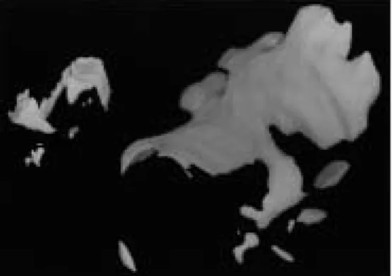
(6) FLUID EXTRAVASATION OF TMJ ARTICULAR CAPSULE. Fig. 9 Lateral view of the upper joint space. Extravasation at the lateral region (arrow).. Fig. 10. Posterior view of the upper joint space of the same case as Fig. 9. Extravasation at the lateral region (arrow).. that could be detected during arthroscopy. We think that a thin articular capsule is associated with extravasation and that pumping or perfusion pressure may cause a tiny perforation or infiltration leakage.. Fig. 11. Fig. 12. 241. Extremely thin articular capsule (arrowhead) at the anterior region of the upper joint space in the same case as Figs. 3, 4 and 5.. The lateral pterygoid muscle is invisible through extremely thin articular capsule (arrow head) after few applications of the balloon pumping ablation technique in the same case as Fig. 11.. These values are not low, so we must take care during the perfusion of arthroscopic surgery and arthrocentesis. This is still a hypothesis, but there are no other explanations, and the subject requires further study.. CONCLUSION ACKNOWLEDGEMENT Among our total of 38 cases and 43 joints, extravasation was recognized in 9 joints (20.9%). We found thin capsules in the anteromedial region of most of these articular capsules, and most of the leakage was also in this area. Perfusion pressure may cause a small perforation or leakage of the articular capsule.. The author would like to thank Mr. H. Somazawa and Mr. K. Amano for their help in this study. Presented at the 45th Japanese Society of Oral & Maxillofacial Surgeons Meeting, held in Chiba, Japan, in October of 2000..
(7) 242. K. SASAKI et al. REFERENCES. 1) Green, M.W. and Van Sickel, E. (1989). Survey of TMJ arthroscopy in oral and maxillofacial surgery residency programs. J Oral Maxillofac Surg 47, 574–576. 2) Kakudo, K., Kotani, J., Suzuki, S., Kim, K.S., Kawabata, T. and Shirasu, R. (1995). Lateral pharyngeal edema following arthroscopic surgery —Report of a case. J Jpn Soc TMJ 7, 16–20. (in Japanese) 3) Nitzan, D.W., Mahler, Y. and Simkin, A. (1992). Intra-articular pressure measurements in patients with suddenly developing, severely limited mouth opening. J Oral Maxillofac Surg 50, 1038–1042. 4) Ohnishi, M. (1975). Arthroscopy of the temporomandibular joint. Kokubyo Gakkai Zasshi 42, 207–213. (in Japanese) 5) Sasaki, K. (1997). A balloon pumping technique for treatment of internal derangement of the temporomandibular joint. J Oral Maxillofac Surg 55, 947–952. 6) Sasaki, K., Tamura, H., Watahiki, R. and Somazawa, F. (1999). Contrast-enhanced 3D-. CT of the temporomandibular joint; One special picture. Quintessence 18, 29. (in Japanese) 7) Tamura, H., Sasaki, K., Watahiki, R. and Ogura, M. (2001). A clinical study of contrastenhanced 3D-CT of upper joint compartment with closed lock. Jpn J Oral Maxillofac Surg 47, 400–404. 8) Watahiki, R., Sasaki, K. and Tamura, H. (2000). Evaluation of adhesions by contrastenhanced 3D-CT and arthroscopic observation of the upper joint compartment of TMJ. Bull Tokyo dent Coll 41, 195–201. 9) White, R.D. (1989). Retrospective analysis of 100 consecutive surgical arthroscopies of the temporomandibular joint. J Oral Maxillofac Surg 47, 1014–1021. Reprint requests to: Dr. Kenichi Sasaki Department of Oral and Maxillofacial Surgery, Kameda General Hospital, 929 Higashi-cho, Kamogawa, Chiba 296-8602, Japan.
(8)
図

関連したドキュメント
The input specification of the process of generating db schema of one appli- cation system, supported by IIS*Case, is the union of sets of form types of a chosen application system
The edges terminating in a correspond to the generators, i.e., the south-west cor- ners of the respective Ferrers diagram, whereas the edges originating in a correspond to the
H ernández , Positive and free boundary solutions to singular nonlinear elliptic problems with absorption; An overview and open problems, in: Proceedings of the Variational
So, the aim of this study is to analyze, numerically, the combined effect of thermal radiation and viscous dissipation on steady MHD flow and heat transfer of an upper-convected
Keywords: Convex order ; Fréchet distribution ; Median ; Mittag-Leffler distribution ; Mittag- Leffler function ; Stable distribution ; Stochastic order.. AMS MSC 2010: Primary 60E05
Making use, from the preceding paper, of the affirmative solution of the Spectral Conjecture, it is shown here that the general boundaries, of the minimal Gerschgorin sets for
We show that a discrete fixed point theorem of Eilenberg is equivalent to the restriction of the contraction principle to the class of non-Archimedean bounded metric spaces.. We
In Section 3, we show that the clique- width is unbounded in any superfactorial class of graphs, and in Section 4, we prove that the clique-width is bounded in any hereditary
