STUDY ON RESPIRATORY PROPERTIES, CHARACTERISTICS
AND FUNCTIONS OF SUBSTRATE OXIDATIONS IN THE
PURIFIED MITOCHONDRIA ISOLATED FROM CRASSULACEAN
ACID METABOLISM (CAM) PLANTS
(数種の
CRASSULASEAN ACID METABOLISM (CAM) 植物から単離した
ミトコンドリアの呼吸特性とその基質酸化機能に関する研究)
HOANGTHI KIM HONG
DOCTOR DISSERTATION
Submitted on Partial-Fulfillment of the Requirement for
the degree of
Doctor of Philosophy
ACKNOWLEDGMENTS
This research work was supported by Ministry of Education, Science, Sports and Culture, Japanese Government and it was conducted in Laboratory of Tropical Crops Improvement Science, Faculty of Agriculture, Saga University under the directly guidance of Prof. Dr. Akihiro Nose. I wish to take this opportunity to acknowledge the financial support, and contributions of all members and institutes who have given me a great change to attend and complete this academic course.
I would like to express my sincere appreciation to my principal supervisor Prof. Dr. Akihiro Nose, for his wholehearted guidance and full supports throughout my study. Without his continuous encouragement and support, it would be impossible for me to finish this work on time. From my heart, I wish to express my thanks for his painstaking instruction and supplying with delicate equipment and good research conditions in my study.
I would like to extend my thanks to Prof. Dr. Yosuke Tashiro, Plant Cell Engineering Laboratory, Saga University and Associate Professor Sakae Agarie, Laboratory of Tropical Crop Improvement Science, Faculty of Agriculture, Saga University as my supervisors, for their time and support during my research.
I wish to express my gratitude to Prof. Dr. Shigeto Tominaga, Kagoshima University and Associate Professor Yoshinobu Kawamitsu, Ryukyus University, and all members of my Advisor Committee for their valuable suggestions and critical comments in the completion of this study.
I sincerely appreciate to Mr. Kenzo Oshima, Laboratory of Tropical Crop Improvement Science, Faculty of Agriculture, Saga University for his valuable helps,
share and support during this research course. Many thanks to my tutor Mr Okimoto and all friends in Laboratory of Tropical Crops Improvement Science, Faculty of Agriculture, Saga University who has spent a lot of time to share, help and make my stay here so enjoyable.
I wish to express sincere thanks to Professor Thomas E. Elthon (School of Biological Sciences, University of Nebraska-Lincoln, USA) for his generous gift of AOX antisera, Professor Ian Max Møller (Risø National Laboratory, Denmark) and Professor Hans Lambers (University of Western Australia) for their criticalreading of my manuscripts and their valuable comments regarding my work.
I would like to extend my profound gratitude to all of my colleagues in my Biology Department, Hue Sciences Faculty, Hue University, Vietnam who are willing share and do all my teaching work during my long absence from University and always supported me with spiritual encouragement.
Personally, I wish to express my whole hearted gratitude to my own parent and my parent in-laws; all of the sisters and brother for their love and kindly take care my son for me in some time when I send him to Vietnam. Their support and inspiration never ceased. Especially, I wish to present this dissertation to my beloved husband Ho Duc Hung and my son Ho Duc Nhat Dan as a spiritual gift to express my deep gratitude for their love with an unbounded sympathy, sharing and moral support in my academy work and daily life.
Hoang Thi Kim Hong Saga University, Japan March, 2005
TABLE OF CONTENTS Pages ACKNOWLEDGEMENTS...………. TABLE OF CONTENTS...……… LIST OF FIGURE………...……… LIST OF TABLE ...………... ABBREVIATION ......………
CHAPTER 1. General Introduction
1.1. Plant Mitochondria...……...…. 1.2. Characterization of mitochondrial substrate oxidations…... 1.3. Mitochondrial respiration in CAM plants………
1.3.1. General introduction about CAM plants………. 1.3.2. Studies on mitochondria of CAM plants ………
CHAPTER 2. Oxidation of substrates in Percoll-purified mitochondria isolated from Kalanchoë daigreemontian
2.1. Introduction. ...………..
2.2. Materials and Methods...
2.2.1. Plant materials...………... 2.2.2. Preparation of mitochondria……… 2.2.3. Preparation of leaf extraction for enzyme assays……… 2.2.4. Preparation of mitochondria for enzyme assays... 2.2.5. Mitochondrial activity ...………... 2.2.6. Enzyme activity ...………...
2.3. Results...………...
2.3.1. Purity and integrity of mitochondria...
i iii vii x xi 1 3 5 5 6 13 14 14 14 15 15 15 17 17 17
2.3.2. Enzyme activities ...………... 2.3.3. Respiratory properties of K. daigremontiana mitochondria...
2.4. Discussion ...……… 2.5. Summary ...………. CHAPTER 3. Oxidations of various substrate and effects of the inhibitors on purified mitochondria isolated from Kalanchoë pinnata
3.1. Introduction. ...……… 3.2. Materials and Methods...
3.2.1. Plant material and mitochondria respiration... 3.2.2. Preparation of leaf extraction and mitochondria for enzyme assays………. 3.2.3. Preparation of mitochondria for enzyme assays... 3.2.4. Enzyme assays...
3.3. Results ...……….
3.3.1. Enzyme activity...…. 3.3.2. Respiratory properties of K. pinnata mitochondria with the single substrates……… 3.3.3. Respiratory properties of K. pinnata mitochondria with the multiple substrates………. 3.3.4. Effect of KCN, SHAM and rotenone on oxidation of various substrates………... 3.4. Discussion... 3.5. Summary... 20 20 26 28 29 30 30 31 31 31 32 32 32 36 40 45 52
CHAPTER 4. Respiratory properties and malate metabolism in Percoll-purified mitochondria of Ananas comosus (L.) Merr. cv. smooth cayenne (pineapple)
4.1. Introduction ...……… 4.2. Materials and Methods...
4.2.1. Plant materials... 4.2.2. Isolation of mitochondria... 4.2.3. Oxygen uptake and protein determination……….. 4.2.4. Enzyme assays...
4.3. Results...
4.3.1. Purity of mitochondria………... 4.3.2. Enzyme activities... 4.3.3. Respiratory properties of A. comosus mitochondria...
4.4. Discussion... 4.5. Summary... CHAPTER 5. Effects of KCN, SHAM and Oxygen Concentrations on Respiratory Properties of Purified Mitochondria Isolated from Ananas
comosus (Pineapple) and Kalanchoë daigremontiana
5.1. Introduction ...……… 5.2. Materials and Methods... 5.3. Results...
5.3.1. Effects of individual KCN or SHAM... 5.3.2. Effects in combinations of KCN and SHAM... 5.3.3. Effects of KCN and SHAM on the concurrent oxidation of
53 55 55 55 56 57 58 58 60 60 73 82 83 86 88 88 92
succinate and NADH……...……….. 5.3.4. Effects of oxygen conditions on mitochondrial succinate oxidation...
5.4. Discussion... 5.5. Summary... CHAPTER 6. General Discussion
6.1. Method to isolate and purify mitochondria of three CAM species... 6.2. General characteristics of substrate oxidations in mitochondria of three CAM species………...
6.2.1. Individual succinate, NADH and NADPH oxidations... 6.2.2 Enzyme activities and individual malate oxidation.……... 6.2.3. General characteristics of mitochondrial alternative respiration in CAM species………...… 6.2. 4. Characteristics of simultaneous substrate oxidations………. 6.2.5. Effect of low oxygen concentration on mitochondrial substrate oxidations………...
6.3. The role of mitochondria in three CAM species……….. Future Progress... Summary... References……… Abstract in Japanese………... 94 97 97 109 110 111 112 113 117 118 121 121 125 128 130 143
List of Figure
No. Content Pages
1.1. 1.2. 2.1. 2.2. 2.3. 2.4 3.1. 3.2. 3.3.
Diagram of the structural membrane organization of a plant mitochondrion. (Siedow and Day, 2000)………. Malate metabolisms in cytosol and mitochondria of malic enzyme (ME)- Crassualacean acid metabolism (CAM) and phosphoenolpyruvate carboxykinase (PCK)-CAM plants……….. Methods to isolate and purify mitochondria of three investigated CAM species………...………... Malate dehydrogenase activities before (A) and after (B) lysis with Triton X-100 in Percoll-purified mitochondria isolated from K.
daigremontiana...
Individual oxidation of succinate (A), NADPH (B), NADH (C) and malate (D) in Percoll-purified K. daigremontiana mitochondria…………. Cooperative oxidation of two substrates in Percoll-purified K.
daigremontiana mitochondria………..
Effect of KCN (A), SHAM (B), KCN in the presence of SHAM (C), and SHAM in the presence of KCN (D) on the succinate oxidation by
K. pinnata mitochondria………
Effect of rotenone (A), KCN (B) and SHAM (C) on malate oxidation at pH 6.8 in the presence of 0.5 mM CoA and 1 mM NAD by K. pinnata mitochondria……… Effect of KCN (A), KCN and SHAM (B) and SHAM and KCN (C) on
2 12 16 19 22 25 37 41
3.4. 3.5. 4.1. 4.2. 4.3. 4.4. 4.5. 4.6. 4.7. 4.8. 4.9.
the malate oxidation at pH 6.8 in the presence of 1 mM TPP……….. Effect of KCN and SHAM in the presence of rotenone on the malate oxidation at pH 6.8 in the presence of 1 mM TPP with other substrates…. Organization of malate-oxidizing systems in cytosol and mitochondria of
K.pinnata……….
Respiratory properties of A. comosus mitochondria with 10 mM succinate (A), 1 mM NADH (B), 2mM NADPH (C) and 2mM NADPH with 1
mM Ca2+(D)……….
Effects of NADPH concentrations on state 3 in NADPH oxidation with 1 mM Ca2+ by A. comosus mitochondria………. Malate oxidation at pH 6.8 with CoA (A), CoA and TPP (B), and CoA, TPP and NAD (C)………... Malate oxidation at pH 7.2 with malate (A), malate and NAD (B), malate and glutamate (C), and malate, CoA, TPP and NAD (D)……… Malate oxidation at pH 7.6 with malate (A), malate and NAD (B), malate and glutamate (C), and malate with glutamate and GOT (D)……….. The OAA appearance outside A. comosus mitochondria. Experiment was assayed according to Pastore et al. (2003)………... The import of externally added OAA in A. comosus mitochondrial matrix to inhibit the succinate oxidation………..………... Aspartate oxidation (A), α-ketoglutarate oxidation (B), α-ketoglutarate oxidation in the presence of CoA, TPP and NAD (C), and malate oxidation with α-ketoglutarate (D)……….. Organization of malate-oxidizing system in A. comosus mitochondria...
42 43 51 62 65 66 67 70 72 74 75 80
5.1. 5.2. 5.3. 5.4. 5.5. 5.6 6
Effects of KCN or SHAM on succinate and NADH oxidations in mitochondria of A. comosus and K. daigremontiana……… Effects of both KCN and SHAM on succinate oxidations in mitochondria of A. comosus (A) and K. daigremontiana (B)………. Effects of both KCN and SHAM on succinate oxidations in
K. daigremontiana mitochondria………..………..
Effects of both KCN and SHAM on oxidations of both succinate and NADH in mitochondria of A. comosus and K. daigremontiana…………. Effect of low oxygen on succinate oxidation in A. comosus mitochondria (A) and K. daigremontiana mitochondria (B)……….. Electron transport chain during NADH and succinate oxidation in
mitochondria of A. comosus (A) and K. daigremontiana (B)…………... Comparison of substrate metabolisms in cytosol and mitochondria of A.
comosus (A) and Kalanchoë (B)………..
89 93 95 96 98 102 115
List of Table
No. Content Pages
2.1. 2.2. 2.3. 3.1 3.2. 3.3. 3.4. 3.5. 4.1. 4.2. 4.3. 5.1. 5.2.
Enzyme activities in leaf extract and purified K. daigremontiana mitochondria………... Respiratory properties of Percoll-purified K. daigremontiana mitochondria... Respiratory properties of Percoll-purified K. daigremontiana mitochondria with two substrates……… Respiratory properties of Percoll-purified K. daigremontiana mitochondria with two substrates………..…………... Rubisco and PEPC activities in leaf extract and mitochondria of K. pinnata... Enzyme activities in leaf extract and in mitochondria of K. pinnata……….. Individual substrate oxidation in K. pinnata mitochondria……… Simultaneous substrate oxidation in K. pinnata mitochondria…... Effect of KCN on individual and simultaneous substrate oxidation by K.
pinnata mitochondria………...
Rubisco and PEPC activities in leaf extract and Percoll-purified A. comosus mitochondria……….. The comparison of enzyme activities in A. comosus mitochondria with mitochondria of K. daigremontiana and K. pinnata……….. Effect of KCN and SHAM on succinate and NADH oxidations in mitochondria of A. comosus and K. daigremontiana………. Effects of oxygen on respiratory property and alternative respiration on succinate oxidation in mitochondria of K. daigremontiana and A. comosus…
18 21 24 33 34 35 38 39 59 61 64 90 99
Abbreviations ADP Alt AOX ATP Ca2+ CAM CoA COX CRR Cyt ETC GOT KCN MDH ME Mp NAD NADH NADP NADPH NDin (NADH) NDin (NADPH) adenosine diphosphate alternative alternative oxidase adenosine triphosphate calcium
crassulacean acid metabolism coenzyme A
cytochrome oxidase cyanide resistant respiration cytochrome
electron transport chain
glutamate-oxaloacetate transaminase potassium cyanide
malate dehydrogenase malic enzyme
purified mitochondria
nicotinamide adenine dinucleotide (oxidised form) nicotinamide adenine dinucleotide (reduced form)
nicotinamide adenine dinucleotide phosphate (oxidised form) nicotinamide adenine dinucleotide phosphate (reduced form) matrix facing rotenone-insensitive NADH dehydrogenase matrix facing rotenone-insensitive NADPH dehydrogenase
O2 OAA PCK PEP PEPC PMSF PPDK RCR RuBP SHAM TPP UQ oxygen oxaloacetate phosphoenolpyruvate carboxykinase phosphoenolpyruvate phosphoenolpyruvate carboxylase phenylmethylsulfonyl fluoride pyruvate orthophosphate dikinase respiratory control ratio
ribulose 1,5-bisphosphate salicylhydroxamic acid thiamine pyrophosphate ubiquinone
Chapter 1
General introduction
1.1. Plant Mitochondria
Mitochondria are small (0.5 µm x 2 µm) semi-autonomous organelles varying in shape from spherical to filamentous. Their number per cell varies depending on the type and size of the cell and the extent of cellular differentiation (Douce, 1985, McCabe et al., 2000). Thought mitochondria occupy as little as 1% of the total volume of the cell, they play a pivotal role in energy metabolism and in the maintenance of cellular homeostasis. Mitochondria contain two set of membranes that divide the organelle into four compartments; the outer membrane, the region between the two membranes (inter-membrane space), a highly invaginated inner membrane, which gives rise to structures known as cristae, and the aqueous phase contained within the inner membrane (mitochondria matrix) (Siedow and Day, 2000; Fig. 1.1).
The mitochondrion is the principal organelle of eukaryotic respiration. This respiration includes three processes. Two of these process of respiration are the citric acid (TCA) cycle and electron transfer/oxidative phosphorylation, which occurs within the mitochondrion, whilst the third, glycolysis, occurs in the cytoplasm and supplies the mitochondria with substrates such as pyruvate from the oxidation of carbohydrate (Bowsher and Tobin, 2001). Glycolysis involves a series of soluble cytosolic enzymes that oxidize sugars to organic acids. The TCA cycle serves as an important source of carbon skeletons for biosynthetic processes. The reducing equivalents, synthesized during glycolysis and TCA cycle are oxidized by the mitochondrial electron transport chain (ETC) that ultimately donates electrons to molecular oxygen producing water.
Fig. 1.1. Diagram of the structural membrane organization of a plant mitochondrion
Respiration requires the operation of an ETC that converts the reducing equivalents NAD(P)H into energy. As a result, oxygen is reduced to water and ATP is synthesized via oxidative phosphorylation. The ETC in the inner mitochondrial membrane is composed of four complexes: complex I oxidize matrix-produced NADH and transfers electrons to ubiquinone, complex II catalyses the oxidation of succinate, transferring electrons to the ubiquinone pool, complex III oxidizes ubiquinone and transfers electrons to cytochrome C and complex IV oxidizes reduced cytochrome C and electrons are transferred to molecular oxygen, producing water. In addition, plant mitochondria also possess four additional NAD(P)H dehydrogenases (Roberts et al., 1995, Melo et al., 1996, Agius, 2001), characterized by their insensitivity towards the complex I inhibitor rotenone, and a quinol oxidase providing an alternative, non-phosphorylating route for the reduction of molecular oxygen to water (Siedow and Day, 2000; Agius, 2001). Two of these additional NAD(P)H dehydrogenases are found on the outer surface of the inner membrane facing the intermembrane space and two on the inner surface facing the matrix like the standard mitochondrial NADH dehydrogenase (Agius, 2001).
1.2. Characterization of mitochondrial substrate oxidations
One of the unique features of plant mitochondria is their ability to oxidize cytosolic NAD(P)H directly with NAD(P)H dehydrogenases on the outer surface of the inner membrane. Apart from the capacity of cytosolic NAD(P)H oxidations, plant mitochondria normally oxidize some main other substrates as succinate, malate, etc. Added external succinate was oxidized by succinate dehydrogenase in mitochondria matrix. Electron transport by succinate dehydrogenase to ubiquinone proceeds with no major decrease in the redox potential. Therefore, no energy is gained in the electron transport from succinate to ubiquinone (Heldt, 1997). The addition of malate to isolated plant
ADP increased oxygen consumption, with the concomitant oxidation of matrix NADH (state 3), followed by a re-reduction of matrix NAD during state 4 conditions (Neuburger
et al., 1984). In isolated mitochondria, malate dehydrogenase (MDH) catalyses the
oxidation of malate to oxaloacetate (OAA), with the concomitant reduction of NAD. The NADH produced is rapidly oxidized by internal NADH dehydrogenases, and OAA is excreted from the mitochondrial matrix. This product removal allows for continuous oxidation of malate, until OAA accumulates in the matrix, decreasing the equilibrium concentration of NADH and rate of malate oxidation (Palmer et al., 1982, Neuburger et al., 1984). This inhibition however, can be circumvented by the participation of NAD malic enzyme (NAD-ME). NAD-ME also oxidizes malate, but differs from MDH in that it is able to provide a constant source of NADH, despite equilibrium conditions of MDH (Palmer and Moller, 1982). Therefore, NAD-ME increases the intramitochondrial concentration of NADH, which in turn can be used to reduce previously accumulated OAA. When all excess OAA has been removed, NADH is once again available to the respiratory NADH dehydrogenases (Agius, 2001). In addition, plant mitochondria have overflow mechanisms, which oxidized surplus NADH without synthesis of ATP. Moreover, mitochondria possess an alternative (Alt) oxidase (AOX) by which electrons can be transferred directly from ubiquinon to oxygen, and this way passed cytochrome (Cyt) C oxidase (COX). AOX allows reduction of oxygen without the generation of a proton-motive-force. The contribution of AOX to respiration can be determined by inhibiting oxidative phosphorylation. Salicylhydroxamic acid (SHAM) is an inhibitor of AOX that can be used to determine the quantitative contribution of AOX to respiration. The AOX is found in plants, many algae and fungy, and some protozoa. Electron flow through the AOX is insensitive to classis inhibitors of COX as cyanide, azide, and carbon monoxide; however, it can be specifically inhibited by SHAM, and n-propylgallate
(Siedow and Day, 2000).
In the last decade, mitochondrial substrate oxidations were studied very detail in many species of C3 and C4 species. And the role of mitochondria in cell biochemistry of
these plants has been the subject of intensive investigation. In mitochondria of these species, oxidative phosphorylation has been suggested to be the main process supplying ATP for the cytosol both in light and in dark. On the other hand, mitochondria can regulate both the redox and the energy state of the cell by virtue of the existence of a number of metabolite carries and of a respiratory chain (Pastore et al., 2003). It has been also indicated that mitochondria play the important roles in producing ATP as energy source for the cell and in supplying the carbon compounds necessary for many biosynthetic pathways in many species of C3 and C4 plants. Many recent observations on mitochondrial
respiration also provide the evidence that respiration and photosynthesis are mutually beneficial and that respiration maintains a high photosynthesis rate necessary for maximal growth (Laloi, 1999). However, mitochondrial substrate oxidations and the role of mitochondria are not fully understood in Crassulacean acid metabolism (CAM) species.
1.3.Mitochondrial respiration in CAM plants 1.3.1. General introduction about CAM plants
Crassulacean acid metabolism is a photosynthetic system in which the operation
of C3 and C4 carboxylases occur within a common cell, with enzyme activity separated
temporally (Dodd et al., 2002). CAM represents one of three major photosynthetic modes, together with C3 and C4 pathways. They occurs in approximately 6%of higher plant
(Winter and Smith, 1996) yet suffersfrom being perceived as a minor photosynthetic pathway, restrictedto a small number of highly specialized desert-dwelling plants. CAM is a complex adaptation that allows photosynthesis to remain active in the virtual absence of
accumulated in the vacuoles of mesophyl cells at night as a result of fixation of CO2 by
phosphoenolpyruvate carboxylase (PEPC). During the day, malic acid is decarboxylated, an released CO2 is refixed in C3 cycle (Kondo et al., 1998). During noctural CO2 fixation
in CAM plants, the rate of malate production in the cytoplasm must be closely linked to its removal into the vacuole across the tonoplast. However, it is still ignorant of the mechanism of malate efflux from vacuole during light period, and of the way in which the transport processes are regulated during the CAM cycle to prevent futile cycling of malate into and out of the vacuole.
Crassulacean acid metabolism plants are characterizied by the accumulation of malate in the vacuoles at night and releasion malate into cytoplasm at day. In CAM plants, malate decarboxylation is one of the most important metabolisms during CAM phase III. Based on the malate metabolism, CAM plants can be divided into two groups, malic enzyme (ME)-CAM and phosphoenolpyruvate carboxykinase (PCK)-CAM plants. ME-CAM plants contain significant activities of ME without PCK, and they use ME to decarboxylate malate, generating pyruvate and CO2. By contrast, PCK-CAM plants
contain significant activities of PCK with lower levels of ME, they require the operation of MDH to convert malate to oxaloacetate (OAA), and then OAA is further converted to PEP and CO2 by cytosol PCK (Dittrich et al., 1973; Winter and Smith, 1996; Cuevas and
Podestá, 2000). Generally, the understanding on CAM activity during Phase III most comes from the reports on intact leaf of CAM plants. And, these reports have provided important insights into the control of carbon metabolism in photosynthesis tissues. This knowledge in mitochondria of CAM plants, in other hand, is still limited.
1.3.2. Studies on mitochondria of CAM plants
In fact, the method to isolate mitochondria from CAM plants has been firstly reported in some different species such as Mesembryanthemum crystallinum (Von Willert
and Schwöel, 1978), Sedum praealtum (Arron et al., 1979) and Kalanchoë daigremontiana (Day et al., 1980). After that, Rustin and his co-workers have been improved this method by using Percoll to purified mitochondria after isolating from K. blossfeldiana (Rustin and Queiroz-Claret, 1985; Rustin and Lance, 1986). Recently, Percoll was further used to purify mitochondria after isolated from some other species in ME-CAM plants as K.
fedtschenkoi (Cook et al., 1995) or in one species in PCK-CAM plants as Crassula lycopodioides (Peckmann and Rustin, 1992). These studies have been indicated that
mitochondria isolated from these CAM species readily oxidized the main substrates as succinate, NADH or NADPH with typical respiration rate similarly as other plant mitochondria. Also, Percoll-purified mitochondria normally showed high level of intactness and they oxidized malate, succinate, NADH, and NADPH with respiratory control rate and ADP/O ratios better than those observed in washed mitochondria. Though from 1980, Day (1980) has been reported the method to isolate and characterize the main respiratory properties of K. daigremontiana mitochondria; however this report has been conducted on washed mitochondria, not Percoll-purified mitochondria. For the present, the using Percoll to purify mitochondria for studying on the characteristic respiration of
Kalanchoë and A. comosus species are unwell- known.
Rustin and Queiroz-Claret (1985) have been reported that similarly as other plants, cyanide resistant oxidation was activated in both intact leaf and mitochondria of K.
blossfeldiana. During CAM development of K. blossfeldiana, all substrate oxidations, and
particularly malate oxidation, are activated and became more resistant to cyanide, indicating that CAM induction is accompanied to Alt pathway. By using the inhibitor method, these authors have been observed that cyanide resistant oxidation of the leaf respiration was increased during the first daylight hours, when malate was oxidized to
resistant oxidation can be ascribed to the operation of an Alt electron transport pathway. The main feature of this pathway is its non-coupling to phosphorylation processes. In addition, by using novel oxygen-isotope-discrimination technique, Robinson et al. (1992) showed that the flux through the Alt pathway was increased in phase III tissue in parallel with the rate of malate decarboxylation. This confirmed a similar observation of Rustin and Queiroz-Claret (1985) that a large proportion of the respiratory property in CAM-induced
K. blossfeldiana was cyanide resistant. Recently, the previous studies in our laboratory also
indicated that the intact leaves of the typical ME-CAM species as K. daigremontiana and K.
pinnata, or typical PCK-CAM species as A. comosus showed Alt respiration in CAM phase
III, however K. daigremontiana was found to have a clear requirement of O2 whereas the
O2 requirements in A. comosus were low (Nose et al., 1999, Nose and Takashi, 2001).
Clearly that Alt respiration was activated during leaf respiration in some investigated CAM plants; however, what controls the activity of Alt pathway in vivo is still not fully understood, and physiological role for the Alt pathway in Phase III metabolism of CAM plants is not readily apparent (Winter and Smith, 1996). In addition, it has been suggested that ME may be linked to non-phosphorylating electron transport pathways in plant mitochondria (Lance and Rustin, 1984; Lance et al., 1985). The oxidation of malate via ME essentially proceeds through the cyanide-insensitive Alt electron transport pathway, whereas the oxidation of malate via MDH appears strongly linked to the cyanide sensitive Cyt pathway (Rustin et al., 1980). However, it has been argued that there is no structural link between these enzymes, since under appropriate conditions other NAD-linked enzyme, in addition to ME, can be liked to the rotenone-insensitive bypass and AOX (Day et al., 1987).
In fact, the knowledge on respiratory property and Alt respiration in CAM plants were mainly collected from the reports which were mainly investigated with only one of
the respiratory substrates. While many experimental evident in other plant mitochondria indicated that mitochondrial respiratory properties and Alt respiration were significantly increased in simultaneously oxidation of two or three substrates, suggesting that during the
in vivo plant respiration, the mitochondrial electron transport chain was not saturated with
only one respiration substrate (Shugaev and Vyskrebentseva, 1999). Furthermore, the effect of O2 concentrations on plant respiration and Alt respiration has been recently reported by
some researchers. Ricard et al. (1994) showed that low O2 concentrations in root tissue
resulting from an anaerobic environment allowing for limited COX activity, while the behaviors of AOX is unknown. More recent, Szal et al. (2003) has been also reported that the O2 concentrations may be one of the control mechanisms of AOX activity in barley
roots. Noticeably, all of these problems were not probed in mitochondria isolated from CAM plants.
Generally, malate oxidation in the mitochondria isolated from other plants proceeds through the activities of two enzymes: MDH and NAD-ME. The balance between the two enzyme systems competing for malate oxidation may be controlled by various internal or external factors, such as intramitochondrial pH or addition of cofactors (Rustin
et al., 1980). The major products of malate oxidation by these mitochondria in most cases
were found to be pyruvate and CO2, indicated that their malate oxidation proceeds mainly
through NAD-ME rather than NAD-MDH. In CAM plants, it is acknowledged that during the deacidification in ME-CAM phase III, malate entering the mitochondrion can be oxidatively decarboxylated by NAD-ME, but it is not clear how much of the resultant pyruvate subsequently enters the TCA cycle. At that time, they had no direct information on how much malate passes through the mitochondrion during the deacidification phase in the light period. This was linked to the still unresolved question of the relative contribution
ME-CAM plants. Attempts to determine how much malate is oxidized in the mitochondria during phase III have been made by measuring the rates of oxygen uptake in the dark by leaf discs of K. daigremontiana sampled at different time during the day-night cycle. By comparing the observed rates of O2 uptake (in the dark) with those of malate consumption
(in the light), Robinson et al. (1992) calculated that up to 58% of the malate decarboxylated in phase III may be oxidized in the mitochondria by NAD-ME to pyruvate and CO2 and also implied that significantly more would have to be provided by
photophosphorylation. For PCK-CAM plants, the question that whether NAD-ME or NADP-ME activities might also be controlled by reversible covalent modification during the CAM cycle in a manner equivalent to PEPC has not yet been determine (Winter and Smith, 1996).
For the present, the pathways of malate decarboxylation in cytosolic of CAM plants have been studied in some detail in both ME and PCK-CAM plants. And the malate metabolism has been also investigated in mitochondria of some ME-CAM species. In contrast, the malate metabolism in mitochondria of PCK-CAM plants has received comparatively little attention. Among typical PCK-CAM plants, A. comosus is a species in which MDH plays an important role in the interconversion between malate and OAA, catalyzing the reductive reaction during the night period, while it oxidized malate during daytime deacidification (Cuevas and Podestá, 2000). Recently, it has been found that A.
comosus leaves contained high levels of cytosolic MDH (cMDH). This cMDH in A. comosus leaf extracts was very active, and OAA reduction by this cMDH enormously
exceeded. However, the question that where cMDH is engaged in malate oxidation at daytime, and which malate metabolism operates during phase III in A. comosus mitochondria is still not completely understood. In addition the role of mitochondria isolated from A. comosus as well as other PCK-CAM plants during CAM phase III is
completely unknown. So far, associated with these distinctive pathways of malate (or OAA) decarboxylation, different properties of intact mitochondria are suspected (Fig. 1.2). Surprising from 1996, when referring to the problem of CAM-current and perspective, Winter and Smith did emphasize that in fact, one important area that clearly warrants more detailed study is the role of the mitochondrion in CAM pathway. To determine what malate metabolism occurs in mitochondrial matrix of PCK-CAM species and discover the role of the mitochondrion in CAM pathway would be an important question for future work in this area. However in recent years, based on the reports at the 3rd International Congress on CAM, Holtum (2002) again concluded that two unwell known features of CAM are the role of mitochondria and regulation of decarboxylation during the light. It is clearly that till now these remained problems are still unsolved in mitochondria of CAM plants and they required being study as soon as possible to bring full understanding on the characteristics, functions and regulation of mitochondria in CAM plants.
In this study, at first, we tried to find out the suitable method for isolating and using Percoll to purify mitochondria of two typical ME-CAM species, K. daigremontiana and K. pinnata, and one typical PCK-CAM species, A. comosus. Then, we investigated their mitochondrial respiration in both individual and simultaneous substrate oxidations in which we especially focused on the different characteristics in enzyme activity and mitochondrial malate metabolism of two Kalanchoë species and A. comosus. Concurrently, we investigated the the effects of KCN, SHAM and low oxygen concentration on the mitochondrial respiration in these CAM species. Our study was conducted to gain insight into the respiratory property, characteristics and the role of mitochondria in the respiration process and find out their relationships with photosynthesis in CAM phase III.
CAM Plants
ME-CAM
PCK-CAM
Fig. 1.2 Malate metabolisms in cytosol and mitochondria of ME-CAM and PCK-CAM plants.
Cytosol
Malate
Pyruvate + CO2 NAD(P)-MDHOAA PEP + CO
2 NAD(P)-MEPCK
Mitochondria
Other
Malate
Pyruvate + CO
2 NAD-MDHOAA PEP + CO
2 NAD-ME?
?
?
PCK
?
?
Cytosol
Malate
NAD(P)-ME
Pyruvate + CO
2Mitochondria
Malate
OAA
NAD-ME
Pyruvate + CO
2 NAD-MDH?
?
Chapter 2
Oxidation of substrates in Percoll-purified mitochondria isolated from
Kalanchoë daigremontiana
2.1. Introduction
Mitochondria play a central role in energy and carbon metabolism of eukaryotic cell, being the site of both the tricarboxylic acid cycle and oxidative phosphorylation pathways (Siedow and Day, 2000; Vanlerberghe et al., 2002). Over the past 20 years, investigation of researchers concerning the mitochondria of plants mainly concentrated in C3 and C4 species. There is very little research that is conducted in mitochondria of CAM
species. Recently, based on the reports at the 3rd International Congress on Crassulacean Acid Metabolism (2001), Holtum (2002) has been concluded that most of the research in CAM plants concentrated on three broad categories: circadian rhythms and biological clocks, carbon flux and metabolic control, and phenotypic plasticity. Two features of CAM that are not well understood are the role of mitochondria and regulation of decarboxylation during the light (Holtum, 2002). So that, studying on mitochondria in CAM plants becomes more necessary for providing a completely understanding in whole plant mitochondrial biology.
The respiratory property of K. daigremontiana mitochondria with malate is reported by Day (1980), and Wiskich and Day (1982). However, they use washed mitochondria that are not purified on a Percoll gradient. In this study, we used Percoll-purified mitochondria of K. daigremontiana to investigate the differences in the respiratory properties with succinate, malate, NADH and NADPH in both oxidation of simple substrate and cooperative substrates. Another parts of this work were to investigate
the levels of purify and intactness of the inner membrane mitochondria, and activities of MDH, NAD-ME and NADP-ME in leaf and in the mitochondria.
2.2. Materials and methods 2.2.1. Plant materials
Experimental K. daigremontiana plants were transferred to a growth chamber (KG-50 HLA, Koito Industrial Co., LTD., Japan) with a photoperiod of 12 h light and 12 h dark. Conditions in the growth chamber were 35oC during the light period at a light intensity of 420 to 450 µmol m-2 s-1 at the top of the plant, and 25oCduring the dark period,
and a relative humidity of 70% during both periods. The leaves were harvested at 6 to 7 h after the beginning of the light period. The harvested leaves were transported to the laboratory, rinsed thoroughly with distilled water and used for isolating of mitochondria.
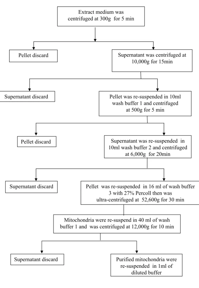
2.2.2. Preparation of mitochondria
The midrib and margins of K. daigremontiana leaves were excised and discarded, and the remaining tissue was cut into small pieces and ground in a blender (National MX-X1, Japan) with 150 mL of ice-cold isolation buffer (350 mM manitol, 250 mM sucrose, 0.1% (w/v) BSA, 1% PVP-40, 5 mM KH2PO4, 5 mM EDTA-KOH (pH 7.4), 1
mM DTT and 100 mM HEPES-KOH (pH 7.4). After filtration through four layers of sterile Mira-cloth (Calbiochem-Novabiochem, La Jolla, CA, USA) the homogenate was centrifuged at 300 g (Tomy CX-250 refrigerated centrifuge, Japan) for 5 min. The resulting supernatant was centrifuged at 10,000 g for 15 min to pellet the mitochondria. The pellets were resuspended in approximately 10 mL of wash buffer (400 mM sucrose, 5 mM KH2PO4, 5 mM EDTA-KOH (pH 7.4), and 50 mM HEPES-KOH (pH 7.4) and then
centrifuged at 500 g for 5 min. The supernatant was again resuspended in 10 mL of wash buffer and centrifuge at 6,000 g for 20 min to pellet mitochondria. The pellets were resuspended in 2.5 mL of wash buffer and then further purified in 16 mL Percoll 27% by
centrifugation at 4oC, 52,600 g (P28S rotor, CP75β ultracentrifuge, Hitachi Koki Co., Ltd, Japan) for 30 min. The mitochondria were found in a band of the lower half of the centrifuge tube, and were extracted from the gradient by pipette. The mitochondria were resuspended in 40 mL of wash medium and pelleted by centrifuging at 12,000 g for 10 min. Final pellets were resuspended in 1mL of assay buffer contained 400 mM sucrose, 0.1% BSA and 40 mM HEPES-KOH (pH 7.4) (Fig. 2.1).
2.2.3. Preparation of leaf extraction for enzyme assays
The tissue frozed by liquid nitrogen (0.5 g fresh weigh) was homogenized using a mortar and pestle with 40 mg of polyvinylpolypyrrolidone in 4mL of ice-cold extraction buffer. The extraction buffer for MDH, NAD-ME and NADP-ME contained 50 mM Tris-HCl of pH 7.8, 8 mM MgCl2, 1 mM EDTA-KOH (pH 7.4), 5 mM DTT, 0.2% (w/v)
BSA and 0.02% (w/v) Triton X-100. The homogenate was filtered through one layer of Mira-cloth and the filtrate was centrifuged at 10,000 g for 10 minutes at 4oC. The supernatant was desalted by passing through 5 mL Sephadex G-25 column (PD-10 column, Pharmacia Biotech AB Uppsala Sweden) previously equilibrated with the enzyme extraction medium. The desalting extract was used immediately for determination of enzyme activity.
2.2.4. Preparation of mitochondria for enzyme assays
The Percoll-purified mitochondria were filtered at room temperature on a column of Sephadex G-25 (PD-10 column, Pharmacia Biotech AB Uppsala Sweden) previously equilibrated with the suspending buffer (400 mM sucrose, 0.1% BSA and 40 mM HEPES-KOH (pH 7.4); thereafter, MDH, NAD-ME and NADP-ME were assayed in mitochondria after lysis with 0.1% (v/v) Triton X-100.
Pellet discard Supernatant was centrifuged at 10,000g for 15min
Pellet was re-suspended in 10ml wash buffer 1 and centrifuged
at 500g for 5 min Supernatant discard
Pellet discard Supernatant was re-suspended in
10ml wash buffer 2 and centrifuged at 6,000g for 20min
Supernatant discard Pellet was re-suspended in 16 ml of wash buffer 3 with 27% Percoll then was
ultra-centrifuged at 52,600g for 30 min
Mitochondria were re-suspend in 40 ml of wash buffer 1 and was centrifuged at 12,000g for 10 min
Supernatant discard Purified mitochondria were re-suspended in 1ml of
diluted buffer Extract medium was
centrifuged at 300g for 5 min
CAM specie.
2.3.2. Enzyme activities
Table 2.1 showed the activities of malate dehydrogenase (MDH), NAD-malic enzyme (NAD-ME) and NADP-malic enzyme (NADP-ME) in leaf tissue and in mitochondria isolated from K. daigremontiana leaves. The activities of NAD-ME, NADP-ME and MDH were detected in leaf extract and in Percoll-purified mitochondria. NAD-ME activity was higher than NADP-ME activity in leaf extracts. This result was similar with the observation of Winter and Smith (1996) who showed that in K.
daigremontiana, the development of CAM in the course of leaf ontogeny was associated
with an increase in NAD-ME activity but a significant declined in NADP-ME activities. In mitochondria of K. daigremontiana, MDH activity was higher than that in leaf. Although NAD-ME was predominantly located in the mitochondria, a small amount of NADP-ME was also detected in these mitochondria (Table 2.1).
2.3.3. Respiratory properties of K. daigremontiana mitochondria
Discontinuous Percoll density gradients had been used for the purification of mitochondria. In our experiment, concentration of 27% Percoll was found to be ideal for purifying K. daigremontiana mitochondria. Percoll-purified K. daigremontiana mitochondria all readily oxidized succinate, malate, NADH and NADPH as substrates (Fig. 2.3). Total respiration rates, respiratory control values (RCR) and ADP/O ratios differed for the substrates (Table 2.2). The results in Fig. 2.3 showed typical electrode traces for the oxidation of substrates by K. daigremontiana mitochondria. The total respiration rates in succinate oxidation (Fig. 2.3A) were near similar with that of NADPH oxidation (Fig. 2.3B), were slightly lower than that with NADH oxidation (Fig. 2.3C) and near double of that with malate oxidation. The ADP/O ratios with succnate, NADH and NADPH
Table 2.2. Respiratory properties of Percoll-purified K. daigremontiana mitochondria.
Oxygen uptake was measured as described in “Materials and Methods”. Concentrations used were: 10 mM succinate, 1 mM NADH, 1 mM NADPH, 10 mM malate, 400 nmol ADP with succinate as a substrate, and, 240 nmol ADP with all another substrates. State 3 refers to the respiration rate of O2 uptake in the presence of ADP; state 4
refers to the rate upon depletion of ADP. Respiratory control ratio (RCR) was calculated as the ratio of state 3 to state 4 rates. Each value was the average of four or five independent experiments.
Respiration rate (nmol O2 min -1mg -1protein)
Substrates State 3 State 4 RCR ADP/O Succinate NADH NADPH Malate 142 ± 18 137 ± 28 124 ± 16 75 ± 12 67 ± 6 60 ± 34 60 ± 12 43 ± 11 2.1 ± 0.2 2.4 ± 0.7 2.0 ± 0.4 1.7 ± 0.3 1.6 ± 0.3 1.5 ± 0.4 1.7 ± 0.2 2.5 ± 0.2
A B
Fig. 2.2. Malate dehydrogenase activities before (A)and after (B) lysis with Triton X-100
in Percoll-purified mitochondria isolated from K. daigremontiana. Assay conditions were shown in ‘Materials and methods’.
CAM specie.
2.3.2. Enzyme activities
Table 2.1 showed the activities of malate dehydrogenase (MDH), NAD-malic enzyme (NAD-ME) and NADP-malic enzyme (NADP-ME) in leaf tissue and in mitochondria isolated from K. daigremontiana leaves. The activities of NAD-ME, NADP-ME and MDH were detected in leaf extract and in Percoll-purified mitochondria. NAD-ME activity was higher than NADP-ME activity in leaf extracts. This result was similar with the observation of Winter and Smith (1996) who showed that in K.
daigremontiana, the development of CAM in the course of leaf ontogeny was associated
with an increase in NAD-ME activity but a significant declined in NADP-ME activities. In mitochondria of K. daigremontiana, MDH activity was higher than that in leaf. Although NAD-ME was predominantly located in the mitochondria, a small amount of NADP-ME was also detected in these mitochondria (Table 2.1).
2.3.3. Respiratory properties of K. daigremontiana mitochondria
Discontinuous Percoll density gradients had been used for the purification of mitochondria. In our experiment, concentration of 27% Percoll was found to be ideal for purifying K. daigremontiana mitochondria. Percoll-purified K. daigremontiana mitochondria all readily oxidized succinate, malate, NADH and NADPH as substrates (Fig. 2.3). Total respiration rates, respiratory control values (RCR) and ADP/O ratios differed for the substrates (Table 2.2). The results in Fig. 2.3 showed typical electrode traces for the oxidation of substrates by K. daigremontiana mitochondria. The total respiration rates in succinate oxidation (Fig. 2.3A) were near similar with that of NADPH oxidation (Fig. 2.3B), were slightly lower than that with NADH oxidation (Fig. 2.3C) and near double of that with malate oxidation. The ADP/O ratios with succnate, NADH and NADPH oxidations were less than 2 indicating that these oxidations were coupled to twoproton -
Table 2.2. Respiratory properties of Percoll-purified K. daigremontiana mitochondria.
Oxygen uptake was measured as described in “Materials and Methods”. Concentrations used were: 10 mM succinate, 1 mM NADH, 1 mM NADPH, 10 mM malate, 400 nmol ADP with succinate as a substrate, and, 240 nmol ADP with all another substrates. State 3 refers to the respiration rate of O2 uptake in the presence of ADP; state 4
refers to the rate upon depletion of ADP. Respiratory control ratio (RCR) was calculated as the ratio of state 3 to state 4 rates. Each value was the average of four or five independent experiments.
Respiration rate (nmol O2 min -1mg -1protein)
Substrates State 3 State 4 RCR ADP/O Succinate NADH NADPH Malate 142 ± 18 137 ± 28 124 ± 16 75 ± 12 67 ± 6 60 ± 34 60 ± 12 43 ± 11 2.1 ± 0.2 2.4 ± 0.7 2.0 ± 0.4 1.7 ± 0.3 1.6 ± 0.3 1.5 ± 0.4 1.7 ± 0.2 2.5 ± 0.2
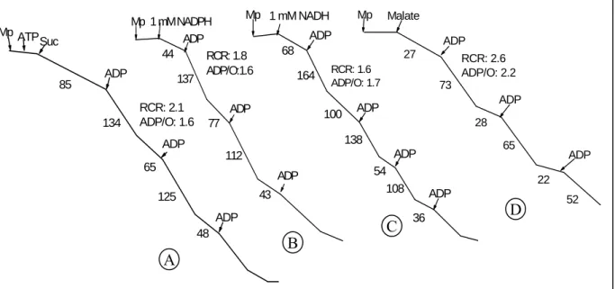
Fig. 2.3. Individual oxidation of succinate (A), NADPH (B), NADH (C) and
malate (D) in Percoll-purified K. daigremontiana mitochondria.
Assay conditions were shown in the legend of Table 2.2. Numbers along the traces refer to nmol O2consumed min-1mg-1protein.
Suc RCR: 2.1 ADP/O: 1.6 48 125 65 134 ADP ADP 85 ADP ATP Mp RCR: 1.8 ADP/O:1.6 43 112 77 137 44 ADP ADP ADP 1 mM NADPH Mp 100 RCR: 1.6 ADP/O: 1.7 36 108 54 138 164 68 ADP ADP ADP ADP 1 mM NADH Mp 22 52 65 28 73 27 RCR: 2.6 ADP/O: 2.2 ADP ADP ADP Malate Mp A B C D
extrusion sites. These ADP/O values were similar to the ADP/O values in washed mitochondria of the other CAM plants such as: Sedum praealtum (Arron et al., 1979), K.
daigremontiana (Day, 1980) and K. blossfeldiana (Rustin and Queiroz-Claret, 1985).
NADH and NADPH were readily oxidized by K. daigremontiana mitochondria in the absence of Ca2+, an activator of external NAD(P)H dehydrogenase. The mitochondria were capable of oxidizing exogenous NADH and NADPH with near similar way, but the respiration rates with NADH were slightly higher than those with NADPH oxidation (Fig. 2.3B and 2.3C). NADPH oxidation by Percoll purified K. daigremontiana mitochondria showed the respiration rate rather higher than that by washed mitochondria of K.
blossfeldiana (104 nmol min-1 mg-1 protein, Rustin and Queiroz-Claret, 1985) and by
washed mitochondria of S. praealtum (28.8 nmol min-1 mg-1 protein, Arron et al., 1979). And the respiration rates of the NADH and NADPH oxidations by Percoll purified mitochondria in our study were near double of that with washed mitochondria of K.
daigremontiana which is previous reported by Day (1980). Fig. 2.3D shows typical O2
uptake patterns obtained with K. daigremontiana mitochondria oxidizing malate as a substrate. The mitochondria isolated from K. daigremontiana oxidized malate (pH 6.8) at significant rates, and the ADP/O ratios in this oxidation were greater than 2 (Table 2.2) indicated that three proton-extrusion sites were utilized. K. daigremontiana mitochondria oxidized malate without any cofactors; however, their oxidation rate was slower than that with other substrates (Table 2.2 and Fig. 2.3D). These results were near similar with those in malate oxidation by washed mitochondria of S. praealtum and sunflower (Arron et al., 1980). In general, before adding ADP, these mitochondria slowly oxidized malate. Upon ADP addition, high rates of oxygen consumption were measured (Fig. 2.3D).
Table 2.3. Respiratory properties of Percoll-purified K. daigremontiana mitochondria in
simultaneous substrate oxidation of succinate and NAD(P)H.
Concentrations used were: 10 mM succinate, 1 mM NADH, 1 mM NADPH, 10 mM malate, 240 nmol ADP for NADH plus NADPH, and 320 nmol ADP for succinate plus with NADH or NADPH. Each value was the average of four or five independent experiments.
Respiration rate (nmol O2 min -1mg -1protein)
Substrates State 3 State 4 RCR ADP/O Succinate + NADH Succinate + NADPH NADH + NADPH 223 ± 23 231 ± 19 134 ± 20 127 ± 19 141 ± 23 52 ± 21 1.8 ± 0.1 1.7 ± 0.2 2.9 ± 1.4 1.6 ± 0.2 1.5 ± 0.4 1.8 ± 0.2
RCR: 1.68 ADP/O: 1.47 68 166 107 204 249 145 128 216 84 ADP ADP ADP ADP ADP Suc + NADH Mp RCR: 1.71 ADP/O: 1.17 145 115 221 161 232 146 249 79 ADP ADP ADP ADP ADP Suc + NADPH Mp 114 27 RCR: 1.89 ADP/O: 1.83 129 126 65 157 63 34 ADP ADP ADP ADP ADP ADP NADH / NADPH Mp
Fig. 2.4. Cooperative oxidations of succinate and NAD(P)H in Percoll-purified
K. daigremontiana mitochondria.
Assay conditions were shown in Table 2.3. Numbers along the traces refer to nmol O2consumed min-1mg-1protein.
oxidation gave ADP/O ratios of lower than 2 indicated that two proton- extrusion sites were utilized. The respiratory rates with simultaneous oxidation of succinate and NADH or succinate and NADPH always were lower than the sum of the individual respiration rates with each substrate but higher than the individual respiration rates with single substrate (Fig. 2.4). These simultaneous oxidation rates were about 80% and 87% of the sum of the individual respiration rates with succinate plus NADH and succinate plus NADPH, respectively. In contract, combination of NADH and NADPH did not increase the respiratory rates and this rate was about 51% of the sum of the individual rates and was near the same rate with NADH or NADPH individual oxidation (Fig. 2.4 and Table 2.3).
2.4. Discussion
In contrast to mitochondria from animal tissues, plant mitochondria can also oxidize cytosolic NADH and in some cases cytosolic NADPH (Heldt, 1997). Our experiments showed that K. daigremontiana mitochondria were able to oxidize both of NADH and NADPH. These results differed from previous report for mitochondria isolated from aged beetroot slices which cannot oxidize exogenous NADPH (Arron et al., 1979). The respiratory properties of mitochondria isolated from K. daigremontiana with NADH (70 nmol min-1 mg-1 protein) and NADPH (64 nmol min-1 mg-1 protein) was reported by Day (1980), using washed mitochondria. Compared with these results, our results showed near double rate (137 ± 28 nmol min-1 mg-1 protein for NADH and 124 ± 16 nmol min-1 mg-1 protein for NADPH), possibly because we used Percoll-purified mitochondria. Percoll gradient markedly decreased the degree of contamination and showed high rate (Fredlund
et al., 1991).
The property of absorbing malate has been detected in many plant mitochondria in which malate can be decarboxylated in the mitochondrial matrix through the action of NAD-ME to produce pyruvate (Artus and Ewards, 1985), which is oxidized by the TCA
cycle. These data showed that K. daigremontiana mitochondria possessed a larger ME activity (Table 2.1), and readily oxidized malate (Table 2.2, Fig. 2.3). This phenomenon fits with previous results on malate oxidation by mitochondria isolated from K.
daigremontiana (Day, 1980) and K. blossfeldiana (Rustin and Lance, 1986).
Previous experiments with other plants mitochondria have shown that the Cyt chain was not saturated by oxidation of any one substrate. For example, in Iris bulb mitochondria the respiration rates with the combination of succinate and NADH were always significantly lower than the sum of the single rate (Hemrika-Wagner et al., 1986). In our research, for K. daigremontiana mitochondria, when the second substrate (NADH or NADPH) was simultaneously present with the first substrate (succinate), the overall rate of respiration was always significantly higher than the individual rates. However, the respiration rate in simultaneous oxidation of NADH and NADPH was not higher than the individual rates (Fig. 2.4 and Table 2.3). This result was deferred with potato mitochondria in which the oxidation of two substrates NADPH and NADH leads to increase the respiration rate (Arrabaca et al., 1992). It may due to in K. daigremontiana, mitochondrial electron flow from NAD(P)H oxidation to UQ in the individual oxidation of NAD(P)H was saturated, however the mitochondrial electron flow from succinate oxidation to UQ in the individual succinate oxidation was unsaturated. In the simultaneous oxidation of succinate and NADH or NADPH, the NAD(P)H dehydrogenases may also contributed in transporting ETC from succinate oxidation to UQ, increasing the number of electron to UQ and Alt pathway, leading to an significant increase not only in the respiration rate but also in Alt respiration. Therefore, it seems that succinate dehydrogenase and NAD(P)H dehydrogenases could together contributed to catalyzed substrates in the simultaneous oxidation of K. daigremontiana mitochondria, but this function is for future study.
2.5. Summary
Percoll-purified mitochondria isolated from K. daigremontiana showed a rather high degree of purify and intactness of the inner membrane. These mitochondria posses malate dehydrogenase (MDH) and NAD(P)-malic enzyme (ME) activities in which MDH activity was about twenty five folds higher than that with NAD-ME. The K.
daigremontiana mitochondria already oxidized succinate, malate, NADH and NADPH
without any cofactor. The highest respiration rate was found with succinate, followed by NADH, NADPH and malate. The respiration rate in succinate oxidation by K.
daigremontiana mitochondria was near twice as far as the rate by malate oxidation.
Mitochondria of K. daigremontiana oxidized NADH and NADPH with the same way; however the respiration rates in NADH oxidations was slightly higher than that with NADPH. The cooperative oxidation of succinate and NADH or NADPH normally leads to increase the respiratory rates that were lower than the sum of the individual rates but higher than the individual rates.
Chapter 3
Oxidations of various substrate and effects of the inhibitors on purified
mitochondria isolated from Kalanchoë pinnata
3.1. Introduction
Mitochondrial respiration of plants differs from that of animals by the presence of an alternative (Alt) pathway in the electron-transport chain (ETC). It branches from the cytochrome (Cyt) pathway at ubiquinone (Q) and donates electrons directly to oxygen to form water. The Alt pathway is inhibited by SHAM and the Cyt pathway is inhibited by KCN. In spite of extensive investigation among higher plants, fungi, yeasts and protozoa, the physiological role of the Alt pathway in ETC is not fully understood. For CAM plants, it has been shown that there was an increase in cyanide-resistant leaf respiration in the phase III of K. blossfeldiana (Rustin and Queiroz-Claret, 1985), and K. daigremontiana (Robinson, 1992). However, the Alt capacity in CAM mitochondria is probably not great enough to support in vivo rates of malate decarboxylation (Wiskich and Day, 1982).
Kalanchoë pinnata is a ME type CAM plant. In the phase III, under closure of the
stomata, malate is released from the vacuole and oxidatively decarboxylated by NAD(P)-ME to generate pyruvate and CO2. Pyruvate is phosphorylated to
phosphoenolpyruvate (PEP) by catalysis of pyruvate orthophosphate dikinase (PPDK), and then it is conserved in gluconeogenesis (Kondo et al., 1998). Recently, the experiment results in our laboratory indicated that PPDK is distributed both in chloroplast and cytosol in K. pinnata mesophyll cell (Kondo et al., 1998). Under low oxygen conditions, these plants lost phase III in CAM-type diurnal gas-exchange activity (Nose et al., 1999). There was an increase of the Alt pathway activity in CAM phase III of K. pinnata leaf (Tsuchiya
et al., 2001). These results suggest that not only ME but also Alt pathway plays an
important role during the malate decarboxylation in the phase III of K. pinnata intact leaf and raise some further questions about K. pinnata mitochondria. How do the mitochondria contribute to total malate decarboxylation during the phase III? How to reduce the NADPH produced from malate decarboxylation via cytosol NADP-ME? How does the cytosolic PPDK activity involve to the mitochondrial ATP synthesis? Is there any relationship between the malate oxidation and the Alt pathway in mitochondria? Are these activities in mitochondria involved with those in intact leaf of K. pinnata or not? Based on these view points, we investigated respiratory properties with various substrates and effects of the inhibitors on the Alt pathway in K. pinnata mitochondria.
3.2. Materials and methods
3.2.1. Plant material and mitochondria respiration
Kalanchoë pinnata were vegetatively propagated and grown in plastic pots in a
greenhouse with heater under natural light. Ten days before the experiments, the 3 month old plants were transferred to a growth chamber (KG-50 HLA, Koito Industrial Co., LTD., Japan) with a photoperiod of 12 h light and 12 h dark. The temperature in the growth chamber was maintained at 35oC during the light period and 25oCduring the dark period with photosynthetically active radiation at the mid-plant height of 420 to 450 µmol m-2 s-1.
The fifth to seventh leaves, numbered from the apex, were used for the experiments. The leaves were harvested at 6 to 7 h after the beginning of the light period. The harvested leaves were transported to the laboratory, rinsed thoroughly with distilled water and used for isolating of mitochondria.
Mitochondria of K. pinnata were isolated and purified on Percoll gradients as described previously (see Method in chapter 2, Hong et al., 2004). Oxygen consumption was measured using a liquid type oxygen electrode (Rank Brothers England) at 25oC in 2
mL of reaction medium [(300 mM mannitol, 10 mM KH2PO4, 5 mM MgCl2, 10 mM KCl,
100 mM HEPES-KOH (pH 7.4)] and pH was adjusted from 6.8 to 7.8 with KOH. The O2
concentration in air-saturated medium was taken as 258 µM. RCR and ADP/O ratios value were calculated according to Estabrook (1967). The protein content was measured by the method of Bradford (1976) using BSA as the standard. Chlorophyll content was determined according to Arnon (1949).
3.2.2. Preparation of leaf extraction and mitochondria for enzyme assays.
The leaf sample (0.5 g fresh weight) was homogenized using a mortar and pestle with 0.2 g sea sand and 40 mg PVP in 4 mL of ice-cold extraction buffer. The extraction buffer for MDH, NAD-ME and NADP-ME contained 50 mM Tris-HCl of pH 7.8, 8 mM MgCl2, 1 mM EDTA-KOH (pH 7.0), 5 mM DTT, 0.2% (w/v) BSA and 0.02% (w/v) Triton
X-100. The homogenate was filtered through one layer of Miracloth (Calbiochem-Novabiochem, La Jolla, CA, USA). Part of the homogenate was taken for determination of chlorophyll content; the other homogenate was centrifuged at 10,000 g for 10 minutes at 4oC. The supernatant was desalted by passing through a Sephadex G-25 (PD-10 column, Pharmacia Biotech AB Uppsala Sweden) that had been equilibrated with the enzyme extraction medium. The desalting extract was used immediately for determination of enzyme activities.
3.2.3. Preparation of mitochondria for enzyme assays
The mitochondria were filtered at room temperature on a column of Sephadex G-25 (PD-10 column, Pharmacia Biotech AB Uppsala Sweden) previously equilibrated with the suspending buffer contained 400 mM sucrose, 0.1% BSA and 40 mM HEPES-KOH (pH 7.4), thereafter, MDH, NAD-ME and NADP-ME were assayed in mitochondria after lysis with 0.1% (w/v) Triton X-100.
MDH (L-malate: NAD oxidoreductase, EC 1.1.1.37) and NAD-dependent ME (EC 1.1.1.39) were assayed according to Pastore et al. (2001). NADP-dependent ME (EC 1.1.1.40) was assayed according to Kondo et al. (2000). Rubisco was assayed according to Du et al. (1996). PEPC was assayed according to Shaheen et al. (2002).
3.3. Results
3.3.1. Enzyme activities
PEPC and Rubisco were localized unambiguously in the cytosol and chloroplast, respectively, of K. pinnata mesophyll cells (Kondo et al., 1998), so that their activities can be used as the indicators of mitochondrial purity. Rubisco activity was null and PEPC activity in mitochondria was approximate by 4.4% of that in cytosol (Table 3.1). These results indicated that the mitochondria suspensions did not contain chloroplast components and the cytosol contamination of the mitochondria was rather low.
Activities of NAD-ME, NADP-ME and MDH were detected in leaf extract and in
K. pinnata mitochondria. NAD-ME activity was higher than NADP-ME activity in leaf
extracts of K. pinnata (Table 3.2). Although NAD-ME was predominantly located in the mitochondria, a small amount of NADP-ME was also detected in K. pinnata mitochondria. MDH activity in mitochondria was higher than that in leaf of K. pinnata.
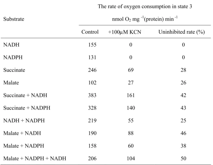
3.3.2. Respiratory properties of K. pinnata mitochondria with the single substrate
Respiratory properties with several different single substrates were shown in Table 3.3. The highest respiration rates were recorded with succinate. They exceeded those for NADH, NADPH and malate (pH 6.8) in the presence of 1 mM TPP, 1.6, 1.9, and 1.8 times, respectively. The ADP/O ratios in succinate oxidation were less than 2, indicating that their oxidations were coupled to twoproton-extrusion sites. K. pinnata mitochondria were able to oxidase external NADH and NADPH with near by similar way; however, the rates of NADH oxidation were rather higher than those of NADPH oxidation (Table 3.3). Malate
Table 3.1. Rubisco and PEPC activities in leaf extract and mitochondria of K. pinnata.
Results shown are means ± SE (n= 4-5) of separate preparations. Rubisco showed initial activity. ND, not detectable.
Enzyme Leaf * Leaf ** Mp **
PEPC 12.97 ± 1.04 0.48 ± 0.04 0.021 ± 0.011 Rubisco 5.99 ± 2.15 0.17 ± 0.03 ND
Enzyme activities are expressed as: * µmolmg-1(chlorophyll) min-1,
Table 3.2. Enzyme activities in leaf extract and in mitochondria of K. pinnata.
Results shown are means ± SE (n= 4-5) of separate preparations. Enzyme activities are expressed as µmolmg-1(protein) min-1.
Enzyme Leaf Mp NADP-ME 0.05 ± 0.01 0.096 ± 0.013 NAD-ME 0.11 ± 0.06 0.95 ± 0.09
Table 3.3. Individual substrate oxidation in K. pinnata mitochondria.
Assay conditions were 10 mM succinate, 1 mM NADH, 1 mM NADPH, 10 mM malate, 1 mM TPP, 0.1 mM CoA, 0.5 mM NAD, and 0.16 mM ADP. State 3 refers to the respiration rate of O2 uptake in the presence of ADP; state 4 refers to the rate upon depletion of ADP.
Respiratory control ratio (RCR) was calculated as the ratio of state 3 to state 4 rates. Results shown are means ± SE (n= 4-5) of separate preparations. ND, Not detectable.
Respiration rate nmol O2 mg –1(protein) min -1
Substrates State 3 State 4 RCR ADP/O Succinate NADH NADPH Malate (pH 7.6, NAD)
Malate (pH 7.2, glutamate, NAD ) Malate (pH 6.8)
Malate (pH 6.8, CoA, NAD) Malate (pH 6.8, TPP) 256 ± 30 155 ± 41 131 ± 23 24 ± 7 41 ± 8 62 ± 16 105 ± 11 139 ± 25 119 ± 26 71 ± 14 60 ± 32 ND 32 ± 6 31 ± 11 74 ± 9 90 ± 23 2.2 ± 0.4 2.1 ± 0.5 2.5 ± 1.0 ND 1.3 ± 0.3 2.0 ± 0.5 1.4 ± 0.4 1.5 ± 0.2 1.5 ± 0.4 1.4 ± 0.3 1.4 ± 0.4 ND 2.2 ± 0.1 2.4 ± 0.3 2.4 ± 0.2 2.3 ± 0.2
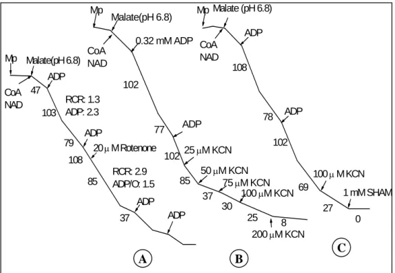
oxidation was investigated under three different pH conditions; at pH 7.6, where only MDH was active, at pH 7.2 where both ME and MDH were active and at pH 6.8 where only ME was active (Agius et al., 1998, Day et al., 1988). NAD was included in all cases to stimulate the optimum ME and MDH activities. K. pinnata mitochondria oxidized malate with varied levels depending on pH assay conditions. The rates of malate oxidation were very low at pH 7.6, whereas they rather increased at pH 7.2 (Table 3.3). K. pinnata mitochondria were able to oxidize malate without any cofactors at pH 6.8; however, its rate was slower than that in the presence of TPP (a pyruvate dehydrogenase activator) or CoA (a ME activator). Adding TPP or CoA and NAD stimulated malate oxidation via ME. The highest rates of malate oxidation were observed at pH 6.8 in the presence of TPP. In general, before adding ADP, K. pinnata mitochondria slowly oxidized malate. Upon ADP addition, high rates of oxygen consumption were measured. In all cases, the ADP/O ratios of malate oxidation were greater than 2 (Table 3.3), indicating that three proton-extrusion sites were utilized.
3.3.3. Respiratory properties of K. pinnata mitochondria with the multiple substrates
The respiratory properties with two or three substrates of succinate, NADH, NADPH and succinate were shown in Table 3.4. Generally, simultaneous oxidation of these substrates normally increased the respiration rates that were lower than the sum of the individual rates but higher than the individual rates, and it was more marked when succinate was the first substrate. The simultaneous oxidations of malate as a first substrate with other substrates were investigated at pH 6.8 in the presence of TPP (Table 3.4 and Fig. 3.1) or in the presence of CoA and NAD (Table 3.5). Addition of NADPH or NADH as the second substrates mainly stimulated the respiration rate that was significantly higher than individual rate, and then addition of NADH as a third substrate did not further increase the simultaneous rate (Table 3.4 and Fig. 3.1). In our experiments, all of the simultaneous
Fig. 3.1. Effects of KCN and SHAM in the presence of rotenone on the malate oxidation
at pH 6.8 in the presence of 1 mM TPP with other substrates. Malate and NADH (A), malate and NADPH (B) and malate, NADH and NADPH (C).
Assay conditions were: 20 mM rotenone, 10 mM malate, 1 mM NADH, 1 mM NADPH, 0.16 mM ADP (first and second adding) and 0.32 mM ADP (last adding). Numbers along the traces refer to nmol O2consumed min-1mg-1protein.
9% 194 17 147 263 ADP/O: 1.2 RCR: 1.8 ADP/O: 1.4 RCR:1.2 215 265 120 50 µ M KCN Rotenone ADP ADP ADP NADH Malate Mp 13% 23 180 101 222 ADP/O:1.5 RCR: 2.2 ADP/O: 1.3 RCR: 1.3 155 209 87 50 µ M KCN Rotenone Malate NADPH ADP ADP ADP Mp 35% ADP/O: 1.1 RCR: 1.6 ADP/O: 1.4 RCR: 1.2 0 67 191 129 203 172 206 83 1 mM SHAM 50 µ M KCN Rotenone ADP ADP ADP Malate NADH NADPH Mp A B C
Table 3.4. Simultaneous substrate oxidation in K. pinnata mitochondria.
Results shown are means ± SE (n= 4-5) of separate preparations. Assay conditions were 1 mM NADH, 1 mM NADPH, 0.16 mM ADP, 10 mM malate at pH 6.8 in the presence of 1 mM TPP and 10 mM succinate in the presence of 10 mM ATP.
Respiration rate nmol O2 mg -1(protein) min -1
Substrates State 3 State 4 RCR ADP/O Succinate + NADH Succinate + NADPH NADH + NADPH Malate + NADH Malate + NADPH
Malate + NADPH + NADH
364 ± 22 329 ± 10 231 ± 12 249 ± 49 204 ± 22 216 ± 17 202 ± 12 218 ± 16 112 ± 23 181 ± 32 128 ± 23 139 ± 31 1.8 ± 0.3 1.6 ± 0.2 2.1 ± 0.8 1.4 ± 0.6 1.7 ± 0.7 1.6 ± 0.4 1.5 ± 0.2 1.2 ± 0.2 1.5 ± 0.2 1.3 ± 0.3 1.5 ± 0.2 1.3 ± 0.2