Hedgehog Signal and Genetic Disorders
23
0
0
全文
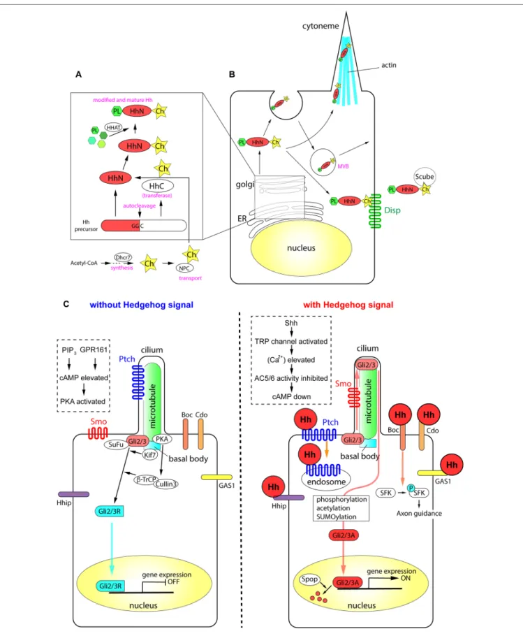
(2) Sasai et al.. Recent Progress on Hedgehog Research. FIGURE 1 | The processing of Hh proteins and their intracellular signaling pathways. (A) Cleavage of Hh polypeptides and their modifications in ER and golgi apparatus. (B) Processing, modifications and transport of Hh proteins in the producing cells. (C) Active signaling pathways in absence and in presence of Hh molecules. Schemas in dotted rectangles represent regulation of cAMP level in the cilia. The figure was made based on the information in (Varjosalo and Taipale, 2008; Balaskas et al., 2012; Moore et al., 2016; Parchure et al., 2018).. Frontiers in Genetics | www.frontiersin.org. 2. November 2019 | Volume 10 | Article 1103.
(3) Sasai et al.. Recent Progress on Hedgehog Research. Production and Secretion of Hh Proteins. Membrane Receptors in Hh-Responding Cells. All Hh polypeptides produced are transported into the endoplasmic reticulum (ER) and Golgi apparatus, where they undergo autoprocessing (Jeong and McMahon, 2002; Cai and Liu, 2016). The polypeptides are cleaved into two parts, the amino-terminal domain, which functions as a signaling molecule, and the carboxyl-terminal domain, which functions in autoprocessing regulation (Mann and Beachy, 2004). The amino-terminal part is further modified with palmitate and cholesterol (Porter et al., 1996; Chamoun et al., 2001; Lewis et al., 2001; Mann and Beachy, 2004; Chen et al., 2004a; Chen et al., 2011a; Li et al., 2006; Hardy and Resh, 2012). The carboxyl ends of the initial polypeptides act as cholesterol transferases (Porter et al., 1996), and palmitoylation is mediated by skinny Hh (Ski) acyltransferase (also known as Hh acyltransferase or HHAT), a transmembrane acyltransferase located in the ER (Chamoun et al., 2001; Dennis et al., 2012; Konitsiotis et al., 2015). The modified Shh protein was recently purified, and the fatty acids bound to the amino terminus were identified, which revealed that other unsaturated fatty acids in addition to palmitate are involved in the modification (Long et al., 2015). These lipid modifications are essential for the stability of the protein in the extracellular matrix (Callejo et al., 2006) and during long-range transport (Lewis et al., 2001; Zeng et al., 2001). The NiemannPick C (NPC1/2) proteins transport cholesterol from the endosome to the ER (Loftus et al., 1997; Tukachinsky et al., 2012; Formichi et al., 2018), and play an essential role in cholesterol modification of Hh. The membrane protein dispatched (Disp) and the secreted protein Scube2 bind to the cholesterol moiety of the modified Hh proteins (Tukachinsky et al., 2012) and release them from the cell membrane. Some of the Hh proteins at the cell surface can be recycled. After recycling, the Hh proteins are released by lipid bilayer membrane vesicles called exosomes (Vlassov et al., 2012; Parchure et al., 2018). Exosomes are formed at the multivesicular body (MVB) in the cytosol, and bud from the plasma membrane with a size of 30–100 nm containing functional molecules (Parchure et al., 2018). For exosome formation, membrane proteins collectively called endosomal sorting complexes required for transport (ESCRT) are essential (Juan and Furthauer, 2018). A specialized filopodial structure, the cytoneme, which forms in Hh-producing cells, is important for transmission of the Hh signal. The cytoneme was initially identified in flies (Bischoff et al., 2013; Gonzalez-Mendez et al., 2017), and later reported in the chick embryonic limb bud (Panman and Zeller, 2003; Sanders et al., 2013). The presence of microvesicles and the cytoneme, together with the palmitoylation and cholesterol modification of Hh proteins, mediate the transport of the Hh signal to distant parts in the tissues (Chen et al., 2004a; Sanders et al., 2013; D'Angelo et al., 2015). Overall, the efficient production and secretion of active Hh proteins involves the following: (i) processing of the polypeptides by autocleavage, (ii) modification of the polypeptides by cholesterol and palmitate, (iii) recycling and packaging of the proteins in the microvesicles, and (iv) the presence of cytosolic structures including cytonemes.. Frontiers in Genetics | www.frontiersin.org. When Hh proteins reach the surface of a responding cell, the signals are introduced into the cell by two membrane proteins, Ptch and Smo. Ptch, a receptor protein that directly interacts with Hh proteins (Marigo et al., 1996; Motoyama et al., 1998b), is a 12-span membrane protein that localizes to the ciliary membrane in the absence of Hh (Rohatgi et al., 2007). Ptch binds to Hh, forming a complex that moves from primary cilia to the plasma membrane. In the case of Shh, the Ptch-Shh complex undergoes endocytosis, which requires the membrane-remodeling GTPase dynamin (Incardona et al., 2000; Ferguson and De Camilli, 2012) and the HECT-domain ubiquitin E3 ligases Smurf1/2 (Yue et al., 2014), and leads to degradation in the lysosome. Once Ptch is cleared from the ciliary membrane, the G proteincoupled receptor (GPCR) Smo is translocated from the plasma membrane to the cilium (Corbit et al., 2005). During this process, Smo is phosphorylated at specific serine residues in the carboxyl intracellular region by casein kinase 1α (CK1α) and G proteincoupled receptor kinase 2 (GRK2) (Chen et al., 2004b; Chen et al., 2011b), and interacts with β-arrestin 2 (Chen et al., 2004b). Smo further forms a complex with the kinesin-like protein Kif3a, and is translocated into primary cilia (Kovacs et al., 2008). β-Arrestin 2 is essential for the translocation of Smo into cilia (Kovacs et al., 2008), and contributes to the dynamin-dependent internalization of Smo (Chen et al., 2004b). The role of β-arrestin 2-mediated internalization of the signaling molecule (Ma and Pei, 2007), together with its involvement in the translocation of Smo, suggests that β-arrestin 2 regulates the activity and the stability of the Smo protein. The Ptch polypeptide contains a sterol sensing domain (Martin et al., 2001; Strutt et al., 2001) and binds to cholesterol. The interaction of Ptch and cholesterol is necessary for pumping out cholesterol molecules from the endosome or from the plasma membrane (Bidet et al., 2011). In the absence of Hh, Ptch pumps out the cholesterol molecule (Denef et al., 2000; Khaliullina et al., 2009; Blassberg and Jacob, 2017). By contrast, when cells are exposed to Hh, Ptch is transported into the endosome and degraded (Incardona et al., 2000; Yue et al., 2014), and Smo is modified with cholesterol. Modified Smo activates cytosolic phospholipase A2 to release a fatty acid, arachidonic acid, and the interaction between arachidonic acid and Smo promotes the ciliary localization of Smo through allosteric changes (Arensdorf et al., 2017). Altogether, the cholesterol modification of Smo is essential for its complete activity and for the localization of the protein (Blassberg et al., 2016; Huang et al., 2016; Luchetti et al., 2016).. Intracellular Signaling Pathways and Gli Transcription Factors. Activation of the Hh signal at the membrane level leads to its transduction into the nucleus by the transcription factors Gli2/3. Gli2/3 are Krüppel-like transcription factors containing zincfinger DNA-binding domains with dual activity: the amino terminus contains a transcriptional repressor domain and the. 3. November 2019 | Volume 10 | Article 1103.
(4) Sasai et al.. Recent Progress on Hedgehog Research. carboxyl terminus has an activator domain. In the absence of the extracellular Hh ligand, the synthesized Gli2/3 polypeptides form complexes with protein kinase A (PKA), CK1, and glycogen synthase kinase 3 (GSK3), the latter two of which are serinethreonine protein kinases (Tempe et al., 2006). This complex interacts with the scaffold protein SuFu in the absence of the Hh signal. The role of SuFu is to stabilize the Gli2/3 proteins (Chen et al., 2009; Humke et al., 2010); in the absence of SuFu, both Gli2/3 proteins are drastically destabilized and become undetectable by western blotting (Chen et al., 2009; Humke et al., 2010). Activation of the Hh signaling pathway disrupts the interaction between SuFu and Gli (Tukachinsky et al., 2010). In the absence of an extracellular Hh ligand, the calcium level in the cytosol and in the cilium is relatively high (Moore et al., 2016). Gli2/3 are phosphorylated by PKA and subsequently targeted by the Skp, cullin, F-box (SCF) ubiquitin ligase complex, which is composed of the scaffold protein cullin 3 and the E3-type ubiquitin ligase β-Trcp (Bhatia et al., 2006; Wang and Li, 2006). The Gli2/3 proteins are cleaved at the amino terminus, and the truncated proteins with the transcriptional repressor domains (Gli2/3R; R for "repressive") are translocated into the nucleus to bind to DNA target sites (Svard et al., 2006; Chen et al., 2009). As Smo couples with Gi-type G proteins (Riobo et al., 2006), the exposure of cells to Hh (i.e., activation of Smo) reduces PKA activity (Moore et al., 2016), which decreases cAMP levels. Thus, the newly synthesized Gli2/3 polypeptides are not cleaved, and the full-length polypeptides are maintained. These Gli2/3 polypeptides are further modified by sumoylation, phosphorylation, and acetylation (Cox et al., 2010; Humke et al., 2010; Coni et al., 2013; Niewiadomski et al., 2014), and translocated into the nucleus to act as transcriptional activators (Gli2/3A; A for “active”). In the nucleus, the representative DNA sequence for Gli2/3 is GACCACCCA/TGGGTGGTC (Sasaki et al., 1997). However, recent chromatin immunoprecipitation (ChIP) and computational prediction studies identified several other sequences with different affinities (Vokes et al., 2007; Vokes et al., 2008; Oosterveen et al., 2012; Peterson et al., 2012). Compared with Gli2/3R, Gli2/3A are relatively labile, and are ubiquitinated at the nucleus by the E3 ubiquitin ligase adaptor speckle-type POZ protein (Spop) (Humke et al., 2010; Cai and Liu, 2017). Target genes of the Hh signaling pathway are cell typedependent. In most cells responsive to Hh, the common target genes are Ptch and Gli1 (Stamataki et al., 2005; Cohen et al., 2015; Tickle and Towers, 2017). In the developing neural tube, the Nkx6.1, Olig2, Nkx2.2, and FoxA2 genes (Vokes et al., 2007; Kutejova et al., 2016), which confer ventral neural fates, have Gli-binding sites in their regulatory regions (mentioned in detail later).. proteins with similar affinity to Ptch (Chuang and McMahon, 1999). Although the downstream pathway remains to be fully elucidated, Hhip overexpression blocks Shh signaling activity, suggesting that Hhip is a negative regulator of Hh signaling (Bishop et al., 2009; Kwong et al., 2014). However, this effect occurs in a non-cell autonomous manner. The Hhip protein is released from producing cells and directly binds to Shh to block signaling (Kwong et al., 2014). On the other hand, in Shh-producing cells, Hhip gene expression is induced by the Hh signal, and the Hhip protein is internalized and degraded upon Smo activation (Kwong et al., 2014), thereby enabling cell autonomous Hh signaling. This complex regulatory loop may explain the temporal changes in Hh signaling activity (Dessaud et al., 2007; Balaskas et al., 2012). Other Hh receptor proteins identified include the glycosylphosphatidylinositol-anchored cell-surface protein growth arrest-specific 1 (Gas1), the membrane protein cell-adhesion molecule-related, downregulated by oncogenes (Cdo), and brother of Cdo (Boc) (Tenzen et al., 2006; Zhang et al., 2006; Allen et al., 2007; Allen et al., 2011). Cdo and Boc have immunoglobulin and fibronectin type III (FNIII) repeats in the extracellular domain, and possess structural properties similar to those of the axon guidance receptors Robo and deleted in colorectal cancer (TessierLavigne and Goodman, 1996; Okada et al., 2006). Cdo and Boc function in axon guidance (Okada et al., 2006). In the developing chick limb, Cdo and Boc localize to discrete microdomains along extensive filopodial structures in the signal-receiving cells, and these receptor proteins interact with the filopodia (cytonemes) of Shh-producing cells (Sanders et al., 2013). Hhip, Gas1, Cdo, and Boc bind to Shh directly. However, in contrast to Ptch, which localizes to cilia in the absence of Hh, these membrane proteins localize to the plasma membrane including filopodial structures. The distinct localization of the proteins may account for the variation in temporal and/or spatial Hh signaling activity. In summary, receptors in cilia and the plasma membrane interact with Hh proteins and transduce the signals into the nucleus, where Gli transcription factors act as effectors of Hh signaling.. FUNCTIONS OF HEDGEHOG PROTEINS AND THEIR RELATED GENES IN TISSUES Hh plays an important role in many biological events, and mutant mice deficient in Hh-related genes therefore show various and severe phenotypes. Mutations, deficiencies, and abnormal expression levels of Hh proteins and related genes cause malformations, hyperplasia, and growth retardation in tissues, especially in the central nervous system (CNS), craniofacial structures, limbs, and skeleton at developmental stages. Conversely, persistent or aberrantly high Hh activation leads to tumorigenesis in organs at postnatal stages. In the next sections, we discuss the tissue distribution of Hh proteins, especially Shh, and the related genes. In addition, we provide data on the critical roles of these molecules in development and tissue homeostasis, which are mostly derived from studies in model mice. The principal gene knockout or mutant phenotypes in mice are summarized in Table 1.. Other Hh Receptors. Additional Hh receptors that act in concert with Ptch and/or Smo have been identified and characterized. Hh-interacting protein (Hhip) is a single-pass membrane glycoprotein with epidermal growth factor (EGF)-repeat domains that binds to the three Hh. Frontiers in Genetics | www.frontiersin.org. 4. November 2019 | Volume 10 | Article 1103.
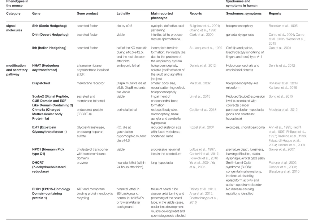
(5) Phenotypes in the mouse. Sasai et al.. Frontiers in Genetics | www.frontiersin.org. TABLE 1 | Main phenotypes and symptoms caused by the mutations and deficiencies of the Hh-related genes in mice and in humans. Syndromes and symptoms in human. Category. Gene. Gene product. Lethality. Main reported phenotype. Reports. Syndromes; symptoms. Reports. signal molecules. Shh (Sonic Hedgehog). secreted factor. die by e9.5. Roessler et al., 1996. secreted factor. viable. Bulgakov et al., 2004; Chiang et al., 1996 Clark et al., 2000. holoprosencephary. Dhh (Desert Hedgehog). cyclopia, defective axial patterning infertile; fail to produce mature spermatozoa. gonadal dysgenesis. Ihh (Indian Hedgehog). secreted factor. Cleft lip and palate, brachydactyly (shortning of fingers and toes) type A-1. a transmembrane acyltransfrase localised at ER. Dennis et al., 2012. Holoprosencephaly and craniofacial defects. Dennis et al., 2012. Dispatched. membrane receptor. Ma et al., 2002. holoprosencephaly-like microform. Roessler et al., 2009; Kantarci et al., 2010. Scube2 (Signal Peptide, CUB Domain and EGF Like Domain Containing 2) Chmp1a (Charged Multivesicular body Protein 1a). secreted and membrane-tethered. Lin et al., 2015. perinatal lethal. Reduced Scube2 expression level is associated with colorectal cancer pontocerebellar hypoplasia (pons and cerebellar hypoplasia). Song et al., 2015. endosomal protein (ESCRT-III). Ext1 (Exostosin Glycosyltransferase 1). Glycosyltransferase, producing heparan sulfate. KO: die at gastrulation hypomorphic mutant: die e14.5. incomplete forelimb formation. Perinatally die due to the problem of the respiratory system holoprosencephaly, acrania (malformation of the skull) and agnathia (no jaw) smaller body size, neural patterning defect, holopocensephaly Impairment of endochondral bone formation reduced body size, microcephaly, basal ganglia and cerebellar hypoplasia reduced skeleton size with fused vertebrae, shortened limbs. St-Jacques et al., 1999. HHAT (Hedgehog acyltransferase). half of the KO mice die during e10.5-e12.5, and the rest die soon after birth embryonic lethal. Canto et al., 2004; Canto et al., 2005; Werner et al., 2015 Gao et al., 2001. Koziel et al., 2004. exostosis, chondrosarcoma. NPC1 (Niemann Pick type C1). cholesterol transporter with transmembrane domains enzyme. viable. progressive neuronal loss in the cerebellum. neonatal lethal (within 24 hours after birth). lung hypoplasia. Loftus et al., 1997; Canterini et al., 2017; Formichi et al., 2018 Yu et al., 2004; Yu et al., 2005. ATP and membrane binding protein; endocytic recycling. prenatal lethal in B6 background; normal in 129/SvEv or SwissWebster background. failure of neural tube closure, axial turning and patterning of the neural tube; in the viable cases, ocular lens development, muscle development and spermatogenesis affected. premature death; lumsiness, learning difficulties, ataxia, dysphagia,vertical gaze palsy Smith-Lemli-Opitz syndrome (SLOS); congenital malformations, intellectual disability, epileptiform activity and autism spectrum disorder No disease-causing mutations identified. Ahn et al., 1995; Hecht et al., 1997; Philippe et al., 1997; Raskind et al., 1998; Faiyaz-Ul-Haque et al., 2004; Heinritz et al., 2009 Garver et al., 2007. modification and secretory pathway. 5. EHD1 (EPS15-Homology Domain-containing protein 1). Coulter et al., 2018. Rainey et al., 2010; Arya et al., 2015; Bhattacharyya et al., 2016. Mochida et al., 2012. Patrono et al., 2002; Cooper et al., 2003; Blassberg et al., 2016. (Continued). Recent Progress on Hedgehog Research. November 2019 | Volume 10 | Article 1103. DHCR7 (7-dehydrocholesterol reductase). DispA mutants die at e9.5; DispB mutants are viable viable.
(6) Phenotypes in the mouse Category. membrane receptors and their associated proteins. Sasai et al.. Frontiers in Genetics | www.frontiersin.org. TABLE 1 | Continued Syndromes and symptoms in human Gene product. Lethality. Main reported phenotype. Reports. Syndromes; symptoms. Reports. GPC3 (Glypican-3). Heparan sulfate proteoglycan. can be born, but die before weaning. pre/postnatal overgrowth. Cano-Gauci et al., 1999. Neri et al., 1998; Capurro et al., 2008. Ptch1 (Patched1). 12-span transmembrane protein. die by e10.5. Marigo et al., 1996; Goodrich et al., 1997. Ptch2 (Patched2). 12-span transmembrane protein. viable, fertile. Motoyama et al., 1998b; Nieuwenhuis et al., 2006. basal cell nevus syndrome (BCNS), Gorlin syndrome, medulloblastoma. Fujii et al., 2013. Smo (Smoothened). G-protein coupled receptor. die by e9.5. Zhang et al., 2001a. active mutations: basal cell carcinoma, Curry-Jones Syndrome. Xie et al., 1998; Twigg et al., 2016. Hhip (Hedgehoginteracting protein 1). membrane protein. neonatal lethal. the neural tube fails to close completely. Overgrown head folds, hindbrain, and spinal cord KO; alopecia and skin lesions mutant; defect in opic cup morphogenesis, and optic fissure and stalk formation fail to turn, arresting at somite stages with a small, linear heart tube, an open gut and cyclopia; similar to the phenotypes in Shh/Ihh double KO lung and endochondral skeleton development is affected. Simpson-Golabi-Behmel overgrowth syndrome (SGBS), pre-and postnatal overgrowth with visceral and skeletal anomalies basal cell nevus syndrome (BCNS), Gorlin syndrome, medulloblastoma. Chuang and McMahon, 1999; Chuang et al., 2003. Zhou et al., 2012. Gas1 (Growth-arrestspecific 1). GPI-anchored cell surface protein. die within 3 days after birth. Lee et al., 2001; Liu et al., 2001; Allen et al., 2007; Seppala et al., 2007. Cdo/Cdon (Cell-adhesion molecule-related, downregulated by oncogenes). a membrane protein of Immunoglobulin superfamily. perinatal lethal; 60% die within 21 days after birth. eye, cerebellar and limb deficiencies; midfacial hypoplasia, premaxillary incisor fusion, and cleft palate mild holoprosencephaly; midline structures lost. decreased expression of Hhip found in chronic obstructive pulmonary disease (COPD) patients Holoprosencephaly. Holoprosencephaly. Bae et al., 2011. Boc (Brother of Cdo). a membrane protein of Immunoglobulin superfamily, similar to Cdo single-span receptor. viable. Cole and Krauss, 2003; Tenzen et al., 2006; Zhang et al., 2006; Allen et al., 2007 Okada et al., 2006. modifying the expressivity of holoprosencephaly. Hong et al., 2017. Willnow et al., 1996; Christ et al., 2012; Baardman et al., 2016 Elyada et al., 2011. Donnai-Barrow syndrome; HPE-like phenotypes. Kantarci et al., 2007. overexpressed in colorectal cancer. Richter et al., 2018. 6. Gene. serine/threonine kinase. perinatal lethal; 60% die within 21 days after birth die before e6.5. unknown. Ribeiro et al., 2010; PinedaAlvarez et al., 2012. (Continued). Recent Progress on Hedgehog Research. November 2019 | Volume 10 | Article 1103. LRP2/megalin/GP330 (Low density lipoproteinrelated protein 2) casein kinase 1a (CK1α). misguidance of commissural axons towards the floor plate. defective forebrain develoipment. Goodrich et al., 1997.
(7) Phenotypes in the mouse Category. signal mediators. Syndromes and symptoms in human Gene. Gene product. Lethality. Main reported phenotype. Reports. Syndromes; symptoms. Reports. G protein-coupled receptor kinase 2 (GRK2). serine/threonine kinase. die at e9–12. marked cardiac abnormalities. Jaber et al., 1996; Penela et al., 2010. Penela et al., 2010. β-Arrestin2. GPCR modulator. viable. Bohn et al., 1999; Thathiah et al., 2013. Kif3a. kinesin-like protein. die at e10.5. sensitivity to pain change; reduced generation of amyloid-b peptide randomization of laterality in heart looping (sinus inversus). GRK2 is upregulated in granulosa cell tumours and in thyroid carcinoma reduced level of amyloid-β peptide, hepatocellular carcinoma. Lefevre et al., 2008; Sipe and Lu, 2011. Costal2/Kif7. Kinesin-4 family, with an N-terminal motor domain. die at the end of gestation. preaxial polydactyly, exencephaly, and microphthalmia. SuFu (Suppressor of Fused). scaffold protein. die at e9.5. ventralised neural tube (similar phenotypes to those of Ptch1 mutant). Endoh-Yamagami et al., 2009; Liem et al., 2009; Hsu et al., 2011; He et al., 2014a Svard et al., 2006; Chen et al.,. 2009. maybe involved in Usher's syndrome (photoreceptor degeneration), but no case reports have been given fetal hydrolethalus and acrocallosal syndromes. Svard et al., 2006; De Mori et al., 2017. Spop (Speckle-type POZ protein). E3 ubiquitin ligase adaptor. neonatal lethal. perturbation of skeletal development. Cai and Liu, 2016. Gorlin syndrome, JBTS32 congenital ataxia, cerebellar vermis hypoplasia, craniofacial dysmorphisms, polydactyly. serous tumors, prostate cancer. Gli1. transcription factor (Krüppel-like zinc finger). viable and fertile. no apparent phenotype. Park et al., 2000. Gli2. transcription factor (Krüppel-like zinc finger). die at birth. Mo et al., 1997; Ding et al., 1998; Motoyama et al., 1998b. Gli3. transcription factor (Krüppel-like zinc finger). viable and fertile. foregut defects, stenosis of the oesophagus and trachea, hypoplasia and lobulation defects of the lung. No floor plate in the neural tube enlarged maxillary arch, a reduced external nasal process, poorly developed eyes, misplaced ears, anomalous numbers of mystacial and supraorbital hair. Takeda et al., 1999. 7 transcption factors. Sasai et al.. Frontiers in Genetics | www.frontiersin.org. TABLE 1 | Continued. Greig cephalopolysyndactyly syndrome (GCPS), postaxial polydactyly. Putoux et al., 2011. Zapata et al., 2001: Le Gallo et al., 2012; Zuhlke et al., 2014 Peng et al, 2013; PalenciaCampos et al., 2017. Roessler et al., 2003. Pettigrew et al., 1991; Wild et al., 1997. Recent Progress on Hedgehog Research. November 2019 | Volume 10 | Article 1103. Hui and Joyner, 1993; Litingtung and Chiang, 2000. Ellis–van Creveld syndrome (short limbs and ribs, postaxialpolydactyly, teeth and nail defects) substitute mutations: pituitary anomalies and holoprosencephaly-like features. Thathiah et al., 2013; Sun et al., 2016.
(8) Sasai et al.. Recent Progress on Hedgehog Research. Expression and Importance of Shh in Neural Tube and Limb Bud Development. construction of these tissues. Mice deficient in the Shh gene have holoprosencephaly (smaller head) with cyclopia (single eye), and lack ventral cell types within the neural tube of the spinal cord and in most of the ribs. Consequently, knockout mice usually die by e10.5 (Chiang et al., 1996; Bulgakov et al., 2004). Shh is also expressed in the lung, which is an endodermderived organ, and Shh is expressed in the distal epithelium at least from e10.5 to e16.5. Shh is detected in epithelial cells both in peripheral and conducting airways (Miller et al., 2001). Analysis of Shh knockout mice with a mild phenotype shows defects in foregut development and lung branching morphogenesis (Litingtung et al., 1998; Pepicelli et al., 1998).. Hh genes are expressed in a variety of tissues throughout developmental and postnatal stages (Petrova and Joyner, 2014), and play critical roles in the development and homeostasis of these tissues. Among the three vertebrate Hh genes, Shh is highly expressed starting at early developmental stages and shows strong activity; therefore, it has been analyzed extensively. Shh expression is initially detected in the axial mesoderm in the head and caudal areas at embryonic day 7.5 (e7.5) (Echelard et al., 1993; Marti et al., 1995; Blaess et al., 2014). At e8.5, Shh expression is detected in the neural tube (i.e., the embryonic CNS). Shh expression in the neural tube is restricted to the ventral region. At the trunk level, Shh is expressed in the floor plate domain, which is located at the ventral-most part of the neural tube. In addition to its expression in the nervous system and the axial mesoderm, Shh is expressed in the zone of polarizing activity (ZPA) of the fore- and hind-limb starting at e9.5 (Echelard et al., 1993). This expression pattern is maintained until late developmental stages (Echelard et al., 1993). One important characteristic of Shh is that it acts as a morphogen. Morphogens are a group of signaling (secretory) molecules that determine different cell fates in a concentrationdependent manner (Wolpert, 1969; Cohen et al., 2013; Matise, 2013; Briscoe and Small, 2015; Tickle and Towers, 2017). Shh is expressed in the floor plate in the neural tube, and its levels reach a concentration gradient along the dorsal-ventral axis, assigning different neural identities in a concentrationdependent manner (Alaynick et al., 2011). This is experimentally proven by incubating neural progenitor cells with different concentrations of Shh, which results in the production of different neural subtypes after a certain time period (Dessaud et al., 2007; Dessaud et al., 2010). However, the concentration of Shh in the neural tube changes dynamically over time during embryogenesis, and the distribution of the Shh protein does not correspond to intracellular Shh activity or Gli activity (Balaskas et al., 2012). Cells responsive to Shh show a rapid increase in activity followed by a gradual decrease. Multiple mechanisms for this negative feedback (termed "temporal adaptation") (Dessaud et al., 2007; Cohen et al., 2015) have been proposed. One mechanism involves the expression of Ptch (Dessaud et al., 2007); the Ptch gene is induced by Shh, and the accumulation of Ptch in the ciliary membrane blocks the Shh signal (Dessaud et al., 2007). Moreover, Gli2 transcription in the neural tube gradually decreases during development, and this decrease downregulates the Shh signal transduction over time (Cohen et al., 2015). In addition, the new regulator GPR17, which plays an essential role in this temporal adaptation, was recently isolated (Yatsuzuka et al., 2019). In the limb bud, a classical experiment shows that transplantation of ZPA into the anterior side of the limb bud induces an extra number of fingers, which is called "polydactyly" (Tickle and Towers, 2017). Therefore, the regulation of Shh activity in the limb bud is critical for proper digit formation. Shh is expressed in the CNS and limb bud starting at early stages, and it plays essential roles in the differentiation and. Frontiers in Genetics | www.frontiersin.org. Adult Neural Stem Cells and Shh. Although Shh plays essential roles in various developmental stages, it is also expressed and plays important roles at postnatal stages (Alvarez-Buylla and Ihrie, 2014; Petrova and Joyner, 2014), particularly in brain neural stem cells. In the adult brain, two regions undergo continuous neurogenesis: the ventricular subventricular zone (V-SVZ) and the subgranular zone (SGZ) of the dentate gyrus (Ahn and Joyner, 2005; Alvarez-Buylla and Ihrie, 2014). After these regions are established (Ahn and Joyner, 2005), both quiescent and transit-amplifying neural stem cells respond to Shh and start to expand (Ahn and Joyner, 2005; Alvarez-Buylla and Ihrie, 2014). Shh is required for the maintenance of stem cells and migrating newborn neurons (Balordi and Fishell, 2007a). Analysis of tamoxifen-inducible Smo mutants shows that both proliferation and neurogenesis of neural stem cells are compromised (Balordi and Fishell, 2007b). Therefore, Shh is required for the maintenance, proliferation, differentiation, and migration of adult neural stem cells.. Ihh and Dhh. Ihh is mainly expressed in the developing skeletal system. The Ihh mRNA can be detected in cartilage at e11.5, and later in prehypertrophic chondrocytes (Bitgood and McMahon, 1995; St-Jacques et al., 1999). In developing chondrocytes, parathyroid hormone-related peptide (PTHrP) is one of the target genes of Ihh. PTHrP in turn antagonizes Ihh and sequesters the Ihh signal, and thereby proceeds differentiation. Half of the Ihh knockout mice die between e10.5 and e12.5, probably because of circulatory abnormalities. The rest of the mutant mice remain alive until the end of gestation, but die at birth from respiratory failure. The mutant embryos are smaller in size and have short forelimbs with reduced chondrocyte proliferation (St-Jacques et al., 1999; Karp et al., 2000; Levi et al., 2011). PTHrP expression is downregulated in Ihh knockout mice (Karp et al., 2000). However, constitutive expression of PTHrP in the Ihh knockout background cannot rescue all Ihh knockout phenotypes, suggesting the existence of a PTHrP-independent pathway involved in the short-limb phenotype or decreased chondrocyte proliferation. Although Ihh knockout mice are embryonic-lethal, the postnatal function of Ihh was elucidated by conditional gene disruption in a tamoxifen-inducible system (Zhou et al.,. 8. November 2019 | Volume 10 | Article 1103.
(9) Sasai et al.. Recent Progress on Hedgehog Research. embryo stage (Ma et al., 2002), and DispA mutant embryos die at e9.5 with defects in head structure and abnormal neural patterning (Ma et al., 2002). In mutant mice, the Hh modification is intact, but the secretion process is perturbed (Ma et al., 2002). Scube2 encodes a secreted and membrane-tethered protein with complement C1r/C1s, Uegf, Bmp1 (CUB)- (Bork and Beckmann, 1993) and EGF-like domains, and plays essential roles in chondrocyte differentiation and cell proliferation (Lin et al., 2015). Detailed analysis revealed that Scube2 is a strong promoter of Ihh activity (Lin et al., 2015). After Hh proteins are released from producing cells, their stability and tissue distribution depend on the extracellular environment. For instance, vitronectin is an extracellular matrix protein that acts synergistically with Shh during motor neuron differentiation (Pons and Marti, 2000). By contrast, the heparan sulfate (HS) chain, synthesized by the glycosyltransferase exostosin1 (Ext1), restricts the spread of Hh (Koziel et al., 2004; Callejo et al., 2006). Ext1 acts in the Golgi apparatus and catalyzes the elongating reaction of the HS tetrasaccharide chain, thereby contributing to the production of HS proteoglycan (HSPG) (Koziel et al., 2004). HSPG binds to Ihh and restricts Ihh signaling in chondrocytes. In Ext1 hypomorphic mutants (mutants in which the Ext1 expression is significantly reduced), HS synthesis is suppressed and hypertrophic chondrocyte differentiation is delayed, presumably because Ihh is spread in the tissue (Koziel et al., 2004). In a similar process, deficiency of the Ext2 gene results in the reduction of HS synthesis and early embryonic lethality (i.e., embryos die around e6.5) (Stickens et al., 2005). One of the glypican family proteins, glypican-3 (GPC-3), is expressed on the surface of Hh-receiving cells and directly binds to Hh proteins (Capurro et al., 2008), thereby suppressing Hh signaling (Capurro et al., 2008). This activity involves endocytosis and degradation of the GPC-3-Hh complex, and low-density lipoprotein receptor-related protein-1 plays an essential role in this process (Capurro et al., 2012). The interaction between GPC-3 and Hh competes with the Ptch-Hh interaction. Therefore, the phenotypes of GPC-3 mutant mice are related to excessive Hh signaling, and are characterized by embryonic overgrowth (heavier embryos) with expanded ventral identity in the spinal cord, and renal and lung anomalies (Cano-Gauci et al., 1999; Chiao et al., 2002).; Capurro et al., 2008). 2014). In these conditional knockout mice, surgically induced osteoarthritis (OA) was attenuated, whereas wild-type mice showed OA progression (Zhou et al., 2014). This result provides possible novel therapeutic strategies to prevent OA progression. In the mouse, Dhh is expressed in Sertoli cells immediately after sex determination at e11.5 (Bitgood et al., 1996) and in Schwann cells, which are a component of the peripheral nervous system, at e14.5 (Bitgood and McMahon, 1995). Dhh expression lasts until the postnatal stages (Bitgood and McMahon, 1995; Bitgood et al., 1996). Dhh-deficient mice are viable, and show sex-specific phenotypes. Female knockout mice do not exhibit an obvious phenotype and are fertile, whereas mutant male mice show defects in spermatogenesis and are therefore infertile (Bitgood et al., 1996; Clark et al., 2000). In the adult testis, Dhh is expressed in Sertoli cells, and Dhh knockout mice lack Ptch1 expression in neighboring Leydig cells, suggesting that Leydig cells are the target of Dhh (Bitgood et al., 1996). In the peripheral nervous system, detailed analysis of Dhh mutant mice showed a thin perineurium and abnormal formation of perineural tight junctions (Parmantier et al., 1999). These findings suggest that Dhh is also important for the development of peripheral nerve sheaths. Taken together, these studies indicate that Hh signaling is critical not only during embryogenesis, but also for postnatal biological processes.. Essential Functions of Genes Involved in the Hh Secretion Process. The acyltransferase HHAT is essential for embryonic development (Dennis et al., 2012). Deficiency of HHAT function perturbs long-range Hh transport in tissues, and affects the activity of other signaling molecules of FGF and BMP as a secondary effect, which results in holoprosencephaly and craniofacial defects (Dennis et al., 2012; Konitsiotis et al., 2015). Chmp1a is an ESCRT-III component that mediates Shh secretion through the endosome. Shh secretion is impaired in Chmp1a knockout mice, and mutant mice show a small body size and microcephaly (small brain), and die soon after birth (Coulter et al., 2018). This phenotype can be reversed through crossing with a Ptch1 heterozygote. Ptch per se antagonizes the Hh signal by blocking Smo activity, suggesting that Hh signaling is activated in the Ptch mutant (Goodrich et al., 1997). The reversal of the phenotype by the heterozygotic reduction of Ptch expression under the Chmp1a-deficient background suggests that the phenotypes caused by single Chmp1a gene knockout are caused by decreased Hh function (Coulter et al., 2018). The ESCRT-II subunit Vps25 (Handschuh et al., 2014) does not directly affect Shh secretion. However, degradation of the FGF receptor is perturbed in Vps25 hypomorphic mutants. This increases FGF activity and accelerates the feedback loop between FGF and Shh involved in limb bud development. Deficiency of the Vps25 gene therefore represents aberrant upregulation of Shh, and results in the polydactyly phenotype (Handschuh et al., 2014). Two proteins of Disp and Scube2 are related to the Hh protein releasing process. DispA is detected from e7.5 throughout the. Frontiers in Genetics | www.frontiersin.org. Membrane Receptors. The main function of Ptch1 (Goodrich et al., 1997) and Ptch2 (Alfaro et al., 2014; Zhulyn et al., 2015) in Hh signaling is to inhibit Smo activity, thereby maintaining the quiescence of the Hh signaling pathway. Knockout of the Ptch1 gene causes constitutive activation of Smo and its downstream Hh pathway, and the phenotype is reminiscent of that caused by overactivation of the Hh pathway. For instance, Ptch1 knockout mice, which die by e10.5, show a ventralized neural tube (Goodrich et al., 1997). Ptch2 plays redundant roles during embryogenesis (Motoyama et al., 1998b; Zhulyn et al., 2015), and the phenotypes of Ptch2 mutants are manifested at postnatal stages; the mutation causes alopecia (hair loss) and epidermal hyperplasia (Nieuwenhuis et al., 2006).. 9. November 2019 | Volume 10 | Article 1103.
(10) Sasai et al.. Recent Progress on Hedgehog Research. receptor system consisting of Ptch/Smo need to be addressed in the future.. Studies of another membrane protein, Hhip, show that Hhip knockout mice die at neonatal stages because of lung and endochondral skeleton defects (Chuang et al., 2003). Conversely, transgenic mice in which Hhip is overexpressed in cartilage show a shortened skeleton, which is similar to the phenotype of Ihh knockout mice (Chuang and McMahon, 1999), suggesting that Hhip is a negative regulator of the Hh signal. During the translocation of Smo into primary cilia, phosphorylation of the serine residues at the carboxyl-terminal region is essential for its full function. Although CK1α and GRK2 are essential for the phosphorylation and translocation of Smo into cilia, they have a wide variety of substrate proteins, namely, Smo is not the only substrate for these kinases. Therefore, the phenotypes associated with mutations of each gene are more severe or distinct from the ones expected from the deficiency of Hh signaling molecules (Jaber et al., 1996; Elyada et al., 2011). β-Arrestin 2 also has a variety of partner proteins. In zebrafish, mutation of the Smo gene and knockdown of β-arrestin 2 produce similar phenotypes characterized by defects in muscle and ventral neural tube differentiation. Moreover, the phenotypes caused by the Smo mutation can be rescued by overexpression of β-arrestin 2, suggesting that there are strong epistatic connections between Smo and β-arrestin 2 (Wilbanks et al., 2004). However, β-arrestin 2 mutant mice show different phenotypes involving changes in pain sensitivity (Bohn et al., 1999) and the generation of amyloid β-peptide, which is involved in Alzheimer's disease (AD) (Thathiah et al., 2013), in postnatal stages. Therefore, the dependency on β-arrestin 2 during development may be speciesspecific, and is partly redundant with other genes in the mouse. When Smo is translocated into cilia, the endocytic recycling regulatory protein EPS15-homology domain-containing protein 1 (EHD1) (Rainey et al., 2010; Arya et al., 2015; Bhattacharyya et al., 2016) directly interacts with Smo, and is co-trafficked into primary cilia. EHD1-null cells show alterations in the shape of cilia, and the embryonic phenotype is the ventralized neural tube, which is caused by hyperactivation of Hh signaling (Bhattacharyya et al., 2016). This suggests that EHD1 affects the localization or the stability of the Smo protein. Gas1, Cdo, and Boc are expressed in the dorsal part of the neural tube and in the anterior two-thirds of the forelimb bud in e10.5 mouse embryos, which is complementary to the expression of Shh (Allen et al., 2011). However, overexpression of each of these factors in the neural tube induces the ventralization of the neural tube, suggesting that these factors support Shh signaling (Allen et al., 2011). Cdo and Boc have partially redundant roles in neural tube dorso-ventral pattern formation and digit specification during limb bud development. Cdo/Boc compound mutants show a markedly defective structure, whereas the phenotypes are subtle in each single mutant. By contrast, the Gas1 mutant on its own has a strong phenotype in these tissues (Allen et al., 2011). Triple mutants deficient in the three receptors Gas1, Cdo, and Boc display severe forebrain and cardiovascular defects at e8.5, and later holoprosencephaly, cyclopia, and neural tube pattern defects (Allen et al., 2011), which resemble the Shh/Ihh compound or Smo knockout phenotypes (Zhang et al., 2001b). The mechanisms underlying the coordination of Hh signal reception between Gas1, Cdo, and Boc and the conventional. Frontiers in Genetics | www.frontiersin.org. Intracellular Signaling Mediators and Gli Proteins. Among mediators of the Hh signaling pathway, SuFu and its modulator Kif7 play critical roles in tissue morphogenesis, and the respective mutants therefore exhibit severe phenotypes. SuFu mutant mice exhibit a severely ventralized pattern in the neural tube, suggesting that SuFu is a negative regulator of Hh signaling (Svard et al., 2006; Chen et al., 2009). Kif7, a kinesin-4 family motor protein, localizes to the distal tip of primary cilia in vivo and determines the length of cilia (He et al., 2014a). The Kif7 mutant is characterized by long and twisted cilia (He et al., 2014a). Kif7 binds to Gli2/3, and Gli activity is aberrantly upregulated in Kif7 mutant cells (Cheung et al., 2009). In addition, ventral domains in the neural tube are expanded and polydactyly is observed in the mutant (Endoh-Yamagami et al., 2009; Liem et al., 2009), suggesting that Kif7 acts as a negative regulator of Hh signaling. In addition, Kif7 mutation partially rescues the phenotype caused by Smo knockout, confirming that Kif7 acts as a negative effector in the Hh pathway downstream of Smo (Liem et al., 2009). However, the effect of combined Kif7 knockout and Ptch1 mutation on rescuing the phenotype indicates that Kif7 can also promote the activity of the Shh pathway (Liem et al., 2009). Kif7 functions both as a positive and a negative factor in chondrocyte differentiation also in the growth plate (Hsu et al., 2011). Kif7 plays a positive role for the Hh activity by excluding the Gli-SuFu complex from primary cilia, thereby limiting the repressive effect of Gli. However, in the absence of SuFu, Kif7 inhibits Gli transcriptional activity (Hsu et al., 2011). The dual role of Kif7 may be dependent on other partner proteins forming a complex with SuFu and Kif7. Spop regulates the stability of the Gli2 and Gli3 proteins by ubiquitinating at their carboxyl-terminal domains (Wang et al., 2010). However, the phenotypes in the Spop mutants are more complex than expected. Single Spop deficiency does not severely affect dorsal-ventral pattern formation in the neural tube (Cai and Liu, 2017). On the other hand, Spop and Gli2 compound mutants rescue the loss of ventral neural identity caused by the Gli2 single mutant, suggesting a negative role of Spop in Gli activity (Cai and Liu, 2017). However, during skeletal development, Spop deficiency results in the accumulation of Gli3R, and causes defects in chondrocyte and osteoblast differentiation (Cai and Liu, 2016). Therefore in this context, Spop acts as a positive regulator of Ihh signaling/skeletal development. These findings indicate that the activity of Spop in the Hh signaling pathway is context-dependent. At the transcriptional level, Gli genes play partially overlapping roles in mediating Shh intracellular signaling (Bai and Joyner, 2001; Lei et al., 2004). However, the three genes have different requirements. Gli1-deficient mice do not show apparent phenotypes and are viable and fertile (Park et al., 2000), whereas Gli2 knockout mice die at birth (Motoyama et al., 1998a). Gli3 mutants are viable and exhibit tumorigenic phenotypes. 10. November 2019 | Volume 10 | Article 1103.
(11) Sasai et al.. Recent Progress on Hedgehog Research. with craniofacial anomalies (Hui and Joyner, 1993; Veistinen et al., 2012). Moreover, Gli2 and Gli3 have partially redundant functions in limb bud development (Mo et al., 1997). The different requirements of Gli proteins may be related to spatial differences in the expression of the three Gli proteins.. (Robertson et al., 2007). Gremlin and Hand2, which encode a BMP antagonist and a transcription factor, respectively, are expressed in a region distinct from that of Shh. Nevertheless, they have Gli-binding sites in their regulatory regions and are directly regulated by active Gli (Vokes et al., 2008). As in the neural tube, Shh acts in a long-range and concentrationdependent manner in the limb bud, and target gene expression is regulated both spatially and temporally. In the Ihh pathway, because PTHrP is a target gene of Ihh signaling, disruption of PTHrP or its receptor PTH/ PTHrP-R results in skeletal dysplasia, premature maturation of chondrocytes, and excessive bone formation, and causes a lethal phenotype (Karaplis et al., 1994; Lanske et al., 1996). Overall, the critical roles of Hh molecules are at least partly reflected in the downstream factors and their transcriptional networks.. Fatty Acids, Lipids, and Processing of Hh Molecules. Cholesterol biosynthetic enzymes play indirect but important roles in Hh signaling. Cholesterol and fatty acids are synthesized from acetyl-CoA through several steps. The enzyme 7-dehydrocholesterol reductase (DHCR7) converts 7-dehydrocholesterol to cholesterol in the last step of cholesterol synthesis (Yu et al., 2004; Blassberg et al., 2016). Mice deficient in the DHCR7 gene die immediately after birth from lung hypoplasia (Yu et al., 2004). At the molecular level, full activation of Smo requires cholesterol binding at the extracellular cysteinerich domain (Nedelcu et al., 2013; Luchetti et al., 2016), which induces a conformational change in Smo to its active state (Huang et al., 2016; Myers et al., 2017; Xiao et al., 2017). The NPC1/2 proteins are also critical for Hh modification. NPC1/2 transport cholesterol from the endosome to the ER/Golgi (Urano et al., 2008), and this transport is blocked in NPC1/2deficient mice. This results in the accumulation of cholesterol in the lysosome, which prevents cholesterol modification of Hh proteins as well as Ptch and Smo (Orth and Bellosta, 2012) and the production of active proteins. NPC1/2-deficient mice are characterized by increased cholesterol levels and progressive neuronal loss in the cerebellum (Loftus et al., 1997; Canterini et al., 2017; Formichi et al., 2018). In addition, shortened primary cilia are detected in NPC1 mutant cells (Formichi et al., 2018), suggesting that NPC affects the production of sterols in cilia, and Hh signal transduction is perturbed not only at the secretion level but also at the receptor level.. Non-Canonical Hh Pathways and Their Biological Roles. Although the pathway composed of Ptch, Smo, Gli, and their associated factors plays a major role in Hh function, other pathways that bypass this canonical pathway have been analyzed. Shh functions in axon guidance in commissural axons (Charron et al., 2003; Hammond et al., 2009; Aviles et al., 2013) in collaboration with the long-range diffusible chemoattractant netrin (Kennedy et al., 1994; Charron et al., 2003). Notably, Shhdependent axon guidance is not mediated by Ptch, but Boc (not Cdo) acts as a receptor for Shh (Okada et al., 2006). Boc knockout mice show misplaced axons invading into the motor column (Okada et al., 2006). Shh acts as an attractant before commissural axons cross the midline, whereas it acts as a repellent for post-crossing axons. This is related to changes in the protein composition of the growth cone. Accumulation of the adaptor protein 14-3-3 in a timedependent manner results in repulsive axons (Yam et al., 2012). Because of this change in responsiveness, immediately after crossing the midline the axons are redirected perpendicularly to the rostral direction, where Shh concentration is low (Bourikas et al., 2005). Recently it has been shown that Boc is internalized into early endosomes and this endocytosis is required for the growth-cone turning of neurons (Ferent et al., 2019). Boc is also enriched in ipsilateral retinal ganglion cells (RGCs), and Shh, expressed at the midline, acts as a repellent for the RGCs, which thereby regulates the segregation of ipsilateral and contralateral neurons (Fabre et al., 2010). In chick, Hhip is transiently expressed in the commissural axon at the time when axons turn into the longitudinal axis (Bourikas et al., 2005). However, this mechanism is not detected in mice, suggesting that the mechanism is species-specific. Src family kinases (SFKs) are downstream Boc signaling effectors (Yam et al., 2009). While SFK activation is required for axonal guidance, it is not involved in Gli-mediated transcription. In Shh-mediated chemotaxis, cilia are not essential for pathway activation, suggesting that the role of the Shh pathway in chemotaxis and axon guidance is mediated by other factors than Ptch-Smo-Gli (Bijlsma et al., 2012). The extension of the axon correlates with the Wnt signal, supporting a mechanistic. Mutations in Hh Target Genes. Several target genes are induced by Hh signals. In the spinal cord, the transcription factors that determine neural identity are induced by Shh in a concentration-dependent manner (Dessaud et al., 2008) and form regulatory networks (Briscoe and Small, 2015). The transcription factors FoxA2, Olig2, and Nkx2.2 have Gli-binding sites in their regulatory domains (Vokes et al., 2007; Kutejova et al., 2016). Olig2 and Nkx2.2, which are basic helixloop-helix and homeobox transcription factors, respectively, are induced by Shh and repress each other (Kutejova et al., 2016). In addition, Nkx2.2 (expressed in the ventral p3 domain) and Pax6 (expressed in the intermediate domains) are mutually repressive (Briscoe et al., 2000). Knockout of each of these components perturbs the transcriptional network and alters pattern formation (Balaskas et al., 2012). In limb development, Shh induces a set of genes distinct from those involved in neural development (Vokes et al., 2008; Lewandowski et al., 2015). For instance, the transcription factor Blimp1, which is expressed in the posterior forelimb, is a direct target gene of Shh and is essential for the maintenance of ZPA and other mesodermal tissues in mice. Frontiers in Genetics | www.frontiersin.org. 11. November 2019 | Volume 10 | Article 1103.
(12) Sasai et al.. Recent Progress on Hedgehog Research. Mutation of the Ihh gene causes defects in skeletal development, and is associated with brachydactyly (short fingers) (Gao et al., 2001; Byrnes et al., 2009) and acrocapitofemoral dysplasia (short limbs, relatively large head, and narrow thorax) (Hellemans et al., 2003). In all reported cases, the mutations are located in the amino-terminal active fragment of Ihh (Byrnes et al., 2009). Another Ihh-related growth defect is hereditary multiple exostoses (HME), which is characterized by reduced skeletal size and multiple, cartilage-capped bony outgrowths, accompanied by benign bone tumors (exostoses) in endochondral bones (Wuyts et al., 1998; Beltrami et al., 2016). HME and two other related bone disorders, exostosis and chondrosarcoma, are found in families that have mutations in the Ext1/2 genes (Ahn et al., 1995; Hecht et al., 1997; Philippe et al., 1997; Raskind et al., 1998; Faiyaz-Ul-Haque et al., 2004; Heinritz et al., 2009). Ext1 is essential for HS synthesis and proper Ihh signal distribution (Koziel et al., 2004) in the mouse, and the symptoms of HME are reminiscent of the mutant phenotypes. Mutations in Dhh are associated with congenital disorders. Dhh mutation was found in a patient with premature female genitalia and an immature uterus, regardless of the XY karyotype (Umehara et al., 2000). This patient also suffered from neuropathy (Umehara et al., 2000), which is reasonable considering that Dhh is expressed in the peripheral nervous system (Parmantier et al., 1999).. difference in the Shh pathway from that mediated by the PtchSmo-Gli axis (Aviles et al., 2013).. GENETIC DISEASES CAUSED BY ABNORMALITIES IN HEDGEHOGRELATED GENES Mutations or deletions in genomic regions, either in coding or non-coding regions, cause malfunctions of the translated proteins or alterations in gene expression levels, resulting in genetic disorders. The phenotypic penetrance caused is variable in humans compared with that resulting from gene knockout or disease models in mice, as a pure genetic background and rearing under standardized conditions confer sensitivity to genetic mutations. In human mutations, complete removal of genes is rare, as genomic mutations normally encode proteins with incomplete or submaximal levels of activity. Nevertheless, mutations of the indicated Hh-related genes result in severe hereditary disorders in humans, and in most cases, the effects are similar to those in knockout mice (overall symptoms based on the mutations of the Hh-related genes are summarized in Table 1, and the detailed symptoms are shown in Table 2).. Developmental Disorders. Ciliopathies. Among the three Hh genes, Shh, Ihh, and Dhh, mutations in Shh or its abnormal expression produces severe symptoms in humans, including abnormalities in the development of the nervous system, facial structure, and limbs. Holoprosencephaly (HPE) is a common disorder of brain development with a prevalence of 1 in 250 embryos and in 16,000 live births (Roessler et al., 1997). In HPE, the cerebral hemisphere is not formed, and the patients develop an abnormal brain and facial structure, with frequent midfacial clefts such as cleft lip and palate (Roessler et al., 1997). Severe cases of HPE, which are rare, show cyclopia (single eye) (Sharma et al., 2014). This syndrome is not only associated with mutations in Shh (Roessler et al., 1997; Heussler et al., 2002), but also with those in downstream genes. To date, HHAT (Dennis et al., 2012), Disp (Roessler et al., 2009; Kantarci et al., 2010), Cdo (Bae et al., 2011), Gas1 (PinedaAlvarez et al., 2012), and Gli2 (Roessler et al., 2003) have been reported as causal genes for HPE. Greig cephalopolysyndactyly syndrome (GCPS or Greig syndrome), which is characterized by abnormal development of the limbs, head, and face, is considered a genetic developmental disorder. It shows an autosomal dominant inheritance pattern (Pettigrew et al., 1991; Wild et al., 1997), and Gli3 has been recognized as the causative gene for GCPS and its related disorder, Pallister-Hall syndrome (Kang et al., 1997). In addition Shh-related genes, the genes encoding the transcription factors Zic2, Six3, and TGF-β induced factor homeobox 1 (TGIF 1) are considered major causative genes for HPE (Dubourg et al., 2004; Dubourg et al., 2016). Recent studies using next generation sequencing show that the FGF signalingrelated genes FGF8 and FGF receptor 1 are also causal genes (Dubourg et al., 2016).. Frontiers in Genetics | www.frontiersin.org. Primary cilia act as a cellular "antenna" that receives chemical and mechanical signals from the extracellular environment to transduce them into the cells, and play important roles in development and homeostasis (Sasai and Briscoe, 2012; Wheway et al., 2018). Several transmembrane receptors related to Wnt, Notch, Hippo, and platelet-derived growth factor, as well as the receptors for Hh (i.e., Ptch, Gli; Figure 1) localize to cilia (Sasai and Briscoe, 2012; Malicki and Johnson, 2017; Wheway et al., 2018). Therefore, ciliary dysfunction abrogates signal transduction by these factors, resulting in a group of diseases termed ciliopathies. The symptoms of ciliopathies are malformation of the body, including abnormal neural development (Waters and Beales, 2011; Guo et al., 2015) and situs inversus (major visceral organs are deployed with right and left sides reversed) (Pennekamp et al., 2015), during development, and retinal degeneration (Wheway et al., 2014; Bujakowska et al., 2017), kidney disease (Hildebrandt et al., 2009; Habbig and Liebau, 2015), and male infertility (Inaba and Mizuno, 2016) in the postnatal stages (Nigg and Raff, 2009; Waters and Beales, 2011; Shaheen et al., 2016). To date, more than 400 proteins have been identified and characterized, and many more unknown genes/proteins are expected to exist (Nigg and Raff, 2009; Goetz and Anderson, 2010; Reiter and Leroux, 2017). Ciliary defects cause symptoms similar to those found in Hh signaling mutants such as polydactyly, encephalocele, neural tube defects, and cerebellar vermis hypoplasia (Huangfu et al., 2003; Sasai and Briscoe, 2012; Bangs and Anderson, 2017). Dysfunction of Jbts17 (also known as CPLANE1), a gene mutated in Joubert syndrome, Meckel syndrome, and oral-facial-digital syndrome, causes ciliogenesis and ciliary trafficking defects, and. 12. November 2019 | Volume 10 | Article 1103.
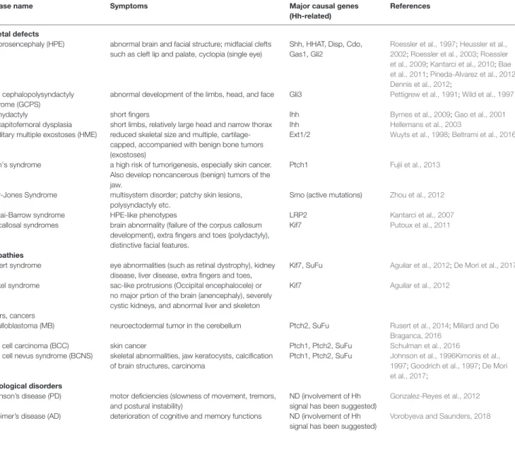
(13) Sasai et al.. Recent Progress on Hedgehog Research. results in decreased Shh target gene expression (Damerla et al., 2015; Toriyama et al., 2016). Disruption of the Cep290 gene, which encodes a centrosomal protein, causes severe renal defects (Hynes et al., 2014), and treatment with purmorphamine, an agonist of Hh signaling (Briscoe, 2006), increases the number of ciliated cells. This suggests that Hh signaling is a potential therapeutic target in some ciliopathies. Similarly, mutations in MKS1 (Weatherbee et al., 2009) and MKS3 (Abdelhamed et al., 2013), which encode centrosome and orphan proteins, respectively, cause systemic defects related to ciliogenesis in association with alterations in Shh, and sometimes Wnt signaling. Blockade of primary cilia normally perturbs the introduction of Hh into cells. However, lack of primary cilia caused by Kif3a mutation increases Hh activity in facial mesenchymal cells and results in craniofacial defects, hypertelorism (abnormal distance between the eyes), and frontonasal dysplasia (FND; developmental disorder with. abnormal facial structure such as distant eyes and flat nose) (Wong et al., 2009; Brugmann et al., 2010). Because the symptoms of ciliopathies are chronic, the patients are normally not easily treatable. Nevertheless, a number of trials have been made to cure these intractable diseases. For instance, gene therapy, where a viral infection of the functional ciliary genes into the body, has been shown to be successful to restore sensory input in the olfactory ciliopathy model mice (Green et al., 2018). In addition, by utilizing the method to make an organoid (a imitated three-dimensional organ differentiated in vitro from stem cells (Rossi et al., 2018)), a three-dimensional retina has been made from the iPS cells derived from the patients of retinitis pigmentosa, a type of ciliopathies. This method is useful because the progress of ciliopathies can be chased in vitro, and gives hints to find the strategies for the treatment (Deng et al., 2018). In addition, a microscopy-based automated system for small molecule screening was designed to search for new drugs that encourage ciliation.. TABLE 2 | Detailed symptoms in the Hh-related disease. ND, not determined. Disease name skeletal defects Holoprosencephaly (HPE). Greig cephalopolysyndactyly syndrome (GCPS) Brachydactyly acrocapitofemoral dysplasia hereditary multiple exostoses (HME). Gorlin's syndrome. Curry-Jones Syndrome Donnai-Barrow syndrome Acrocallosal syndromes. ciliopathies Joubert syndrome Meckel syndrome. tumors, cancers medulloblastoma (MB) basal cell carcinoma (BCC) basal cell nevus syndrome (BCNS). neurological disorders Parkinson’s disease (PD) Alzheimer’s disease (AD). Symptoms. Major causal genes (Hh-related). References. abnormal brain and facial structure; midfacial clefts such as cleft lip and palate, cyclopia (single eye). Shh, HHAT, Disp, Cdo, Gas1, Gli2. abnormal development of the limbs, head, and face. Gli3. Roessler et al., 1997; Heussler et al., 2002; Roessler et al., 2003; Roessler et al., 2009; Kantarci et al., 2010; Bae et al., 2011; Pineda-Alvarez et al., 2012 Dennis et al., 2012; Pettigrew et al., 1991; Wild et al., 1997. short fingers short limbs, relatively large head and narrow thorax reduced skeletal size and multiple, cartilagecapped, accompanied with benign bone tumors (exostoses) a high risk of tumorigenesis, especially skin cancer. Also develop noncancerous (benign) tumors of the jaw. multisystem disorder; patchy skin lesions, polysyndactyly etc. HPE-like phenotypes brain abnormality (failure of the corpus callosum development), extra fingers and toes (polydactyly), distinctive facial features.. Ihh Ihh Ext1/2. Byrnes et al., 2009; Gao et al., 2001 Hellemans et al., 2003 Wuyts et al., 1998; Beltrami et al., 2016. Ptch1. Fujii et al., 2013. Smo (active mutations). Zhou et al., 2012. LRP2 Kif7. Kantarci et al., 2007 Putoux et al., 2011. eye abnormalities (such as retinal dystrophy), kidney disease, liver disease, extra fingers and toes, sac-like protrusions (Occipital encephalocele) or no major prtion of the brain (anencephaly), severely cystic kidneys, and abnormal liver and skeleton. Kif7, SuFu. Aguilar et al., 2012; De Mori et al., 2017. Kif7. Aguilar et al., 2012. neuroectodermal tumor in the cerebellum. Ptch2, SuFu. skin cancer skeletal abnormalities, jaw keratocysts, calcification of brain structures, carcinoma. Ptch1, Ptch2, SuFu Ptch1, Ptch2, SuFu. Rusert et al., 2014; Millard and De Braganca, 2016 Schulman et al., 2016 Johnson et al., 1996Kimonis et al., 1997; Goodrich et al., 1997; De Mori et al., 2017;. motor deficiencies (slowness of movement, tremors, and postural instability) deterioration of cognitive and memory functions. ND (involvement of Hh signal has been suggested) ND (involvement of Hh signal has been suggested). Frontiers in Genetics | www.frontiersin.org. 13. Gonzalez-Reyes et al., 2012 Vorobyeva and Saunders, 2018. November 2019 | Volume 10 | Article 1103.
(14) Sasai et al.. Recent Progress on Hedgehog Research. This system is expected to facilitate drug screening to find good chemicals to treat ciliopathies (Zhang et al., 2019).. their tumorigenesis (Clement et al., 2007; Takezaki et al., 2011). Therefore, it would be possible to cure the gliomagenesis by blocking the Shh signaling pathway, and as mentioned above, candidate chemicals are being tested for clinical applications (Pietrobono and Stecca, 2018). As for therapy of the tumors, Hh antagonist could be used to block the signal. For instance, cyclopamine, which strongly blocks the Hh signal pathway, could have been a candidate chemical to ameliorate the symptoms (Chen et al., 2002). However, poor oral solubility and severe side effects in mice limited further clinical development of cyclopamine (Pietrobono and Stecca, 2018). Several other chemicals, which either bind to the Smo protein or inhibit its ciliary localization, have been developed and are currently under evaluation at preclinical or clinical trial stages (Pietrobono and Stecca, 2018). Among them, two Smo inhibitors, vismodegib and sonidegib, have already been approved by the US Food and Drug Administration (FDA) for treating the advanced BCC (Axelson et al., 2013; Migden et al., 2018; Pietrobono and Stecca, 2018; Niyaz et al., 2019), and clinical trials to apply these drugs for MB are ongoing as of October 2019 (Li et al., 2019). These drugs, especially sonidegib, can cross the blood-brainbarrier (Li et al., 2019) and have the advantage in the accessibility to tumors in the brain. In addition, statins, HMG-CoA reductase inhibitors, have been shown to synergize with Hh pathway inhibitors (e.g., vismodegib) in inhibiting the MB growth (Gordon et al., 2018). As cholesterol biosynthesis is required for Smo activity in the Hh signaling pathway, it is reasonable to decrease the cholesterol level to treat MB. However, because Hh pathway is involved in many biological events, modification of Smo activity could cause a number of negative side effects. Therefore, a recent study proposed targeting a specific protein located in primary cilia (Bay et al., 2018). Knockdown of the Arl13b gene, which encodes a GTPase expressed in cilia, in MB cell lines suppresses only high level of Shh signaling and decreases cell proliferation. Loss of Arl13b in the Ptch1-null background abolishes the tumorigenic effect of loss of Ptch1 (Goodrich et al., 1997), inhibiting MB formation in the developing cerebellum (Bay et al., 2018). In the treatment of Hh-related cancers, GPCRs are considered effective targets because they are easily accessible from the cell exterior and could therefore be potential drug targets (He and Lu, 2015). Hh signal transduction is associated with the downregulation of PKA and decreased cAMP levels; therefore, chemicals that modify the levels of these molecules can be developed as an effective anti-cancer treatment. For instance, Gαs-type GPCRs upregulate intracellular cAMP and thus inhibit Hh signaling (He and Lu, 2015). For example, GPR161 overexpression changes the sensitivity of cells to Hh and is considered a potential drug target (Mukhopadhyay et al., 2013; He and Lu, 2015; Rao et al., 2016; Shimada et al., 2018; Pusapati et al., 2018a).. Medulloblastoma, Basal Cell Carcinoma, and Other Tumorigenic Disorders. Decreased Hh activity causes systemic tissue hypoplasia; however, overactive and persistent Hh signaling also leads to abnormalities in tissue homeostasis (Petrova and Joyner, 2014). The representative phenotype induced by persistent activation of the Hh signal is cancer (Gupta et al., 2010). Two major types of cancer associated with Hh signaling have been extensively studied, namely, medulloblastoma (MB) and basal cell carcinoma (BCC) (Teglund and Toftgard, 2010). MB is a primitive neuroectodermal tumor that most commonly arises from the cerebellum, and is normally diagnosed at a young age, especially in children younger than 10 years (de Robles et al., 2015). Approximately 200–500 cases are diagnosed in the United States every year (https://www.cancer.net/cancertypes/medulloblastoma-childhood/statistics). MB is divided into four types, Wnt-activated, Shh-activated, group 3, and group 4, according to diagnostic symptoms, genetics, and gene expression (Rusert et al., 2014; Millard and De Braganca, 2016). Approximately 30% of all cases, whether sporadic or genetic in nature, that are found in the cerebellar hemispheres (Rusert et al., 2014; Millard and De Braganca, 2016) are the Hh-activated type. In cases of germline mutations (genetic background), the Ptch and SuFu tumor suppressor genes tend to be abnormal (Yin et al., 2019). BCC is a common skin cancer, accounting for >75% of all skin cancers (Zhang et al., 2001a). BCC is detected in 450–490 per 100,000 individuals in the UK (Epstein, 2008), and some BCC is associated with mutation of the Ptch gene (Gailani et al., 1996; Johnson et al., 1996). Analysis of biopsy samples from human BCC patients identified mutations in the p53 and Ptch1 genes in 30–50% and 20–30% of cases, respectively (Zhang et al., 2001a). Its related syndrome, basal cell nevus syndrome (BCNS), is characterized by skeletal abnormalities, jaw keratocysts, and calcification of brain structures in addition to carcinoma (Kimonis et al., 1997), and mutations in the Ptch1/2 and SuFu genes have been found in BCNS patients (Johnson et al., 1996; Goodrich et al., 1997; Kimonis et al., 1997; De Mori et al., 2017). Other types of cancer caused by the abnormal Hh expression include glioma (Gupta et al., 2010). Glioma is induced by the aberrant proliferation of glial cells both in the brain and in the spinal cord (Gupta et al., 2010). A number of studies have shown that Hh, as well as Wnt, BMP, and Notch, pathway acts for the initiation step of glioma, namely gliomagenesis (Palma and Ruiz i Altaba, 2004; Laug et al., 2018). Importantly, human glioma-initiating cells (GICs; also known as glioma stem cells) undergo an indefinite selfrenewal and are resistant to conventional anti-cancer drugs (Clement et al., 2007; Takezaki et al., 2011; Auffinger et al., 2015; Phi et al., 2018). On the other hand, it was demonstrated that overexpression of a C-terminal-truncated form of Gli2, exerted an antagonistic effect on the Gli activity and knocking-down of Gli1 and Gli2 expression prevented proliferation of GICs and. Frontiers in Genetics | www.frontiersin.org. Shh Signaling in Neurodegenerative Disorders. Parkinson's disease (PD) is one of the most common neurodegenerative disorders, with a prevalence of 1–2 in 1,000. 14. November 2019 | Volume 10 | Article 1103.
図
関連したドキュメント
Lemma4.1.. This is not true if f is not positively homogeneous as the following example shows.. Let f be positively homogeneous. We shall give an example later to show that
We then compute the cyclic spectrum of any finitely generated Boolean flow. We define when a sheaf of Boolean flows can be regarded as cyclic and find necessary conditions
The approach based on the strangeness index includes un- determined solution components but requires a number of constant rank conditions, whereas the approach based on
It is well known that an elliptic curve over a finite field has a group structure which is the product of at most two cyclic groups.. Here L k is the kth Lucas number and F k is the
Therefore, with the weak form of the positive mass theorem, the strict inequality of Theorem 2 is satisfied by locally conformally flat manifolds and by manifolds of dimensions 3, 4
On the other hand, from physical arguments, it is expected that asymptotically in time the concentration approach certain values of the minimizers of the function f appearing in
Using the multi-scale convergence method, we derive a homogenization result whose limit problem is defined on a fixed domain and is of the same type as the problem with
[Mag3] , Painlev´ e-type differential equations for the recurrence coefficients of semi- classical orthogonal polynomials, J. Zaslavsky , Asymptotic expansions of ratios of
