http://internmed.jp
【 CASE REPORT 】
Re-recognition of Age-dependent Reference Range for
the Serum Creatinine Level in Teenagers
- A Case of Slowly Progressive Tubulointerstitial
Nephritis which Occurred in an Adolescent
-Hiroyuki Ono
1, Kojiro Nagai
1, Eriko Shibata
1, Motokazu Matsuura
1, Seiji Kishi
1,
Taizo Inagaki
1, Masanori Minato
1, Sakiya Yoshimoto
1, Sayo Ueda
1, Fumiaki Obata
1,
Kenji Nishimura
1, Masanori Tamaki
1, Fumi Kishi
1, Taichi Murakami
1, Hideharu Abe
1,
Yukiko Kinoshita
2, Maki Urushihara
2, Shoji Kagami
2and Toshio Doi
1Abstract:
For the first time, a 15-year-old boy was found to have a slight degree of proteinuria and microscopic he-maturia during annual school urinalysis screening. His kidney function had already severely deteriorated. A kidney biopsy revealed tubulointerstitial nephritis (TIN) with diffuse inflammatory cell infiltration. His medi-cal records showed his serum creatinine level to be 0.98 mg/dL two years ago, which was abnormally high considering his age. Although the etiology of slowly progressive TIN was unclear, glucocorticoid and im-munosuppressant therapy improved his kidney function. This case report suggests that all doctors should rec-ognize the reference range for the serum creatinine level in teenagers.
Key words:serum creatinine level, tubulointerstitial nephritis, N-acetyl-beta-D-glucosaminidase, β2microglobulin
(Intern Med 56: 2187-2193, 2017) (DOI: 10.2169/internalmedicine.8599-16)
Introduction
Adolescents usually see general practitioners, not pediatri-cians. However, their reference ranges for some laboratory tests are different from those of adults. The serum creatinine level is one of the representative blood tests which are de-pendent on age, sex and muscle mass (Table 1) (1, 2). Gen-erally speaking, the reference range for the serum creatinine level in adolescents is lower than that in adults.
Tubulointerstitial nephritis (TIN) leads to renal dysfunc-tion and it is characterized by an infiltradysfunc-tion of inflammatory cells in the renal interstitium. TIN is often induced by drugs and infections (3). TIN is sometimes one of the clinical manifestations in TIN and uveitis syndrome and sarcoido-sis (4) or autoimmune disease such as Sjögren’s syndrome,
IgG4-related disease and systemic lupus erythemato-sus (5, 6). Idiopathic forms of TIN have also been reported. TIN often occurs without any abnormal urinalysis findings, which makes it difficult to identify at an early stage (6). We herein describe the case of an adolescent whose severe renal dysfunction was found during an investigation of a small amount of proteinuria and microscopic hematuria. A renal biopsy revealed the cause of renal failure to be TIN, which had slowly been progressing based on his medical history.
Case Report
A 15-year-old boy was referred to our hospital because of a positive dipstick reading for proteinuria in the presence of microscopic hematuria, which was detected during school urinalysis screening. Laboratory tests revealed an elevation
1Department of Nephrology, Institute of Biomedical Sciences, Tokushima University Graduate School, Japan and2Department of Pediatrics,
In-stitute of Biomedical Sciences, Tokushima University Graduate School, Japan
Received: November 24, 2016; Accepted: December 26, 2016; Advance Publication by J-STAGE: August 1, 2017 Correspondence to Dr. Kojiro Nagai, [email protected]
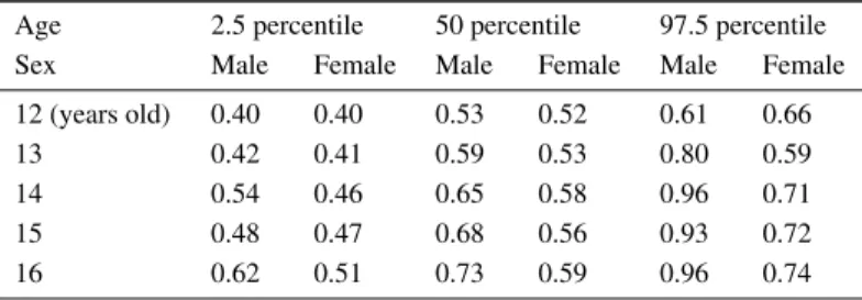
Table 1. Reference Range of Serum Creatinine Level for 12 to 16 Year-old Adolescents (mg/dL).
Age 2.5 percentile 50 percentile 97.5 percentile Sex Male Female Male Female Male Female 12 (years old) 0.40 0.40 0.53 0.52 0.61 0.66
13 0.42 0.41 0.59 0.53 0.80 0.59
14 0.54 0.46 0.65 0.58 0.96 0.71
15 0.48 0.47 0.68 0.56 0.93 0.72
16 0.62 0.51 0.73 0.59 0.96 0.74
(Quoted from reference 1)
of his serum creatinine level to 2.73 mg/dL, and therefore he was hospitalized for further examination.
His past school urinalysis tests had been within the nor-mal range. He had no significant medical history, except for resection of a thyroglossal duct cyst two years before admis-sion. The serum creatinine level of routine preoperative medical testing at that time was 0.98 mg/dL. At that time, he was in a good physical condition with an average phy-sique for his age. He did not take any drugs including sup-plements or Chinese herbal medicines. He did not have any symptoms, such as fever, skin rash, arthralgias, or peripheral eosinophilia suggestive of allergic reaction or infection.
On admission, he had no symptoms. His height was 162 cm and weight was 49.9 kg. His blood pressure was 122/83 mmHg, his pulse was 56/min with a regular rhythm, and his temperature was 36.6 degree centigrade. A physical exami-nation showed no abnormalities in his heart, lungs, or abdo-men. Purpura, skin rash, lymphadenopathy, arthralgia, myal-gia, edema, and peripheral neuropathy were not found.
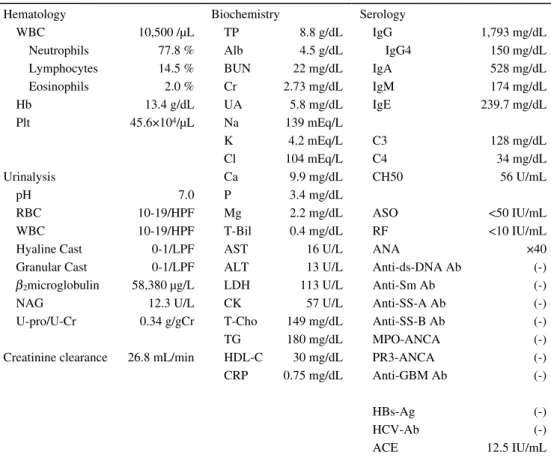
The laboratory data on admission are shown in Table 2. The values of white blood cells and C-reactive protein were slightly increased without eosinophilia. The serum levels of blood urea nitrogen and creatinine were elevated without any abnormalities in electrolytes. Creatinine clearance was 26.8 mL/min. Other biochemical tests were nonspecific ex-cept: the IgG, IgG4, IgA and IgE levels were slightly ele-vated and a urinalysis demonstrated 0.34 g/gCreatinine of proteinuria, microscopic hematuria, high N-acetyl-beta-D-glucosaminidase (NAG) and β2microglobulin (β2MG),
sug-gesting tubulointerstitial injury in the kidneys. In order to rule out the specific diseases that cause tubulointerstitial ne-phritis, serological tests including an autoantibody analysis were performed which resulted in negative studies for an-giotensinogen converting enzyme, anti double-stranded DNA, Sm, SS-A, SS-B, myeloperoxidase anti-neutrophil cy-toplasmic antibody (MPO-ANCA), proteinase (PR)3-ANCA, and glomerular basement membrane antibodies.
The chest radiography and electrocardiogram findings were normal. An abdominal ultrasound examination showed both kidneys to be slightly smaller than usual (Right kidney: 90×41 mm, Left kidney: 92×44 mm), and their margins were irregular, and cortexes were bright, indicating a chronic impairment of the kidney function. Computed tomography, magnetic resonance imaging and gallium scintigraphy
showed no abnormalities, except for the kidney morphology described above. The existence of uveitis was ruled out based on an ophthalmological examination.
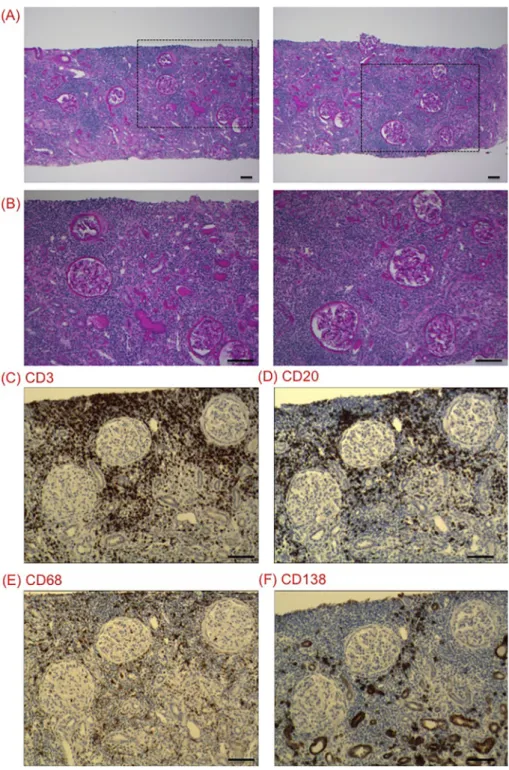
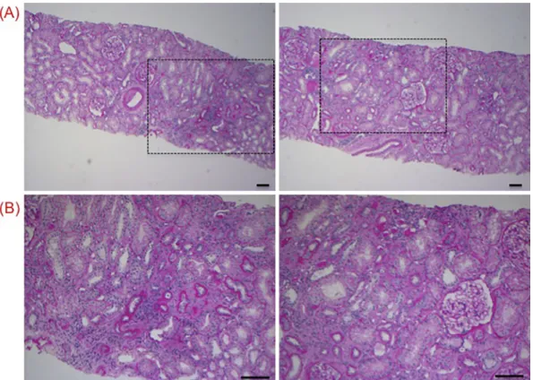
After admission, the first renal biopsy was performed. Six of nineteen glomeruli were globally sclerotic. The rest of the glomeruli had no particular abnormality. Of note, there was diffuse interstitial cell infiltration with interstitial fibrosis and tubular atrophy (Fig. 1). Almost 80% of the tubulointer-stitial area was damaged especially by cell infiltration which mainly consisted of T and B lymphocytes. Some of the in-filtrative cells were IgG4 positive (4-5/high power field), which did not meet the criteria of IgG4 related kidney dis-ease (data not shown). Eosinophils were barely detectable by Hematoxylin and Eosin stained biopsy samples (data not shown). The immunofluorescence results were negative for IgG, IgA, IgM, C3, C4, C1q, and Fib. Thus, renal failure was considered to have been caused by TIN. The patient’s clinical course is shown in Fig. 2. Sixty mg/day of oral prednisolone treatment was started. His renal function gradually improved, and seven weeks after the initiation of prednisolone, his serum creatinine level decreased to 1.49 mg/dL. Then, a second renal biopsy was performed to evaluate the efficacy of corticosteroid therapy. Light micros-copy showed a decrease of inflammatory cell infiltration and an improvement of tubular atrophy (Fig. 3), thus indicating that the corticosteroid therapy was effective. Around 20 to 30% of tubulointerstitial area was affected, especially by fi-brosis. He was then administered methylprednisolone pulse (MP) therapy (a single intravenous drip infusion of 500 mg/ day of methylprednisolone for three consecutive days) and thereafter showed a further improvement. After MP therapy, prednisolone was reduced to 20 mg/day, and 100 mg/day of mizoribine was added. No significant complications associ-ated with prednisolone and mizoribine occurred except for a slight increase in his blood pressure and uric acid levels. He was discharged from our hospital 10 weeks after admission with a creatinine level of 1.47 mg/dL (creatinine clearance: 49.7 mL/min). One year after discharge, he was taking 5 mg of prednisolone and 100 mg/day of mizoribine. In addition, his serum creatinine level was stable at around 1.5 mg/dL.
Discussion
pro-Table 2. Laboratory Findings on Admission.
Hematology Biochemistry Serology
WBC 10,500 /μL TP 8.8 g/dL IgG 1,793 mg/dL
Neutrophils 77.8 % Alb 4.5 g/dL IgG4 150 mg/dL
Lymphocytes 14.5 % BUN 22 mg/dL IgA 528 mg/dL
Eosinophils 2.0 % Cr 2.73 mg/dL IgM 174 mg/dL Hb 13.4 g/dL UA 5.8 mg/dL IgE 239.7 mg/dL Plt 45.6×10⁴/μL Na 139 mEq/L K 4.2 mEq/L C3 128 mg/dL Cl 104 mEq/L C4 34 mg/dL Urinalysis Ca 9.9 mg/dL CH50 56 U/mL pH 7.0 P 3.4 mg/dL RBC 10-19/HPF Mg 2.2 mg/dL ASO <50 IU/mL WBC 10-19/HPF T-Bil 0.4 mg/dL RF <10 IU/mL
Hyaline Cast 0-1/LPF AST 16 U/L ANA ×40
Granular Cast 0-1/LPF ALT 13 U/L Anti-ds-DNA Ab (-) β2microglobulin 58,380 μg/L LDH 113 U/L Anti-Sm Ab (-)
NAG 12.3 U/L CK 57 U/L Anti-SS-A Ab (-)
U-pro/U-Cr 0.34 g/gCr T-Cho 149 mg/dL Anti-SS-B Ab (-)
TG 180 mg/dL MPO-ANCA (-)
Creatinine clearance 26.8 mL/min HDL-C 30 mg/dL PR3-ANCA (-) CRP 0.75 mg/dL Anti-GBM Ab (-)
HBs-Ag (-)
HCV-Ab (-)
ACE 12.5 IU/mL
WBC: white blood cell, Hb: hemoglobin, Plt: platelet, RBC: red blood cell, NAG: N-acetyl-beta-D-glucosaminidase, U-pro/U-Cr: urinary protein/urinary creatinine,TP: total protein, Alb: albumin, BUN: blood urea nitrogen, Cr: creati-nine, UA: uric acid, Na: sodium, K: potassium, Cl: cloride, Ca: calcium, P: phosphate, Mg: magnesium, T-Bil: total bil-irubin, AST: L-aspartate aminotransferase, ALT: alanine aminotransferase, LDH: lactate dehydrogenase, CK: creatine kinase, T-chol: total cholesterol, TG: triglyceride, HDL-C: high density lipoprotein cholesterol, CRP: C-reactive pro-tein, ASO: anti-streptolysin O, RF: rheumatoid factor, ANA: anti-nuclear antibody, ds-DNA: double stranded DNA, Ab: antibody, MPO: myeloperoxidase, ANCA: anti-neutrophil cytoplasmic antibody, PR3: proteinase 3, GBM: glomer-ular basement membrane, Ag: antigen, ACE: angiotensin converting enzyme
posed in Kidney Disease Outcomes Quality Initiative (K/ DOQI) clinical practice guidelines for CKD by the National Kidney Foundation in 2002 (7). The glomerular filtration rate (GFR) estimating equations for Japanese populations are a robust reflection of the response to the CKD guide-lines in Japan (8). It is therefore considered important for both nephrology professionals and medical practitioners to understand the clinical relevance of CKD. However, the es-tablished equations have only been proposed for people 18 years old or older. Adolescents can therefore be a pitfall for doctors, because they are usually healthy and the onset of kidney diseases is rarely predicted. Therefore, not many doctors recognize the reference range for serum creatinine in adolescents. In this case, we want to emphasize the impor-tance of an early detection of renal impairment. 0.98 mg/dL of serum creatinine level two years before admission was abnormal considering his age. The serum creatinine level can be higher if the body muscle is large. However, his height and weight were normal. Probably, the abnormal cre-atinine level was overlooked because the result was within the reference range for adults. Thus, the reference range does not always mean the normal range. The value may be
due to a technical or sample error, but it would have been preferable to re-evaluate this patient to rule out any hidden kidney disease. If that have been done, then this patient could have been treated earlier, and his kidney function would have been better preserved. Notably, serum creatinine-based and cystatin C-based estimated GFR equa-tions in Japanese children and adolescents were established by comparison with renal inulin clearance (Table 3) (9, 10). These equations can therefore help to identify hidden kidney diseases.
We have to know that TIN can represent a frequent cause of kidney injury with either no or only inconspicuous toms. Therefore, the presence of specific extrarenal symp-toms, such as fever, skin rash, arthralgias, and peripheral eosinophilia, plays an important role in making a timely clinical diagnosis (5). In addition, TIN does not always manifest with urinalysis abnormalities, which thus prevents an early detection of kidney disease. Tubulointerstitial injury markers such as NAG andβ2MG are the commercially
avail-able markers for detecting an early stage of TIN, although these markers are not evaluated routinely by medical practi-tioners.
Figure 1. Representative pictures obtained from the first biopsy. (A, B) Diffuse interstitial cell infil-tration was observed with interstitial fibrosis and tubular atrophy. The rectangle area in (A) was magnified to (B). (C, D, E, F) Cell marker staining. The antibodies against CD3 (2GV6) (T lympho-cyte marker) and CD20 (L26) (B lympholympho-cyte marker) were obtained from Ventana Medical Systems (Tucson, USA). The antibodies against CD68 (PG-M1) (Monocyte marker) and CD138 (MI15) (Plas-ma cell (Plas-marker) were purchased from Dako Japan (Tokyo, Japan). Anti-CD138 antibody showed nonspecific staining in the tubule cells. Scale bars represent 100 μm.
The causes of acute TIN included drugs (70%), autoim-mune diseases (20%), and infections (4%) (6). Therefore, idiopathic cases are relatively rare. In our case, the patient was asymptomatic and had no specific physical manifesta-tions to suggest TIN. He did not take any drugs and had no history of recent infections and no serological abnormalities indicating autoimmune diseases. Moreover, sarcoidosis and
IgG4-related kidney disease was excluded by pathological investigations. Thus, this case was diagnosed as idiopathic TIN. Although the optimal treatment for TIN has not yet been established, the most important aspect of therapy of TIN is the removal of any potential causative agents. It has been reported that the cessation of responsible agents or treatment of the underlying infection may lead to an
im-Figure 2. Clinical course of the patient. Coritcosteroid therapy was effective for the treatment of slowly progressive tubulointerstitial nephritis. PSL: prednisolone, mPSL: methylprednisolone, MZB: mizoribine
Figure 3. Representative pictures obtained from the second biopsy. (A, B) Corticosteroid therapy decreased inflammatory cell infiltration and improved tubular atrophy in the kidneys. Rectangle area in (A) was magnified to (B). Scale bars represent 100 μm.
provement of the renal function (11). However, a limited number of studies have reported on the treatment of idi-opathic TIN. In our case, both kidneys were atrophic in ab-dominal ultrasound examinations. Gallium scintigraphy did not show an abnormal uptake in either kidney, probably be-cause the patient’s disease was not during the acute phase, though gallium scintigraphy lacks sensitivity and specificity to identify acute interstitial nephritis (12, 13). However,
dif-fuse interstitial cell infiltration remained, while more than 30% of the total obtained glomeruli were globally screlotic in the first kidney biopsy sample, suggesting not chronic, not acute but slowly progressive TIN. Therefore, we ex-pected that immunosuppressive therapy would be still effec-tive. Glucocorticoid therapy appears to have a positive effect in patients with TIN, especially when treatment is initiated early in the course of the disease (12, 14).
Immunosuppres-Table 3. Proposed Equations in Japanese Children and Adolescents to Detect the Early Phase of Kidney Diseases.
Creatinine-based equation to estimate the glomerular filtration rate eGFR (mL/min/1.73 m2) =
110.2 X (reference serum creatinine (mg/dL) / patient’s serum creatinine (mg/dL)) +2.93
Reference serum creatinine (mg/dL) are shown by the following two equations of body length (BL) (cm):
Male: - 1.259 BL5 + 7.815 BL4 - 18.57 BL3 + 21.39 BL2 - 11.71 BL + 2.628
Female: - 4.536 BL5 + 27.16 BL4 - 63.47 BL3 + 72.43 BL2 - 40.06 BL + 8.778
Applicable in Japanese children and adolescents aged between 2 and 18 years. (Quoted from reference 9)
Cystatin C-based equation to estimate the glomerular filtration rate
eGFR (mL/min/1.73 m2) = 104.1 / serum cystatin C (mg/L) - 7.80
Applicable in Japanese children and adolescents between the ages of 1 month and 18 years
(Quoted from reference 10)
eGFR: estimated glomerular filtration rate
sant drugs were suggested to be a therapeutic option consid-ering the pathogenesis of interstitial nephritis (15) and they have been used effectively in several case reports (16, 17). Among routinely used immunosuppressant drugs to treat kidney diseases, such as cyclophosphamide, cyclosporine A and mizoribine, cyclophosphamide metabolites alkylate DNA bases and preferentially suppress immune responses mediated by B lymphocytes. Cyclosporine A inhibits the phosphatase activity of calcineurin, thereby suppressing the production of interleukin-2 (IL-2) and other cytokines by T lymphocytes (18), while mizoribine is an antimetabolite that exerts its immunosuppressant effect by inhibiting both T and B lymphocyte proliferation (19). As shown in Fig. 1, infil-trating cells in the tubulointerstitial area consists mainly of both T and B lymphocytes. We wanted to reduce the young patient’s disease activity and corticosteroid use as much as possible. Therefore, mizoribine was administered because its suppressive effects on both T and B lymphocytes and its relative lack of toxicity. At first, we used 150 mg/day of mi-zoribine. However, it caused hyperuricemia as a side effect. As a result, we decreased the dosage of mizoribine to 100 mg/day.
Finally, corticosteroid and mizoribine therapy was able to moderately decrease the serum creatinine level. There are no definite criteria to discontinue corticosteroid treatment. We might perform a kidney biopsy again to confirm the lack of infiltrating cells in the interstitial area which would thus al-low us to stop the administration of corticosteroids, though a relapse of interstitial nephritis could still occur even if the previous treatment had been sufficient.
In summary, this is a case of idiopathic slowly
progres-sive TIN, which was found through school urinalysis screen-ing. We need to recognize the reference range for the serum creatinine level, serum creatinine-based and cystatin C-based estimated GFR equations in teenagers and the evaluation of not only routine urinalysis, but also tubulointerstitial injury markers such as NAG andβ2MG are recommended to detect
the presence of early stage kidney diseases including TIN.
The authors state that they have no Conflict of Interest (COI). Acknowledgement
We thank Yoko Okazawa, Akiyo Muramoto (Tokushima Uni-versity) for their valuable clinical assistance.
References
1. Uemura O, Honda M, Matsuyama T, et al. Age, gender, and body
length effects on reference serum creatinine levels determined by an enzymatic method in Japanese children: a multicenter study. Clin Exp Nephrol 15: 694-699, 2011.
2. Ishikura K, Uemura O, Ito S, et al. Pre-dialysis chronic kidney
disease in children: results of a nationwide survey in Japan. Neph-rol Dial Transplant 28: 2345-2355, 2013.
3. Schwarz A, Krause PH, Kunzendorf U, Keller F, Distler A. The
outcome of acute interstitial nephritis: risk factors for the transi-tion from acute to chronic interstitial nephritis. Clin Nephrol 54: 179-190, 2000.
4. Baker RJ, Pusey CD. The changing profile of acute
tubulointersti-tial nephritis. Nephrol Dial Transplant 19: 8-11, 2004.
5. Praga M, González E. Acute interstitial nephritis. Kidney Int 2010 77: 956-961, 2010.
6. Muriithi AK, Leung N, Valeri AM, et al. Biopsy-proven acute
in-terstitial nephritis, 1993-2011: a case series. Am J Kidney Dis 64: 558-566, 2014.
7. National Kidney Foundation. K/DOQI clinical practice guidelines
for chronic kidney disease: evaluation, classification, and stratifica-tion. Am J Kidney Dis 39 (2 Suppl 1): S1-S266, 2002.
8. Mastuo S, Imai E, Horio M, et al. Revised equations for
estimat-ing glomerular filtration rate (GFR) from serum creatinine in Ja-pan. Am J Kidney Dis 53: 982-992, 2009.
9. Uemura O, Nagai T, Ishikura K, et al. Creatinine-based equation
to estimate the glomerular filtration rate in Japanese children and adolescents with chronic kidney disease. Clin Exp Nephrol 18: 626-633, 2014.
10. Uemura O, Nagai T, Ishikura K, et al. Cystatin C-based equation
for estimating glomerular filtration rate in Japanese children and adolescents. Clin Exp Nephrol 18: 718-725, 2014.
11. Buysen JG, Houthoff HJ, Krediet RT, Arisz L. Acute interstitial
nephritis: a clinical and morphological study in 27 patients. Neph-rol Dial Transplant 5: 94-99, 1990.
12. Perazella MA, Markowitz GS. Drug-induce acute interstitial
ne-phritis. Nat Rev Nephrol 6: 461-470, 2010.
13. Rossert J. Drug-induced acute interstitial nephritis. Kidney Int 60:
804-817, 2001.
14. Raghavan R, Eknoyan G. Acute interstitial nephritis - a reappraisal
and update. Clin Nephrol 82: 149-162, 2014.
15. Neilson EG. Pathogenesis and therapy of interstitial nephritis.
Kid-ney Int 35: 1257-1270, 1989.
16. Lee JW, Kim HJ, Sung SH, Lee SJ. A case of tubulointerstitial
ne-phritis and uveitis syndrome with severe immunologic dysregula-tion. Pediatr Nephrol 20: 1805-1808, 2005.
17. Büscher R, Vij O, Hudde T, Hoyer PF, Vester U. Pseudotumor
cerebri following cyclosporine A treatment in a boy with tubu-lointerstitial nephritis associated with uveitis. Pediatr Nephrol 19: 558-560, 2004.
18. Allison AC. Immunosuppressive drugs: the first 50 years and a
glance forward. Immunopharmacology 47: 63-83, 2000.
19. Yoshikawa N, Nakanishi K, Ishikura K, Hataya H, Iijima K,
Honda M; Japanese Pediatric IgA Nephropathy Treatment Study Group. Combination therapy with mizoribine for severe childhood IgA nephropathy: a pilot study. Pediatr Nephrol 23: 757-763, 2008.
The Internal Medicine is an Open Access article distributed under the Creative Commons Attribution-NonCommercial-NoDerivatives 4.0 International License. To view the details of this license, please visit (https://creativecommons.org/licenses/ by-nc-nd/4.0/).
Ⓒ 2017 The Japanese Society of Internal Medicine