INTRODUCTION
Since Trokel and coauthors introduced photorefractive keratectomy (PRK) in 1983 (1), numerous studies have been done on this procedure. The refractive surgery using a 193 nm argon fluoride excimer laser has become a reasonably predictable, effective, and safe method for treating low to moderate myopia (2-7). Its major limitations are postoperative pain, subepithelial haze, and prolonged visual rehabilitation (8, 9). Laser in situ keratomileusis (LASIK) combines lamellar corneal surgery with the accuracy of the excimer laser. It
is a procedure that has evolved from a variety of techniques in refractive surgery. The first LASIK procedure on the human eye was performed by Pallikaris and coauthors in 1991 (10, 11). Since then, this procedure has gradually become more popular, particularly among high-volume refractive surgeons. LASIK offers more comfort, faster visual rehabilitation, and minimal haze, but epithelial ingrowths, corneal-flap-related complications, and corneal ectasia are shortcomings of LASIK (12-15). To date, a number of studies have reported the results that both techniques effectively correct varying degrees of myopia. Although several prospective or retrospective controlled trials have been suggested for the advantages of LASIK over PRK (16-20), several well-designed randomized controlled clinical trials indicated that the efficacy outcomes in the longer term are generally similar between the two procedures for
ORIGINAL
Evaluation of the effectiveness of laser in situ keratomileusis
and photorefractive keratectomy for myopia : A meta-analysis
Xin-Jun Yang, Hong-Tao Yan, and Yutaka Nakahori
Department of Human Genetics and Public Health, Graduate School of Proteomics, The University of Tokushima, Tokushima, Japan
Abstract : Objective: To evaluate the effectiveness of laser in situ keratomileusis (LASIK) and photorefractive keratectomy (PRK) for correcting myopia.
Methods : Study selection, data extraction, and quality assessment were performed by two of authors independently. Summary odds ratios and 95% confidence intervals were calculated by DerSimonian & Laird random-effects model and Mantel-Haenszel (fixed-effects) model. All calculations were based on an intention-to-treat and per protocol analysis.
Results : Five hundred and eighty eyes (476 patients) from 5 randomized controlled trials were included in this study. At≧6 months follow-up, by random-effects model, the pooled odds ratios (OR, for LASIK vs. PRK) of postoperative uncorrected visual acuity (UCVA) of 20/20 or better for all trials were 1.31 (95% CI=0.77-2.22) by per protocol analysis and 1.18 (95% CI=0.74-1.88) by intention-to-treat analysis. In the refractive outcome, the pooled OR of the postoperative spherical equivalent refraction within±0.5 diopter (D) of emmetropia did not show any statistical significance, for which the OR were 0.75 (95% CI=0.48-1.18) by per protocol analysis and 0.70 (95% CI=0.47-1.04) by intention-to-treat analysis.
Conclusions : LASIK and PRK were found to be similarly effective for the correction of myopia from-1.5 to -15.0 D in a greater than 6 month follow-up. J. Med. Invest. 50 : 180-186, 2003
Keywords : laser in situ keratomileusis (LASIK), photorefractive keratectomy (PRK), myopia, meta-analysis
Received for publication May 8, 2003 ; accepted July 23, 2003. Address correspondence and reprint requests to Yutaka Nakahori, M.D., Department of Human Genetics and Public Health, Graduate School of Proteomics, The University of Tokushima Kuramoto-cho, Tokushima. 770-8503, Japan and Fax : +81-88-633-7453.
The Journal of Medical Investigation Vol. 50 2003 180
correcting low to high myopia (21-24).
In this study, we have summarized the published randomized controlled trials of PRK and LASIK, for correcting myopia, to quantitatively evaluate the effec-tiveness of myopia treatment with both PRK and LASIK procedures.
METHODS
Data sources
Studies were identified by MEDLINE and EMBASE searches through October 2002. The terms
photore-fractive keratectomyand laser in situ keratomileusis, and
myopia or refractive myopia, were used for a sensitive search. In addition, we limited publication types to
Randomized Controlled Trial. Bibliographies of retrieved articles were manually searched.
Study selection
We included English-language studies that met all of the following criteria, as judged independently by two investigators (Yang and Yan) : (1) a prospective, random-ized controlled clinical trial, (2) comparison study for effectiveness, safety, and stability of PRK and LASIK for correcting myopia,(3) main outcome measures included manifest refraction, uncorrected and spectacle-corrected visual acuity, predictability and stability of refraction, and complications, (4) original data of every follow-up time was available. Reviews, abstracts and articles published in non-English languages were excluded.
Assessment of study quality
The quality of the included studies was assessed using the criteria proposed by Chalmers et al.(25). This method evaluates the design, implementation, and analysis, of randomized controlled trials. The overall index of trial quality was weighted as follows : trial design and protocol, 0.6 ; statistical analysis, 0.3 ; pres-entation of results, 0.1. The final quality score ranged from 0 (lowest) to 1(highest).
Data extraction
Data extraction was done independently by two authors (Yang and Yan) using a predefined review form. Postoperative outcome of refraction, visual outcome, and loss of spectacle-corrected visual acuity, were calculated by intention-to treat and per protocol analyses. Any discrepancies between the authors in data extraction were resolved through discussion to reach a consensus of opinion.
Statistical analysis
We pooled all the data using the DerSimonian and Laird random-effects model that considers both within-study variance and variability among studies. The Mantel-Haenszel (fixed-effects) model was also used for comparison calculations ; this approach allowed us to verify the validity of the random-effects estimates in some analyses in which the numbers of events were small. All estimated odds ratios (OR) were for the LASIK group compared with the PRK group. Summary esti-mates of effect were calculated with weighting based on the inverse of the study’s variance. Two-tailedP values and a 95 percent confidence interval (95% CI) were used.
The heterogeneity between studies was examined by DerSimonian and LairdQ statistic (26). Egger’s linear regression approach (27) and Begg and Mazumdar’s proposed adjusted rank correlation test (28) were used to measure publication bias. Sensitivity analysis was also performed by comparing the overall effect of different statistical models calculated to assess the reliability of meta-analysis.
All statistic analyses were carried out using Metaview 3.1 in Rev Man 4.04(Cochrane Collaboration, Oxford, England) and SPSS version 11.0 (SPSS, Inc., Chicago, Illinois).
RESULTS
Study characteristics and quality score
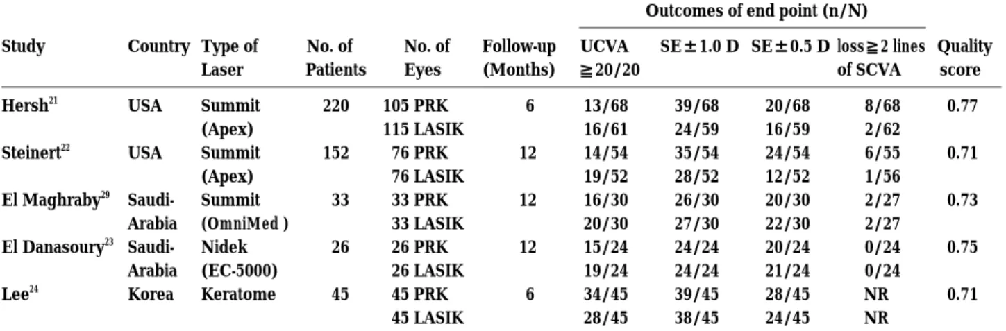
A total of 15 studies were initially identified by two computerized database and manual searches as poten-tially relevant. Ten of these 15 studies were excluded because they did not meet our criteria (Among those, one was comparison of the effect of laser epithelial keratomileusis (LASEK) and PRK and other nine were a paper about the side effects after the operation of the LASIK and PRK). Five prospective, randomized con-trolled trials were included in this meta-analysis (21-24, 29). The characteristics of the studies are presented in Table 1. A total of 580 eyes (476 patients) were enrolled in the five trials. Preoperative manifest spherical equiva-lent refraction ranged from‐1.25 to‐14.38 diopter (D). The follow-up ranged from 6 months to 12 months. The studies had a mean quality score of 0.73 (range, 0.71 to 0.77), which was considered to be high compared with the scores of trials in other clinical domains (30). Pooling all 5 trials, the characteristics of the patients receiving PRK or LASIK had no significant differences (Table 2).
Pooling of Uncorrected Visual Acuity
Figure 1 shows the odds ratios (OR) for postoperative uncorrected visual acuity (UCVA) of 20/20 or better at a follow-up≧6 months. Using both random-effects model and fixed-effects model analyses, the pooled OR were virtually identical, and there were no statistically significant differences between LASIK and PRK. The Q statistic did not indicate significant heterogeneity (Q=5.31, 4 degrees of freedom, P>0.1). Uncorrected visual acuity of 20/20 or better after LASIK was 48% and 42% after PRK. Improvement in uncorrected visual
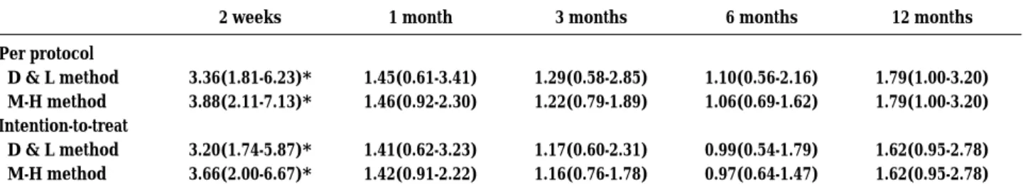
acuity in the LASIK group occurred significantly faster than in the PRK group. The pooled OR of UCVA≧ 20/20 are presented in Table 3 ; at 2 weeks, the ORs showed statistical significances, but after this, the differ-ence was not statistically significant.
Pooled Refractive Outcome
The results of the postoperative spherical equivalent refraction within±1.0 D (SE±1.0 D) and±0.5 D (SE± 0.5 D) of emmetropia were analyzed. At a follow-up of greater than or equal to 6 months, using the random-effects-model analysis, the pooled OR of SE±1.0 D were 0.64 (95% CI=0.41-1.02) for per protocol analysis and 0.63 (95% CI=0.43-0.92) for intention-to-treat analysis ; the latter shows statistical significance. However, no statistically significant differences were found in the pooled OR of SE±0.5 D between LASIK and PRK ; the OR were 0.75 (95% CI=0.48-1.18) for per protocol analysis and 0.70 (95% CI=0.47-1.04) for intention-to-treat analysis (Figure 2). Using fixed-effects model analysis, the same results were achieved (data not shown).
Table 1.Characteristics and quality scores of trials included in the meta-analysis
Outcomes of end point (n/N) Study Country Type of
Laser No. of Patients No. of Eyes Follow-up (Months) UCVA ≧20/20
SE±1.0 D SE±0.5 D loss≧2 lines of SCVA Quality score Hersh21 Steinert22 El Maghraby29 El Danasoury23 Lee24 USA USA Saudi-Arabia Saudi-Arabia Korea Summit (Apex) Summit (Apex) Summit (OmniMed ) Nidek (EC-5000) Keratome 220 152 33 26 45 105 PRK 115 LASIK 76 PRK 76 LASIK 33 PRK 33 LASIK 26 PRK 26 LASIK 45 PRK 45 LASIK 6 12 12 12 6 13/68 16/61 14/54 19/52 16/30 20/30 15/24 19/24 34/45 28/45 39/68 24/59 35/54 28/52 26/30 27/30 24/24 24/24 39/45 38/45 20/68 16/59 24/54 12/52 20/30 22/30 20/24 21/24 28/45 24/45 8/68 2/62 6/55 1/56 2/27 2/27 0/24 0/24 NR NR 0.77 0.71 0.73 0.75 0.71 UCVA= uncorrected visual acuity ; SE= spherical equivalent ; SCVA=spectacle-corrected visual acuity ; NR= not reported.
Table 2.Comparison of LASIK and PRK characteristics
Variables LASIK PRK No. of eyes
Mean age (yrs) Age range (yrs) Male/Female
Preoperative mean SE (D) (Range)
Preoperative mean cylinder (D) With astigmatism (D) 295 37.0 16-64 137/158 -7.51 (-1.25 to‐13.88) 0.70 <2.5 285 37.5 16-59 156/129 -7.52 (-1.25 to -14.38) 0.63 <2.5 SE=spherical equivalent ; D=diopter.
Figure 1.Postoperative UCVA of 20/20 or better for LASIK compared with PRK at 6 to 12 months follow-up (By
intention-to-treat analyses). Shown as OR with 95% confidence interval.
XJ. Yang et al. A meta-analysis of PRK and LASIK for myopia 182
Loss of Spectacle-Corrected Visual Acuity
The following refers to the outcome of loss of spectacle-corrected visual acuity of two Snellen lines or more at ≧ 6 months follow-up, and the statistically significant differences between LASIK and PRK. For the per protocol analysis, the pooled OR were 0.32 (95% CI= 0.11-0.96, D&L method) and 0.30 (95% CI=0.11-0.85, M-H method) ; for the intention-to-treat analysis, the OR were 0.31 (95% CI=0.10-0.89, D&L method) and 0.28 (95% CI=0.10-0.79, M-H method). Therefore, the results suggest a lesser likelihood of loss of spectacle-corrected visual acuity with LASIK compared with that of PRK.
Subgroup analysis
For low to moderate myopia (-1.5 to-8.0 D) (Ref 23, 24, 29), the pooled OR for UCVA≧20/20 and the refractive outcome (SE±1.0 D and SE±0.5D) showed no statistically significant differences between the two procedures. For moderate to high myopia (-6.0 to-15.0 D)(Ref 21, 22), the pooled OR for refractive outcome showed statistical significances, namely PRK-treated eyes had a higher percentage of success than LASIK at≧6 months follow-up. There were no statistically significant differences for the UCVA of 20/20 or better results between LASIK and PRK (Table 4).
Complications
The following refers to the subepithelial haze seen in PRK-treated eyes at 6 and 12 months follow-up. The subepithlial haze decreased trace haze (1+) was 31.9% (36/113), mild haze (2+) was 6.0% (10/167), mod-erate haze (3+) was 4.4% (3/68), and severe haze (4+) was 3.3% (1/30). After LASIK, the subepithelial haze was not seen, but there were 4.4% (13/295) flap-related complications. These events included free cap, stopping of microkeratome in the middle of the pass, and incom-plete, slipped, or dislocated flaps. No other adverse reactions, such as microbial keratitis, endophthalmitis, corneal melting or perforation, corneal ectasia, or retinal lesions, were found in these studies.
Publication bias and sensitivity analysis
The measure of funnel plot asymmetry showed no statistical significance with either the regression test or with the rank correlation test ; the P values were 0.5 and 0.327, respectively.
Sensitivity analysis was performed using the Der-Simonian & Laird random effects model and the Mantel-Haenszel method (fixed effects model) to calculate the overall effects. The results showed that the overall estimates were virtually identical, and that the confidence intervals were similar between the random effects model
Table 3.Pooled OR(95% CI) of postoperative UCVA≧20/20 at different times
2 weeks 1 month 3 months 6 months 12 months Per protocol D & L method M-H method Intention-to-treat D & L method M-H method 3.36(1.81-6.23)* 3.88(2.11-7.13)* 3.20(1.74-5.87)* 3.66(2.00-6.67)* 1.45(0.61-3.41) 1.46(0.92-2.30) 1.41(0.62-3.23) 1.42(0.91-2.22) 1.29(0.58-2.85) 1.22(0.79-1.89) 1.17(0.60-2.31) 1.16(0.76-1.78) 1.10(0.56-2.16) 1.06(0.69-1.62) 0.99(0.54-1.79) 0.97(0.64-1.47) 1.79(1.00-3.20) 1.79(1.00-3.20) 1.62(0.95-2.78) 1.62(0.95-2.78) D & L method = DerSimonian & Laird method ; M-H method = Mantel-Haenszel method ; CI = confidence interval.
*P<0.05
Figure 2.Postoperative spherical equivalent refractive outcome for LASIK compared with PRK at 6 to 12 months
follow-up (By intention-to-treat analyses). Shown as OR and 95% confidence interval. SE=spherical equivalent; Q= Q statistic.
and the fixed-effects model (data not shown). This was explained by the relatively small amount of variation between the trials in this meta-analysis.
DISCUSSION
This meta-analysis combined data from five randomized controlled trials comparing the effectiveness, safety, and stability of correcting myopia with LASIK and PRK. Pooled odds ratios of all data for postoperative UCVA of 20/20 or better in each trial showed that although the improvement in uncorrected visual acuity was more rapid in LASIK than in PRK, the final visual outcome showed no statistically significant differences between the two procedures. At≧ 6 months after surgery, 48% (102/212) LASIK and 42% (93/221) PRK-treated eyes achieved uncorrected visual acuity of 20/20 or better. Uncorrected visual acuity of 20/20 or better is one requirement for patients to be free from distance optical correction, and to be highly satisfied after refractive surgery (31, 32). Therefore, our results demonstrated the efficacy of refractive surgery using either technique. Regarding the refractive outcome, at 6 months after surgery 67.1% of the eyes in the LASIK group, compared with 73.8% of the eyes in the PRK group, were within± 1.0 D of emmetropia, while 45.2% of the eyes in the LASIK group, compared with 50.7% of the eyes in the PRK group, were within±0.5 D of emmetropia. The former showed a statistically significant difference be-tween the two procedures, suggesting a trend toward the lesser likelihood of achieving a spherical equivalent refraction within 1.0 D in LASIK compared with PRK.
This may possibly be interpreted by a preoperative higher refractive error with more undercorrections in LASIK. Although there was no statistically significant difference between the two procedures for an under-correction greater than 1.0 D at 6 to 12 months follow-up, the pooled OR of 1.47 (95% CI, 0.68 to 3.18) suggests a possible trend toward a high likelihood of undercorrec-tion with LASIK compared with PRK. In addiundercorrec-tion, subgroup analysis results indicated that the refractive outcome was significantly different between the two procedures in the moderate to high myopia group (Table 4).
Spectacle-corrected visual acuity is a general indicator of a variety of changes in the optics of the cornea and visual function after refractive surgical procedures (33). In this study, after LASIK, there was a lower percentage of eyes that had lost two or more Snellen lines of spectacle-corrected visual acuity than after PRK, especially in the moderate to high myopia group (>6.0 D). The results suggest that LASIK is reasonably safe for moderate to high myopia, compared to PRK.
Although both PRK and LASIK are satisfying to most patients, optical sequelae, such as glare, halo, and monocu-lar diplopia, remain as troubling side effects for some. A randomized clinical trial (34) reported that there was a somewhat lesser tendency toward postoperative symptoms in LASIK compared with PRK treated eyes. In this study, subepithelial central stromal haze was seen only after PRK, the majority exhibiting trace to mild. Although there was no central corneal haze beneath the epithelium or in the lamellar bed, there were approximately four percent of flap-relation com-plications after LASIK surgery, such as free flap,
incom-Table 4.Pooled OR(95% CI) for postoperative results
Preoperative myopia
‐1.5 to‐8.0 D ‐6.0 to‐15.0 D
Results by PP by ITT by PP by ITT
UCVA≧20/20 Total eyes D & L M‐H SE±1.0 D Total eyes D & L M‐H SE±0.5 D Total eyes D & L M‐H 198 1.16(0.43‐3.16) 1.05(0.58‐1.88) 198 1.00(0.39‐2.58) 1.00(0.39‐2.56) 198 0.95(0.51‐1.77) 0.95(0.52‐1.76) 208 1.11(0.44‐2.76) 1.04(0.59‐1.83) 208 1.00(0.46‐2.19) 1.00(0.46‐2.18) 208 0.96(0.54‐1.71) 0.96(0.54‐1.71) 235 1.57(0.87‐2.83) 1.57(0.87‐2.83) 233 0.56(0.33‐0.95)* 0.56(0.33‐0.95)* 233 0.59(0.25‐1.37) 0.60(0.34‐1.05) 372 1.30(0.75‐2.26) 1.30(0.75‐2.26) 372 0.54(0.35‐0.84)* 0.54(0.35‐0.84)* 372 0.54(0.32‐0.92)* 0.54(0.32‐0.91)* By PP= by per protocol analyses ; by ITT=by intention-to-treat analyses ;
D & L=DerSimonian & Laird method ; M-H=Mantel-Haenszel method ;
UCVA=uncorrected visual acuity ; SE=spherical equivalent ; CI = confidence interval ; * P<0.05
XJ. Yang et al. A meta-analysis of PRK and LASIK for myopia 184
plete flap, flap decentration, or slipped flap. This result was similar to those reported by other authors (35-37). This meta-analysis had several potential limitations. First, the number of trials and enrolled subjects were small, limiting the precision of all estimates. However, the five included trials were of high quality, and no evidence of heterogeneity in the outcomes was found. In addition, in three of the 5 trials, the same surgeon used the same equipment on both eyes of the patient, during the same surgical session, to compare the two procedures. Thus, the study design minimized variability between surgeons and patients, and strengthens the precision of the estimated results.
Second, publication bias is a common problem in meta-analyses, and is more likely to affect small studies. Although we used two statistical methods (a rank correlation test and a regression method) to examine this bias, of which none was found, the power of both methods is low for meta-analyses based on 10 or fewer trialsd (38).
Lastly, a thorough sensitivity analysis is essential to assess the robustness of combined estimates for different assumptions and inclusion criteria (39). Al-though we attempted to perform a sensitivity analysis by comparing the overall effect of the different statistical methods, and to show that the results from this meta-analysis were robust, we did not think it was sufficient as it was not possible to check other factors.
In summary, the results of the meta-analysis showed that both LASIK and PRK have similar efficacy, pre-dictability, and final visual outcome in correcting myopia between-1.5 and-15.0 D at 6 to 12 months follow-up. LASIK has the advantage of faster visual recovery. For moderate to high myopia, LASIK has less likelihood of loss of spectacle-corrected visual acuity, but PRK has a higher percentage of eyes with a refraction within±
1.0 D of emmetropia. The most common complications are related to the flap in LASIK, and to subepithelial haze in PRK.
REFERENCES
1. Trokel SL, Srinivasan R, Braren B : Excimer laser surgery of the cornea. Am J Ophthalmol 96 : 710-5, 1983
2. Seiler T, Wollensak J : Myopic photorefractive keratectomy with the excimer laser. One-year follow-up. Ophthalmology 98 : 1156 -63, 1991 3. Gartry DS, Kerr Muir MG, Marshall J : Excimer
laser photorefractive keratectomy. 18-month follow-up. Ophthalmology 99 : 1209-19, 1992
4. Epstein D, Fagerholm P, Hamberg-Nystrom H, Tengroth B : Twenty-four-month follow-up of excimer laser photorefractive keratectomy for myopia. Refractive and visual acuity results. Ophthal-mology 101 : 1558 - 63 ; discussion 1563-4, 1994 5. Dutt S, Steinert RF, Raizman MB, Puliafito CA : One-year results of excimer laser photorefractive keratectomy for low to moderate myopia. Arch Ophthalmol 112 : 1427-36, 1994
6. Hersh PS, Stulting RD, Steinert RF, Waring GO Ⅲ, Thompson KP, O’Connell M, Doney K, Schein OD : Results of phase Ⅲ excimer laser photore-fractive keratectomy for myopia. The Summit PRK Study Group. Ophthalmology 104 : 1535-53, 1997
7. McDonald MB, Deitz MR, Frantz JM, Kraff MC, Krueger RR, Salz JJ, Kraff CR, Maguen E, Matta CS, Nesburn AB, Piebenga LW: Photorefractive keratectomy for low-to-moderate myopia and astig-matism with a small-beam, tracker-directed excimer laser. Ophthalmology 106 : 1481-8 ; discussion 1488-9, 1999
8. Steinert RF, Bafna S : Surgical correction of moder-ate myopia : which method should you choose? Ⅱ. PRK and LASIK are the treatments of choice. Surv Ophthalmol 43 : 157-79, 1998
9. Bower KS, Weichel ED, Kim TJ : Overview of refractive surgery. Am Fam Physician 64 : 1183-90, 2001
10. Pallikaris IG, Papatzanaki ME, Siganos DS, Tsilimbaris MK : A corneal flap technique for laser in situ keratomileusis. Human studies. Arch Ophthalmol 109 : 1699-702, 1991
11. Pallikaris IG, Papatzanaki ME, Stathi EZ, Frenschock O, Georgiadis A : Laser in situ kera-tomileusis. Lasers Surg Med 10 : 463-8, 1990 12. Farah SG, Azar DT, Gurdal C, Wong J : Laser in situ
keratomileusis : literature review of a developing technique. J Cataract Refract Surg 24 : 989-1006, 1998
13. Salah T, Waring GO 3rd, el Maghraby A, Moadel K, Grimm SB : Excimer laser in situ keratomileusis under a corneal flap for myopia of 2 to 20 diopters. Am J Ophthalmol 121 : 143-55, 1996
14. Gimbel HV, van Westenbrugge JA, Penno EE, Ferensowicz M, Feinerman GA, Chen R : Simultaneous bilateral laser in situ keratomileusis : safety and efficacy. Ophthalmology 106:1461-7;discussion 1467-8, 1999
15. Maldonado-Bas A, Onnis R : Results of laser in situ keratomileusis in different degrees of myopia. Ophthalmology 105 : 606 -11, 1998
16. Pallikaris IG, Siganos DS : Excimer laser in situ keratomileusis and photorefractive keratectomy for correction of high myopia. J Refract Corneal Surg 10 : 498-510, 1994
17. Helmy SA, Salah A, Badawy TT, Sidky AN : Photorefractive keratectomy and laser in situ keratomileusis for myopia between 6.00 and 10.00 diopters. J Refract Surg 12 : 417-21, 1996 18. Wang Z, Chen J, Yang B : Comparison of laser in
situ keratomileusis and photorefractive keratec-tomy to correct myopia from -1.25 to -6.00 diopters. J Refract Surg 13 : 528 -34, 1997
19. Fernandez AP, Jaramillo J, Jaramillo M : Com-parison of photorefractive keratectomy and laser in situ keratomileusis for myopia of -6 D or less using the Nidek EC-5000 laser. J Refract Surg 16 : 711-5, 2000
20. Tole DM, McCarty DJ, Couper T, Taylor HR : Comparison of laser in situ keratomileusis and photorefractive keratectomy for the correction of myopia of -6.00 diopters or less. Melbourne Excimer Laser Group. J Refract Surg 17 : 46-54, 2001
21. Hersh PS, Brint SF, Maloney RK, Durrie DS, Gordon M, Michelson MA, Thompson VM, Berkeley RB, Schein OD, Steinert RF : Pho-torefractive keratectomy versus laser in situ keratomileusis for moderate to high myopia. A randomized prospective study. Ophthalmology 105 : 1512-22, discussion 1522-3, 1998
22. Steinert RF, Hersh PS : Spherical and aspherical photorefractive keratectomy and laser in-situ keratomileusis for moderate to high myopia : two prospective, randomized clinical trials. Summit technology PRK-LASIK study group. Trans Am Ophthalmol Soc 96 : 197-221 ; discussion 221-7, 1998
23. el Danasoury MA, el Maghraby A, Klyce SD, Mehrez K : Comparison of photorefractive keratec-tomy with excimer laser in situ keratomileusis in correcting low myopia (from -2.00 to -5.50 diopters). A randomized study. Ophthalmology 106 : 411-20 ; discussion 420-1, 1999
24. Lee JB, Kim JS, Choe C, Seong GJ, Kim EK : Comparison of two procedures : photorefractive keratectomy versus laser in situ keratomileusis for low to moderate myopia. Jpn J Ophthalmol 45 : 487-91, 2001
25. Chalmers TC, Smith H Jr, Blackburn B, Silverman B, Schroeder B, Reitman D, Ambroz A : A method for assessing the quality of a randomized control trial. Control Clin Trials 2 : 31-49, 1981
26. DerSimonian R, Laird N : Meta-analysis in clinical trials. Control Clin Trials 7 : 177-88, 1986 27. Egger M, Davey Smith G, Schneider M, Minder C :
Bias in meta-analysis detected by a simple, graphical test. BMJ 315 : 629-34, 1997
28. Begg CB, Mazumdar M : Operating characteristics of a rank correlation test for publication bias. Biometrics 50 : 1088 -101, 1994
29. El-Maghraby A, Salah T, Waring GO Ⅲ, Klyce S, Ibrahim O : Randomized bilateral comparison of excimer laser in situ keratomileusis and photore-fractive keratectomy for 2.50 to 8.00 diopters of myopia. Ophthalmology 106 : 447-57, 1999 30. Ioannidis JP, Cappelleri JC, Lau J, Skolnik PR,
Melville B, Chalmers TC, Sacks HS : Early or deferred zidovudine therapy in HIV-infected patients without an AIDS-defining illness. Ann Intern Med 122 : 856-66, 1995
31. Bourque LB, Lynn MJ, Waring GO 3rd, Cartwright C : Spectacle and contact lens wearing six years after radial keratotomy in the Prospective Evaluation of Radial Keratotomy Study. Ophthalmology 101: 421-31, 1994
32. Werblin TP : 20/20 - - how close must we get? J Refract Surg 13 : 300-1, 1997
33. Applegate RA, Howland HC : Magnification and visual acuity in refractive surgery. Arch Ophthalmol 111 : 1335-42, 1993
34. Hersh PS, Steinert RF, Brint SF : Photorefractive keratectomy versus laser in situ keratomileusis : comparison of optical side effects. Summit PRK-LASIK Study Group. Ophthalmology 107 : 925-33, 2000
35. Stulting RD, Carr JD, Thompson KP, Waring GO 3rd, Wiley WM, Walker JG : Complications of laser in situ keratomileusis for the correction of myopia. Ophthalmology 106 : 13-20, 1999 36. Gimbel HV, Penno EE, van Westenbrugge JA,
Ferensowicz M, Furlong MT : Incidence and management of intraoperative and early post-operative complications in 1000 consecutive laser in situ keratomileusis cases. Ophthalmology 105 : 1839-47 ; discussion 1847-8, 1998
37. Lin RT, Maloney RK : Flap complications associated with lamellar refractive surgery. Am J Ophthalmol 127 : 129-36, 1999
38. Sterne JA, Gavaghan D, Egger M : Publication and related bias in meta-analysis : power of statis-tical tests and prevalence in the literature. J Clin Epidemiol 53 : 1119-29, 2000
39. Egger M, Smith GD, Phillips AN : Meta-analysis : principles and procedures. BMJ 315 : 1533-7, 1997 XJ. Yang et al. A meta-analysis of PRK and LASIK for myopia