INTRODUCTION
Leiomyoma of the bladder is a rare benign mesenchymal tumor. About 150 cases have been referred in a Japanese literature1). According to
the report1), some recent cases have been
inci-dentally detected by the prevailing medical checkup ultrasonography. We report a case of bladder leiomyoma which was detected by a medical checkup for employee and correctly evaluated with preoperative magnetic reso-nance imaging, resecting in successful treat-ment with careful transurethral resection.
CASE REPORT
A 47-year-old man was referred to our hospi-tal because he had been found to have an asymptomatic solid mass in the bladder wall by a
routine medical checkup ultrasonography for employee in January, 2004. Urinalysis revealed slight microscopic hematuria without abnormal urine cytology. Abdominal ultrasonography showed a 2 cm-diameter-tumor, in the left later-al wlater-all of the bladder (Fig. 1). Cystoscopy
con-A Case of con-Asymptomatic Submucosal-Type Leiomyoma of the Urinary
Bladder Correctly Diagnosed with Magnetic Resonance Imaging
(MRI) and Successfully Treated by Transurethral Resection
Norifumi S
AWADA, Isao A
RAKI1), Shigekazu I
TO, Shouji K
UDO,
Kenzo N
AKAMURA, and Masayuki T
AKEDA1)Department of Urology, Yamanashi Kousei Hospital, and
1)Department of Urology, University of Yamanashi, Yamanashi, Japan
Abstract: Routine medical checkup is common in Japanese society. We report a case of asympto-matic bladder leiomyoma incidentally detected by the routine medical checkup ultrasonography for employee and diagnosed by typical findings of magnetic resonance imaging (MRI). Under diagnosis of submucosal leiomyoma of the urinary bladder, transurethral resection was the choice of treat-ment, and was successfully performed.
Key words: bladder leiomyoma, urinary bladder, magnetic resonance imaging (MRI), transurethral resection (TUR)
Received August 18, 2005 Accepted September 22, 2005
Yamanashi Med. J. 20(3), 65 ~ 67, 2005
症例報告
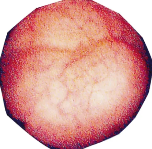
Fig. 1. Cystoscopy showed a smooth surfaced mass,
firmed a smooth, 2 × 2 cm mass protruding into the bladder, with the overlying normal looking mucosa. Computerized tomography showed a 2 cm-diameter-solid mass and homogenous appearance at the left-anterolater-al wleft-anterolater-all of the urinary bladder. T1-weighted images and T2-weighted images revealed homogenous median and low intensity urinary bladder tumor of 2 cm in diameter, respectively (Figs 2,3). Then, superficial and careful resec-tion was successfully performed. Pathological
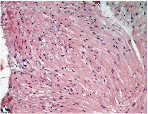
evaluation showed benign smooth muscle bun-dles consistent with leiomyoma, and no mitotic or proliferative changes (Fig 4). The patient remains asymptomatic with normal voiding and there has been no evidence of recurrence for 13 months after operation.
DISCUSSION
Bladder leiomyomas are usually asympto-matic, but can cause storage or voiding
symp-66 Norifumi SAWADAet al.
Fig. 2. MRI showed a homogenous low intensity area which has almost same intensity of muscles
on T1-weighted image in the left. On the Gd-DTPA enhanced T2-weighted image in the right, the tumor showed equal enhancement (arrow ahead).
Fig. 3. MRI showed a homogenous low signal intensity area on T1 and T2-weighted image
toms apart from hematuria2). Although rarely
encountered, they are the most common benign disease of mesenchymal bladder tumor. MRI of the bladder is useful in the diagnosis of this disease3). In the present case, MRI showed a
homogenous low signal intensity area on T2-weighted images, with median intensity area on T1-weighted images. These MRI findings are usually compatible to the findings of urethral leiomyomas3,4). The size of the tumor may be a
key factor for the choice of treatment. The aver-age tumor diameter of the 13 cases with com-plete removal by transurethral resection in Japan was 17.8 mm5). In the present case, the
tumor was 2 cm in diameter, hence we evaluated that complete resection of the tumor was possi-ble and the resection of the tumor was success-fully performed. Conservative treatment as transurethral resection may be recommended
as a safe, definitive way to treat small bladder leiomyomas, because recurrence of the leiomy-oma is very rare.
REFERENCES
1) Ishida K, Yuhara K, Kanimoto Y. Leiomyoma of the urinary bladder: report of three cases. Hiny-oukika-Kiyou. 49: 671–674, 2003.
2) Gaynor-Krupnick DM, Kreder KJ. Bladder neck leiomyoma presenting as voiding dysfunction. J Urol. 172: 249–250, 2004.
3) Sandaram CP, Rawal A, Saltzman B. Characteris-tics of bladder leiomyoma as noted on magnetic resonance. Urology. 52: 1142–1143, 1998. 4) Sugaya S, Hasegawa N, Kawashima A, Shino Y,
Ohishi Y. A case of leiomyoma of the urinary bladder:MRI findings. Jpn J Med Imaging. 21: 57–60, 2002.
5) Hagiwara N, Fujihiro S. Submucosal-type leiomy-oma of the urinary bladder treated by transurethral resection: A case report. Rinshou Hinyoukika. 57: 161–163, 2003.
67 A Case of Asymptomatic Submucosal-type Leiomyoma of the Urinary Bladder Correctly Diagnosed with Magnetic Resonance Imaging (MRI) and Successfully Treated by Transurethral Resection
Fig. 4. Histological examination discloses an increase of spindle cells with interlacing fascicular