CONGENITAL HEART DISEASE
Residual Restrictive Right Ventricular
Physiology after One-and-a-Half Ventricular
Repair Conversion in Pulmonary Atresia with
Intact Ventricular Septum
Kazuki Suruga, MD, Norihisa Toh, MD, PhD, Yasuhiro Kotani, MD, PhD, Hideki Onishi, MD, PhD, Teiji Akagi, MD, PhD, Shingo Kasahara, MD, PhD, and
Hiroshi Ito, MD, PhD,Okayama, Japan
INTRODUCTION
Pulmonary atresia with intact ventricular septum (PA-IVS) is a rare congenital heart defect. Among various surgical procedures that can be performed in early childhood, our institutional strategy is to aim for biventricular repair (BVR) on the basis of evidence of potential right ventricular (RV) growth after adequate creation of RV and pul-monary artery continuity1-3to avert abnormal hemodynamics and late complications in univentricular repair. Despite the relatively good long-term survival in adult patients with PA-IVS who have un-dergone BVR,4one-and-a-half ventricular repair (1.5VR) conversion concomitant with pulmonary valve intervention is required in some adult patients undergoing BVR because of pulmonary regurgitation (PR) and elevated central venous pressure followed by end-organ damage and heart failure. Restrictive RV physiology is a unique feature in patients with PA-IVS after BVR5and can be assessed on pulsed Doppler echocardiography. Although restrictive RV physi-ology is a potential culprit of BVR failure,5changes in Doppler and other parameters after 1.5VR conversion from BVR remain unknown.
CASE PRESENTATION
A 20-year-old woman with PA-IVS who had undergone BVR during infancy presented with progressive shortness of breath on exertion and pedal edema. She was diagnosed with PA-IVS at birth. Her tricuspid valve (TV) Z score from echocardiography was 6, RV end-diastolic volume (RVEDV) from cine angiography was 24% of the predicted normal value, and RVEDV indexed to body surface area (RVEDVi) was 9.5 mL/m2
. Balloon atrioseptostomy was per-formed at birth, followed by pulmonary valvotomy, left modified Blalock-Taussig shunt, and ligation of the patent ductus arteriosus at 2 weeks of age. At the age of 5 months, cardiac catheterization revealed valvular pulmonary stenosis with a peak pressure gradient
of 46 mm Hg and 30% of the predicted normal value of RVEDV (RVEDVi 14 mL/m2). Therefore, balloon pulmonary valvuloplasty was performed, with the expectation of RV growth by means of relief of the pulmonary stenosis. At the age of 1.7 years, the TVZ score from echocardiography was 1, RVEDV from cine angiography was 58% of the predicted normal value, and RVEDVi was 30 mL/m2. Although the right ventricle was shown to be relatively small, it was considered to be large enough to proceed to BVR upon inspection of the right ventricle and TV at surgery. BVR including RV overhaul, RV outflow tract reconstruction with a Gore-Tex monocusp valve, semiclosure of the atrial septal defect, and take-down of a modified left modified Blalock-Taussig shunt were performed at the age of 1.8 years. After definitive surgery, she had been doing well without any symptoms of heart failure or arrhythmia, and RVEDV had enlarged to 73% of the normal predicted value (RVEDVi 42 mL/m2
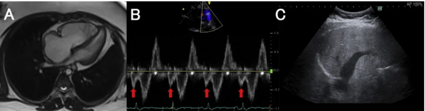
) at the age of 3 years. The patient presented with signs of heart failure at the age of 20 years. Electrocardiography showed normal sinus rhythm with complete right bundle block (Figure 1). Cardiac magnetic resonance imaging revealed an enlarged right ventricle, enlarged right atrium, mild tricuspid regurgitation, and severe PR with a regurgitant fraction of 27% (Figure 2A). Echocardiography showed preserved left ventric-ular ejection fraction of 65%, normal inferior vena cava diameter of 13 mm with poor respiratory change of 38%, and end-diastolic forward flow in the pulmonary artery with a peak velocity of 44 cm/sec (Figure 2B;Videos 1and2), suggesting the presence of restrictive RV physiology.6
Cardiac catheterization showed an elevated right atrial (RA) pres-sure of 11 mm Hg. Liver ultrasound revealed severely dilated hepatic veins, an irregular surface, and a coarse parenchymal texture (Figure 2C). Serum B-type natriuretic peptide level was elevated to 51.0 mg/dL. Therefore, the patient was diagnosed as having severe PR and right heart failure followed by chronic liver damage. A deci-sion was made after multidisciplinary discusdeci-sion to convert to 1.5VR. Intraoperatively, the Gore-Tex monocusp pulmonary valve was severely calcified, and it was replaced with a bioprosthetic valve (25-mm Epic; St. Jude Medical, St. Paul, MN). The RV endocardium was severely scarred as a result of muscle resection at the time of RV overhaul and BVR, and this finding was considered to be closely associated with the presence of restrictive RV physiology. The postop-erative course was uneventful. Three months after 1.5VR and pulmo-nary valve replacement, although restrictive RV physiology was still observed by echocardiography (Figure 3B;Videos 3and4), the pa-tient’s symptoms were dramatically reduced, RV and RA sizes were decreased (Figure 3A), RA pressure was decreased to 5 mm Hg, and serum B-type natriuretic peptide level was decreased to 14.8 mg/dL. Moreover, abdominal ultrasound showed that liver congestion had diminished (Figure 3C).
From the Department of Cardiovascular Medicine (K.S., N.T., T.A., H.I.), Department of Cardiovascular Surgery (Y.K., S.K.), and Department of
Gastroenterology and Hepatology (H.O.), Okayama University Graduate School of Medicine, Dentistry, and Pharmaceutical Sciences, Okayama, Japan.
Keywords: Adult congenital heart disease, Pulmonary atresia with intact ventricular septum, Restrictive right ventricular physiology
Conflicts of interest: The authors reported no actual or potential conflicts of interest relative to this document.
Copyright 2020 by the American Society of Echocardiography. Published by Elsevier Inc. This is an open access article under the CC BY-NC-ND license (http:// creativecommons.org/licenses/by-nc-nd/4.0/).
2468-6441
https://doi.org/10.1016/j.case.2020.08.002
DISCUSSION
Surgical strategies used for patients with PA-IVS vary among institu-tions because of a lack of evidence for achieving optimal physiology in this unique anatomy. Because our institutional strategy is to aim for BVR, our choice of initial palliation includes a modified Blalock-Taussig shunt with pulmonary valvotomy to achieve growth of right-sided heart structures. After the initial surgical palliation, an RV overhaul procedure is performed for patients with RVEDV < 50% of the predicted normal value.1,2 Finally, we generally consider a TV Z score > 3 as an indication for BVR. If the TV Z score is < 8, we consider univentricular repair favorable. Patients with TV Z scores between 8 and 3 are considered
candidates for 1.5VR. In the present case, because RVEDV was a borderline value for BVR, RV overhaul was performed before estab-lishing biventricular circulation.
Although long-term survival is relatively good in BVR patients,4 restrictive RV physiology after BVR is often observed,5and it is asso-ciated with elevated RA pressure, RA dysfunction,7and increased central venous pressure, leading to end-organ damage such as liver congestion. At our institution, according to our accumulated experi-ences1and previous reports,81.5VR conversion is performed for pa-tients with PA-IVS with failed BVR who fulfill the following criteria: (1) heart failure symptoms, (2) significant PR, (3) elevated RA pressure (>10 mm Hg), and (4) liver damage assessed by ultrasound. In the pre-sent case, after successful 1.5VR conversion concomitant with pulmo-nary valve replacement, RA pressure and serum B-type natriuretic peptide were significantly decreased, and liver congestion was mark-edly improved. However, restrictive RV physiology was still observed after 1.5VR conversion, which may be related to either the RV over-haul procedure or unique RV anatomy in patients with PA-IVS. Therefore, close follow-up is mandatory even after 1.5VR conversion from BVR.
CONCLUSION
Restrictive RV physiology is a unique finding in patients with PA-IVS who have undergone BVR. This case demonstrated that restrictive RV physiology persisted even after 1.5VR conversion from BVR.
SUPPLEMENTARY DATA
Supplementary data related to this article can be found athttps://doi. org/10.1016/j.case.2020.08.002.
Figure 1 Electrocardiogram obtained at cardiology visit for heart failure symptoms, showing normal sinus rhythm with complete right bundle block and prolonged QRS duration of 138 msec.
VIDEO HIGHLIGHTS
Video 1: Apical four-chamber window on preoperative transthoracic echocardiography demonstrating a dilated right heart.
Video 2: Parasternal short-axis window on preoperative transthoracic echocardiography demonstrating diastolic flow reversal in branch pulmonary arteries indicating severe PR. Video 3: Apical four-chamber window on postoperative transthoracic echocardiography demonstrating decreased RA and RV volumes compared with those on preoperative trans-thoracic echocardiography.
Video 4: Parasternal short-axis window on postoperative transthoracic echocardiography demonstrating diminished PR.
View thevideo content online at www.cvcasejournal.com
.
524 Suruga et al CASE: Cardiovascular Imaging Case Reports
REFERENCES
1. Kotani Y, Kasahara S, Fujii Y, Eitoku T, Baba K, Otsuki S, et al. A staged decompression of right ventricle allows growth of right ventricle and subse-quent biventricular repair in patients with pulmonary atresia and intact ventricular septum. Eur J Cardiothorac Surg 2016;50:298-303.
2. Sano S, Ishino K, Kawada M, Fujisawa E, Kamada M, Ohtsuki S. Staged bi-ventricular repair of pulmonary atresia or stenosis with intact bi-ventricular septum. Ann Thorac Surg 2000;70:1501-6.
3. Huang SC, Ishino K, Kasahara S, Yoshizumi K, Kotani Y, Sano S. The poten-tial of disproportionate growth of tricuspid valve after decompression of the right ventricle in patients with pulmonary atresia and intact ventricular septa. J Thorac Cardiovasc Surg 2009;138:1160-6.
4. Toh N, Kotani Y, Akagi T, Kuroko Y, Baba K, Otsuki S, et al. Outcomes of patients with pulmonary atresia with intact ventricular septum reaching adulthood. Congenit Heart Dis 2020;15:1-11.
5. Redington AN, Penny D, Rigby ML, Hayes A. Antegrade diastolic pulmonary arterial flow as a marker of right ventricular restriction after complete repair of pulmonary atresia with intact septum and critical pulmonary valvar stenosis. Cardiol Young 1992;2: 382-6.
6. Gatzoulis MA, Clark AL, Cullen S, Newman CG, Redington AN. Right ven-tricular diastolic function 15 to 35 years after repair of tetralogy of Fallot. Restrictive physiology predicts superior exercise performance. Circulation 1995;91:1775-81.
7.To AH, Lai CT, Wong SJ, Cheung YF. Right atrial mechanics
long-term after biventricular repair of pulmonary atresia or ste-nosis with intact ventricular septum. Echocardiography 2016;33: 586-95.
8. Shi JZ, Chow PC, Li W, Kwok SY, Wong WH, Cheung YF. Fifty-five years follow-up of 111 adult survivors after biventricular repair of PAIVS and PS. Pediatr Cardiol 2019;40:374-83.
Figure 3 Postoperative images.(A) Right heart size became smaller after 1.5VR conversion, and RVEDVi was decreased to 54 mL/m2 on cardiac magnetic resonance imaging.(B) An end-diastolic forward flow of 45 cm/sec (red arrow) was still observed even though 1.5VR conversion concomitant with pulmonary valve replacement was successfully performed.(C) The hepatic vein diameter was significantly reduced, indicating that liver congestion was diminished.
Figure 2 Preoperative images.(A) Cardiac magnetic resonance imaging showed a dilated right heart, and RVEDVi was 65 mL/m2.(B) On Doppler echocardiography, an end-diastolic forward flow of 44 cm/sec to the pulmonary trunk (red arrow) was observed, suggest-ing the presence of restrictive RV physiology.(C) Liver ultrasound revealed severely dilated hepatic veins, an irregular surface, and a coarse parenchymal texture.
CASE: Cardiovascular Imaging Case Reports Volume 4 Number 6