T
hermal ablation is a minimally invasive tumor treatment method whose results are comparable to surgical treatment [1-5]. In contrast, percutaneous ablation treatments run a risk of hemorrhage and tumor seeding after needle extraction [6-13]. The incidences of hemorrhage and tumor seeding by percutaneous ablation have been reported to be 1.5-8.1% [6,7] and 0.2-0.5% [11,12], respectively. Radiofrequencyabla-tion (RFA) is less likely to lead to a hemorrhagic com-plication than is cryoablation (CA) [8,14], although the use of a thinner probe could improve the outcome of cryoablation [15]. Needle tract ablation using RFA sys-tems may be a possible method to prevent these events; however, this procedure cannot be performed using a cryoprobe [9,16].
We developed an experimental device for perform-ing needle tract ablation usperform-ing a cryoprobe. The pur-CopyrightⒸ 2021 by Okayama University Medical School.
http ://escholarship.lib.okayama-u.ac.jp/amo/
Original Article
Needle Tract Ablation in Liver Tissue Using a Cryoprobe Combined
with an Electrosurgical Device:
Influence of ex vivo and in vivo Animal Models
Hideo Gobara
a*, Akira Yamamoto
b, Toshiyuki Komaki
c, Toshiaki Kitayama
d,
Jun Sakurai
e, Toshihiro Iguchi
c, Yusuke Matsui
c, Mayu Uka
c,
Koji Tomita
c, Takao Hiraki
f, and Susumu Kanazawa
faDivision of Medical Informatics, cDepartment of Radiology, eCenter for Innovative Clinical Medicine,
Okayama University Hospital, fDepartment of Radiology, Okayama University Graduate School of Medicine,
Dentistry and Pharmaceutical Sciences, Okayama 700-8558, Japan,
bDepartment of Diagnostic and Interventional Radiology, Osaka City University Graduate School of Medicine,
Osaka 545-8585, Japan, dDepartment of Radiology, Otemae Hospital, Osaka 540-0008, Japan
To assess the feasibility of needle tract ablation in liver tissue in ex vivo and in vivo animal models using a cryo-probe and electrosurgical device. The experimental device is made by inserting a cryocryo-probe through an intro-ducer sheath for insulation, with 2-cm of probe tip projecting out. A beagle liver was punctured by the device, and electric current was applied at 30-W with the electrosurgical knife touching the non-insulated device base. The discolored area of cut surface along the device was evaluated in 5 application-time groups (5,10,15,20, or 25 seconds). An ex vivo experiment was performed to determine an ablation algorithm with an appropriate application time by comparison with radiofrequency ablation (RFA) results. Thereafter, an in vivo experiment was performed to verify the algorithm’s feasibility. In the ex vivo model, the cut surface demonstrated different amounts of discolored area according to the application time. The total discolored area in the 20-seconds group was similar to that by RFA. In the in vivo model, the liver did not bleed, the total discolored area was similar to that ex vivo, and coagulation necrosis was confirmed by photomicrograph. Needle tract ablation can be per-formed using the experimental device and electrosurgical device.
Key words: needle tract ablation, cryoablation, electrosurgical device, animal, liver
Received July 10, 2020 ; accepted September 9, 2020.
*Corresponding author. Phone : +81-86-235-7313; Fax : +81-86-235-7316
pose of this study was to assess the feasibility of needle tract ablation in the liver tissue in ex vivo and in vivo animal models using a cryoprobe combined with an electrosurgical device.
Materials and Methods
In this study, an ex vivo experiment was performed to determine an appropriate ablation algorithm. Thereafter, an in vivo experiment was performed to verify the feasibility of tract ablation with the algorithm.
Devices a. RFA system
A 17-gauge internally cooled needle electrode and a generator (Cool-tip RFA system; Covidien, Mansfield, MA, USA) were used as the reference system for needle tract ablation. The generator could produce a radiofre-quency electric current of 472 kHz. The needle we used had a non-insulated tip length of 2 cm; other parts of the needle were fully insulated.
b. Experimental CA device
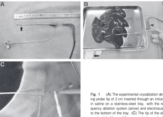
A 17-gauge cryoprobe (Ice-Rod, Galil Medical, Yokneam, Israel) was insulated by inserting it through a 4-French and 10-cm length introducer sheath (Radifocus Introducer II H, Terumo, Tokyo), with
2 cm of the probe tip projecting out (Fig.1A). c. Electrosurgical devices
In the ex vivo model, a Conmed System 5000 elec-trosurgical generator (CONMED, Utica, NY, USA) was used for electrical energy deposition. The generator could produce radiofrequency current at 562 kHz. On the other hand, the ICC 50 generator (Erbe Elektromedizin GmbH, Germany) used in the in vivo model could produce electric current at 510-550 kHz. A monopolar handpiece electrosurgical knife was used in both the ex vivo and in vivo models.
Ex vivo model a. Procedure
The laboratory room temperature was maintained at 20°C. Around 100 ml of saline was poured into a stain-less-steel tray and a beaker, respectively. For the exper-imental CA device system and RFA system, the return electrodes were affixed to the bottom of the tray, and the cable was connected to the generator. Further, an electrosurgical knife or an RFA needle electrode was connected to each correspondent generator. A beagle liver was then placed in the tray (Fig.1B).
The liver was punctured by the RFA needle electrode (depth of puncture >2 cm). After the needle tip place-ment, electric current was applied at 30 W, without
A
B
C
Fig. 1 (A) The experimental cryoablation device: a cryoprobe with a
project-ing probe tip of 2 cm inserted through an introducer sheath. (B) A beagle liver in saline on a stainless-steel tray, with the return electrodes of the radiofre-quency ablation system (arrow) and electrosurgical system (arrowhead) affixed to the bottom of the tray. (C) The tip of the electrosurgical knife touching the non-insulated base of the cryoprobe to conduct electric current.
iced water circulation, until it was automatically shut off when the impedance rapidly increased. This proce-dure was repeated 5 times after repositioning the needle electrode. The experimental CA device was then placed in the same way, and an electric current was applied at 30 W with the electrosurgical knife touching the non- insulated base of the device (Fig.1C). The application times were 5,10,15,20, and 25sec; this procedure was also repeated 5 times, after repositioning the device tip each time. Before each penetration, the RF needle and experimental CA device were dunked in the saline in the beaker to cool the probe tip down to room tempera-ture. After each application, the liver was sectioned along the probe tip.
b. Data collection
We observed a discolored area (ablation zone) on the cut surface in each section and recorded the maximum and minimum width perpendicular to the needle (mm). We also recorded the appearance of the liver during application and appearance of the tip of the introducer sheath after application.
In vivo model a. Animal
The test subject was a female beagle (12.0 kg). The study was approved by the institutional animal ethics committee of Osaka City University (no. 17001: 7/12/ 2017), where the in vivo animal experiment was per-formed.
b. Procedure
All experiments were performed by (A.K. and T.K.) in compliance with the regulations for animal experi-ments. The beagle was anesthetized with an intramus-cular injection of 150 mg of ketamine and 10 mg of midazolam. Anesthesia was maintained intravenously with 25 mg/kg of pentobarbital sodium. The animal could breathe spontaneously throughout the procedure.
This procedure involved exposing the liver surface through laparotomy. The return electrode was affixed to the lower abdominal wall of the beagle. Under direct visual guidance, the device tip punctured the liver (depth >2 cm). The electric current was then applied at 30 W for 20sec; this duration was chosen based on the results obtained from the ex vivo model. After five appli-cations, each followed by a repositioning of the device tip, the liver was harvested and sectioned along the probe tip. A photograph of each cut surface was taken, and specimens from each cut surface were prepared for histologic evaluation.
c. Data collection
The liver was observed for the presence of bleeding from the insertion site after each withdrawal of the device. After the final withdrawal, the discolored area on the cut surface was measured, and the extent of coagulation necrosis was evaluated on the photomicro-graph afterward.
Statistical analyses. In the in vivo model, the
maximum width perpendicular to the device of each application-time group was compared to that of the RFA needle group using an unpaired Student’s t-test. A
P-value of <0.05 was considered to indicate a
statisti-cally significant difference. Statistical Package for the Social Science version 22.0 software (IBM Corp., Armonk, NY, USA) was used for the statistical analy-ses.
Results
Ex vivo model. The beagle liver was easily
punc-tured, although the experimental CA device had caliber difference between the needle tip and introducer sheath. The electric current was successfully passed through both needles. The cut surface along the RFA needle electrode demonstrated a circumferential discolored area around the non-insulated tip. On the other hand, the cut surface along the experimental CA device demonstrated the discolored area only around both ends of the projecting probe tip in the 5-sec and 10-sec groups (Fig.2A, arrowhead) and circumferentially around the tip in the ≥15-sec groups (Fig.2B). In the 25-sec group, vaporing was seen around the insertion site during application; additionally, the tip of the introducer sheath was charred upon withdrawal of the device. The maximum and minimum widths perpen-dicular to the device are summarized in Table 1. The maximum widths in the 15- and 20-sec groups were not significantly different when compared with those in the RFA group.
In vivo model. Electric current was successfully
passed through the beagle liver in the in vivo animal model. After each application followed by the with-drawal of the cryoprobe, the liver did not bleed from the puncture site. The cut surface of the needle tract demonstrated a discolored area around the device tip (Fig.3A). The maximum widths perpendicular to the device were 5.6±0.6 mm. The photomicrograph of the cut surface showed coagulation necrosis correspondent
to about two-thirds of the discolored area (Fig.3B).
Discussion
This experimental device, including the cryoprobe, introducer sheath, and electrosurgical device, was
eas-ily made with commercially available products. We used the introducer sheath as an insulating device for the following reasons. First, an introducer sheath is typically made of resinous material and provides good insulation. Second, the inner diameter of the 4-Fr introducer sheath is about 4.4- or 4.5-Fr (≈ 17-gauge),
A
B
Fig. 2 Ex vivo model: Photographs of the cut surface along the needle: at 10sec of cryoablation (A); at 20sec of cryoablation (B).
A
B
Fig. 3 In vivo model: The cut surface of the liver along the needle shows a discolored area (arrowhead) (A), and a photomicrograph of
the cut surface demonstrates a discolored area (arrow) and coagulation necrosis (arrowhead) along the needle (B).
Table 1 Width of the discolored area with each needle type and application time
Needle type Application time of electric current (sec)
Discolored area width Statistical analyses Max
(mm) (mm)Min (unpaired t-test)P-value
RFA needle * 6.4±0.9 4.8±0.8 † 5 2.2±0.4 0 <0.001 10 4.4±0.9 0 <0.001 Experimental CA device 1520 5.46.8±±0.50.8 2.66.0±±1.70.5 0.060.48 25 8.2±1.3 5.8±0.8 0.03
RFA, radiofrequency ablation; CA, cryoablation.
*time until the electric current is automatically shut off when the impedance is rapidly increased †reference
allowing the passage of a 4-Fr catheter (usually 4.2-Fr in diameter). Third, the caliber difference between the introducer sheath and cryoprobe is minimized because of its tapered tip. Therefore, the introducer sheath in this study was just the right caliber for inserting the cryoprobe, and it easily penetrated the animal liver tis-sue. Although the projecting tip length of 2 cm is shorter than the expected ice-ball long axis diameter, the freezing ability of the cryoprobe may have been affected by the introducer sheath. This problem could be solved by withdrawing the sheath outside the ice-ball area during the freezing procedure and then pushing it through the cryoprobe to the original position.
The term “radiofrequency” applies to coagulation induction from all electromagnetic energy sources within the radiofrequency spectrum (3 kHz to 300 GHz), including “radiofrequency” and “microwave” devices. However, the currently available devices are tradition-ally designed for RFA in the 375-500 kHz range [16]. Like the RFA needle in this study, most devices cur-rently used are monopolar with a single active electrode. On the other hand, electrosurgical devices are used for surgical cutting or to control bleeding by coagulation during surgery. These devices use alternating current at different radiofrequency levels. In a monopolar device, heat is produced when electric current encounters resis-tance from the circuit made by the patient, electrosur-gical generator, active electrode (e.g., electrosurelectrosur-gical knife), and return electrode [18,19]. In this study, the same circuit was replicated in the laboratory using the experimental CA device. Because the introducer sheath provided insulation, the electric current could only flow through the needle tip, which contacted the liver tissue directly without insulation. This insulation allowed only the needle tip to be energized.
Percutaneous CA is a recently developed technique for minimally invasive tumor treatment. Rapid tissue freezing and thawing produces cytotoxic effects by dis-rupting the cellular membranes and inducing cell death [17,20]. CA has several advantages over other heat-based thermal ablation techniques; these include excel-lent visualization of the ablation zone (ice-ball) and absence of pain during the procedure. One of the dis-advantages of CA is a lack of ability to ablate the needle tract. Wu et al. reported in their meta-analysis that the pooled rate of hemorrhage was 4.14% (18 of 435) in the CA group and 1.16% (4 of 346) in the RFA group; there was thus a trend toward higher risk of bleeding by
CA than RFA, despite a lack of statistical significance (OR=1.92, 95% CI: 0.62-5.88) [8].
Needle tract seeding is a rare but serious complica-tion of percutaneous thermal ablacomplica-tion treatment [10,12]. Regarding CA, an incidence rate of 0.2-0.76% has been reported in larger series [11,21,22]. The pos-sible mechanisms of tumor seeding are as follows: adherence of viable cancer cells to the needle, blood reflux along the tract, or cancer cells being forced into the tract because of increased intratumoral pressure [10,22]. Cauterization of the needle may be effective in the prevention of needle tract seeding [6,9,12,22].
Recently, a radiofrequency device for tract ablation after liver biopsy was reported [24]. Additionally, a newly developed CA device, with a non-insulated tip and insulated basal needle, has been made available. Although needle tract cauterization can be performed using this device, it was actually developed for active thawing without helium [25]. Unfortunately, this device is not available in our country; hence, we cannot con-firm the effectiveness of needle tract ablation using this device.
This experimental model has several limitations. First, the same results might not be obtained with nee-dle tract ablation of cold liver tissue after CA, as the temperature of the beagle liver in this experiment was 20°C. Second, the caliber difference may present a serious problem via the percutaneous approach because of puncture resistance. Ideally, the CA probe should be given a thin insulating coating, such as an RFA elec-trode, although the lack of such coating was not of consequence in our experimental study because we performed puncture at the laparotomy site, and liver tissue itself is quite soft. Third, needle tract ablation was performed only in the liver tissue. Finally, although this device was easily made with commercially available products, this constitutes an off-label use of the cryo-probe; therefore, the device cannot be used in humans without the permission of the institutional ethics com-mittee.
In conclusion, needle tract ablation can be performed using our newly developed experimental device com-bined with an electrosurgical device. These results will have important future implications for reducing the risk of bleeding and tumor seeding with CA.
References
1. Chen MS, Li JQ, Zheng Y, Guo RP, Liang HH, Zhang YQ, Lin XJ and Lau WY: A prospective randomized trial comparing percu-taneous local ablative therapy and partial hepatectomy for small hepatocellular carcinoma. Ann Surg (2006) 243: 321-328. 2. Feng K, Yan J, Li X, Xia F, Ma K, Wang S, Bie P and Dong J: A
randomized controlled trial of radiofrequency ablation and surgical resection in the treatment of small hepatocellular carcinoma. J Hepatol (2012) 57: 794-802.
3. Fang Y, Chen W, Liang X, Li D, Lou H, Chen R, Wang K and Pan HM: Comparison of long-term effectiveness and complications of radiofrequency ablation with hepatectomy for small hepatocellu-lar carcinoma. J Gastroenterol Hepatol (2014) 29: 193-200. 4. Hasegawa K, Kokudo N, Makuuchi M, Izumi N, Ichida T, Kudo M,
Ku Y, Sakamoto M, Nakashima O, Matsui O and Matsuyama Y: Comparison of resection and ablation for hepatocellular carcinoma: a cohort study based on a Japanese nationwide survey. J Hepatol (2013) 58: 724-729.
5. Cho YK, Kim JK, Kim MY, Rhim H and Han JK: Systemic review of randomized trials for hepatocellular carcinoma treated with per-cutaneous ablation therapies. Hepatology (2009) 49: 453-459. 6. Goto E, Tateishi R, Shiina S, Masuzaki R, Enooku K, Sato T,
Ohki T, Kondo Y, Goto T, Yoshida H and Omata M: Hemorrhagic complications of percutaneous radiofrequency ablation for liver tumors. J Clin Gastroenterol (2010) 44: 374-380.
7. Takaki H, Yamakado K, Nakatsuka A, Yamada T, Shiraki K, Takei Y and Takeda K: Frequency of and risk factors for complica-tions after liver radiofrequency ablation under CT fluoroscopic guid-ance in 1500 sessions: single-center experience. AJR Am J Roentgenol (2013) 200: 658-664.
8. Wu S, Hou J, Ding Y, Wu F, Hu Y, Jiang Q, Mao P and Yang Y: Cryoablation versus radiofrequency ablation for hepatic malignan-cies: a systematic review and literature-based analysis. Medicine (Baltimore) (2015) 94: e2252.
9. Howenstein MJ and Sato KT: Complications of radiofrequency ablation of hepatic, pulmonary, and renal neoplasm. Semin Intervent Radiol (2010) 27: 285-295.
10. Jaskolka JD, Asch MR, Kachura JR, Ho CS, Ossip M, Wong F, Sherman M, Grant DR, Greig PD and Gallinger S: Needle tract seeding after radiofrequency ablation of hepatic tumors. J Vasc Interv Radiol (2005) 16: 485-491.
11. Mulier S, Mulier P, Ni Y, Miao Y, Dupas B, Marchal G, De Wever I and Michel L: Complications of radiofrequency coagula-tion of liver tumours. Br J Surg (2002) 89: 1206-1222.
12. Livraghi T, Solbiati L, Meloni MF, Gazelle GS, Halpern EF and Goldberg SN: Treatment of focal liver tumors with percutaneous radio-frequency ablation: complications encountered in a multi-center study. Radiology (2003) 226: 441-451.
13. Llovet J, Vilana R, Brú C, Bianchi L, Salmeron JM, Boix L, Ganau S, Sala M, Pagès M, Ayuso C, Solé M, Rodés J, Bruix J and Barcelona Clínic Liver Cancer (BCLC) Group: Increased risk
of tumor seeding after percutaneous radiofrequency ablation for single hepatocellular carcinoma. Hepatology (2001) 33: 1124-1129. 14. Huang YZ, Zhou SC, Zhou H and Tong M: Radiofrequency abla-tion versus cryosurgery ablaabla-tion for hepatocellular carcinoma: a meta-analysis. Hepatogastroenterology (2013) 60: 1131-1135. 15. Lee SM, Won JY, Lee DY, Lee KH, Lee KS, Paik YH and Kim
JK: Percutaneous cryoablation of small hepatocellular carcinomas using a 17-gauge ultrathin probe. Clin Radiol (2011) 66: 752-759. 16. Lencioni R, de Baere T, Martin RC, Nutting CW and Narayanan G:
Image-guided ablation of malignant liver tumors: recommendations for clinical validation of novel thermal and non-thermal technolo-gies - a Western perspective. Liver Cancer (2015) 4: 208-214. 17. Ahmed M, Solbiati L, Brace CL, Breen DJ, Callstrom MR,
Charboneau JW, Chen MH, Choi BI, de Baère T, Dodd GD 3rd, Dupuy DE, Gervais DA, Gianfelice D, Gillams AR, Lee FT Jr, Leen E, Lencioni R, Littrup PJ, Livraghi T, Lu DS, McGahan JP, Meloni MF, Nikolic B, Pereira PL, Liang P, Rhim H, Rose SC, Salem R, Sofocleous CT, Solomon SB, Soulen MC, Tanaka M, Vogl TJ, Wood BJ, Goldberg SN, International Working Group on Image-guided Tumor Ablation; Interventional Oncology Sans Frontières Expert Panel; Technology Assessment Committee of the Society of Interventional Radiology; and Standard of Practice Committee of the Cardiovascular and Interventional Radiological Society of Europe: Image-guided tumor ablation: standardization of terminology and reporting criteria--a 10-year update. Radiology (2014) 273: 241-260.
18. Jones CM, Pierre KB, Nicoud B, Stain SC and Melvin WV: Electrosurgery. Curr Surg (2006) 63: 458-463.
19. Van Way CW and Hinrichs CS: Technology focus: electrosurgery 201: basic electrical principles. Curr Surg (2000) 57: 261-264. 20. Rubinsky B, Lee CY, Bastacky J and Onik G: The process of
freezing and the mechanism of damage during hepatic cryosurgery. Cryobiology (1990) 27: 85-97.
21. Wang C, Wang H, Qu JH, Lu YY, Bai WL, Dong Z, Gao XD, Rong GH, Zeng Z and Yang YP: Tumour seeding after percutane-ous cryoablation for hepatocellular carcinoma. World J Gastroen-terol (2012) 18: 6587-6596.
22. Chang S, Kim SH, Lim HI, Kim SH, Lee WJ, Choi D, Kim YS and Rhim H: Needle tract implantation after percutaneous inter-ventional procedures in hepatocellular carcinomas: lessons learned from a 10-year experience. Korean J Radiol (2008) 9: 268-274.
23. Livraghi T, Solbiati L, Meloni MF, Gazelle GS, Halpern EF and Goldberg SN: Treatment of focal liver tumors with percutaneous radio-frequency ablation: complications encountered in a multi-center study. Radiology (2003) 226: 441-451.
24. Song KD, Rhim H, Lee MW, Kang TW and Lim S: A radiofre-quency device for tract ablation after liver biopsy: a single-institu-tion human feasibility study. Br J Radiol (2018) 91: 20170585. 25. Lau B, Shar TT, Valerio M, Hamid S, Ahmed HU and Arya M:
Technological aspects of delivering cryotherapy for prostate cancer. Expert Rev Med Devices (2015) 12: 183-190.