第2部
人骨骨格図譜(改訂版)作成にあたって
2006 年に「動物考古学の手引き」として、松井章さんの主導のもと、遺跡から出土する哺
乳動物の骨格図譜が世に出された。その後英語版も刊行され、世界の各地で使われることに
なった。その中にはもちろん人骨の図譜も含まれている。図譜は多くの考古学の研究者や発
掘担当者に使われてきた。しかし、2 次元的な図譜には限界もあった。
同定作業にもっとも必要なもの、それは実物の骨格標本である。松井章さんがおられた奈
良文化財研究所の環境考古学研究室は、多くの骨格の比較資料を収集し、各地の研究者から
利用されるようになった。しかし、実物の標本は誰もが手に入れることが出来るわけではな
く、維持・管理も難しい。たとえ資料を持っていても現場に携えることは難しい。そこで、
実物標本との間を埋める 3 次元の骨格データベースが企画されるようになった。これにはい
くつかの課題があった。制作には 3 次元的なデータをとる必要があり、使いやすく、かつ安
い費用でパソコンを介して見られるようにするというハードルを越えなければならなかっ
た。どんなにすぐれていても高額なソフトが必要ならそれはほとんど意味がないデータベー
スである。このハードルは無料ソフトで見られる PDF で配信することで解決された。また、
特定の研究者だけでなく、広く世に出ている必要がある。これには奈良文化財研究所の協
力で、研究所のホームページ https://www.nabunken.go.jp/research/environmental/gaiyo.
html で閲覧が可能になった。既に刊行されている英語版の図譜が海外でも多く利用されて
いることを考えると、今回改訂したものも含めて、3 次元データベースはいっそう利用され
ると期待されるので、国際考古動物学会(ICAZ)のホームページでの公開も目指している。
今回は、人骨部分の改訂をおこなった。形態研究の基礎になるものであり、応用範囲も広
いのでまず人骨を対象とした。人類学者の中橋孝博博士の協力を得られたことも大きな励み
になった。この改訂によって、同定の精度はいっそう高まったといえよう。しかし、まだま
だ課題もある。普及している一部のタブレットで見られないし、他の多くの哺乳動物の 3 次
元画像も追加していかなくてはならない。だが、これらの問題も次第に解決されてゆくであ
ろう。
この計画を立案し、3 次元のデータベース化を最初から推し進めてきた松井章さんを失っ
たことはほんとうに残念である。しかし、その後を継ぐ方々の地道な努力の結果、版を改め
ることが出来たことは、松井さんへの大きな供養とすることが出来るだろう。業績として認
められにくいこのような仕事を継続していくことは、すぐに結果を求められる今の日本では
なかなか難しいことであるが、いっそうの発展を心から期待している。
茂原信生
1. 人骨の特徴
人骨はウシやウマのような大型動物を除く他の動
物骨に比べて,各部位が大きく,長いという特徴が
ある。成人骨は個人差があるものの約206個の骨から
なる。ただし、頭蓋骨のように成長段階で癒合する
部分もあり、その数は変化する。人骨は大きく,頭
蓋骨、体幹骨、上肢骨,下肢骨に分けることができ
る(図1)。人骨を他の哺乳類と比較すると,一般的
な特徴として骨の表面が多孔質であることが挙げら
れる。これは鯨類,鰭脚類などと共通する点である
が,骨の重さは人骨の方が軽く感じられる。上腕骨
や大腿骨などが破片の場合,骨幹部の断面形や筋肉
粗面の形状,骨自体が直線的な点などヒグマ,ツキ
ノワグマと類似する印象を受ける。それに対して,
イノシシ,シカ,カモシカなどは,骨の表面が平滑で,
緻密なことから,人骨と区別することができる。
頭蓋
脳頭蓋
前頭骨(1),頭頂骨(1対)、後頭骨(1),側
頭骨(1対),蝶形骨(1)、篩骨(1)
*側頭骨には耳小骨(ツチ骨、キヌタ骨、アブ
ミ骨)が含まれる
顔面頭蓋
頬骨(1対),上顎骨(1対),鼻骨(1対),口
蓋骨(1対),涙骨(1対),下鼻甲介(1対),
鋤骨(1)
下顎骨,舌骨
体幹
脊柱
頸椎( 7 個-第1頸椎:環椎,第2頸椎:軸椎・
他 5 個)、胸椎(12),腰椎(5),仙骨 (1個:
5個の仙椎が癒合),尾骨(1個:4〜5個の尾
椎が癒合)
肋骨 (12対)
胸骨(1)(胸骨柄、胸骨体、剣状突起からなる)
上肢(各左右1対)
鎖骨、肩甲骨,上腕骨,𣓤骨,尺骨,手根骨(8種:
舟状骨,月状骨,有頭骨,有鈎骨,三角骨,大菱形骨,
小菱形骨,豆状骨),中手骨(5),手の指骨(14)
1. Characteristics of Human Bones
Compared with the bones of other mammals, with the exception of large animals such as horses and cattle, human bones are characteristically larger and longer than those of other animals. An adult human has approximately 206 bones. Some bones, including the cranium, separate in the growth stage, eventually fuse into one, thus changing the total number of bones. The human skeletal system is roughly divided into cranium, axial skeleton (truncus), bones of the upper limbs, and those of the lower limbs (Fig.1). Compared to other mammals, human bones are, generally, characteristically porous on the surface. Although this feature is held in common with cetaceans and pinnipeds, human bones are rather lighter. Observing fragments of human humerus and femur, they are seemingly reminiscent of the brown bear (Ursus arctos) and Asiatic black bear (Ursus thibetanus) in the cross section of the shafts, muscular tuberosities, and the straightness of the bones. On the other hand, the bones of animals such as wild boar (Sus scrofa), sika deer (Cervus nippon), and Japanese serow (Capricornis crispus) are distinguishable from human bones by their smooth and dense surface.Skull
Cranium
【Calvarium:Brain-case】
Frontal (1), parietal (2), occipital(1), temporals (2), sphenoid (1), ethmoid (1).
*three pairs of auditory ossicles (malleus, incus, and stirrup) in the middle ear.
【Face】
Zygomatic (2), maxillae (2), nasal (2), palateine (2), lacrimal (2), inferior nasal concha (2), vomer(1)
Mandible , Hyoid
Trunk
Vertebral column
Cervical (neck) (7) : The 1st(Atlas) and 2nd(Axis) cervical vertebrae are specialized.
Throracic (12) : Have facets for articulation with ribs. Lumber (5)
Sacral (5) : Commonly united to form the sacrum. Coccygeal (4 or 5)
Ribs(12 pairs)
Sternum (Manubrium, Body and Xiphoid precess)
Upper limb (each one pair)
Clavicle, Scapula, Humerus, Radius, Ulna, Carpal bpne (8 each hand: scaphoid, Iunate, triquetral, pisiform, trapezium, trapezoid, capitate, and hamate), Metacarpal bones (5 each hands) , Phalanges(14 each hand).
下肢(各左右1対)
寛骨(腸骨,座骨,恥骨が癒合),大腿骨,膝蓋骨,
𦙾骨,腓骨,足根骨( 7 種:距骨,踵骨,舟状骨、
立法骨、内側楔状骨、中間楔状骨、外側楔状骨),
中足骨(5),趾骨(14)
2. 骨の観察・同定
人骨の端々には,その持ち主の生前の生活史(ラ
イフ・ヒストリー)が凝縮されている。例えば、筋
肉粗面の強弱から,その持ち主の肉体労働の強弱の
程度が類推できる。また、𦙾骨や距骨にみられる蹲
踞面の有無からしゃがむ姿勢をとっていたこと,外
耳道骨腫から潜水に従事していた可能性が高いこと
がわかる。骨病変の有無も,その骨の持ち主の生活
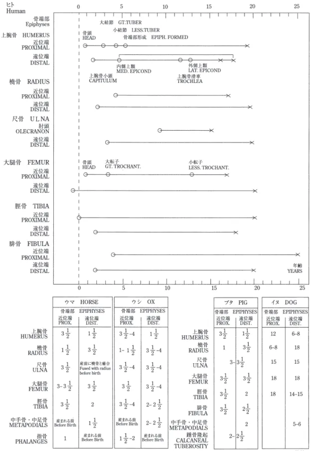
を如実に物語る。たとえば軟X線により四肢骨の骨
端部に残るハリス線を検出することによって,成長
期に栄養障害が生じたこと,頭蓋骨の眼窩上壁が多
孔質になることによって,鉄欠乏による貧血を生じ
ていたことがわかり,出土人骨の生前の生活環境を
知ることができる。骨の物理的損傷は,なぜそのヒ
トが死に至ったのかを物語る証拠となる場合がある。
骨に見られる傷跡が,生前のもので治癒したものか,
死因となったものか,それとも死後に付いたものか
を認識することは,重要な問題である。しかし,こ
うした観察の多くは,専門の形質人類学者,法人類
学者らの裏付けが必要であり,発掘で人骨が出土し
た際,現地でこうした専門家を招聰し,人骨を取り
上げる前に様々な観察を行い,両方の立場から討議
したいものである。
Lower limb (each one pair)
Coxal bones (formed with agglutinated ilium, ischium and pubis)
Femur, patella, Tibia, Fibula, Tarsal bones(7 each limb : talus, calcaneus, navicular, cuboid, medial cuneiform, intermediate cuneiform and lateral cuneiform), Metatarsal bones (5 each foot), Phalanges(14 each foot).
2. Observation/Identification of Human
Bones
Human bones carry the agglomerated life history of the person in various places. For instance, muscular tuberosities suggest the degree of physical labor of the individual. The existence of a squatting facet found at the tibia and talus proves that the person squatted regularly during his or her life, and osteomata of the external auditory meatus indicates the probability that the person was engaged in frequent diving. The pathology of bones is another factor in the telling of one's life history. For example; (i) Harris lines in the epiphyseal regions of the four limbs detected on soft X-ray are evidence of malnutrition during the growth period; and, (ii) the porous texture of the anterior region of the orbital roof proves anemia due to iron and vitamin A deficiency, disclosing the living environments of an ancient person whose bones were unearthed. Furthermore, physical damage to bones may provide evidence of the cause of an individual's death. It is obviously important to identify whether a scar on a bone of an individual was; (i) made and healed during the lifetime; (ii) the cause of his or her death; or, (iii) made after his or her death. However, many such examinations require the technical support of skilled physical or forensic anthropologists. In the event that human bones are unearthed during an excavation, prior to picking up the bones, it is highly desirable to invite such experts to the site to observe the bones from a range of viewpoints and to undertake discussions between the team of archaeologists and anthropologists.
頭蓋骨 Cranium
下顎骨 Mandible
上腕骨 Humerus
骨盤 Pelvis
大腿骨 Femur
膝蓋骨 Patella
腓骨 Fibula
𦙾𦙾骨 Tibia
肩甲骨 Scapula
鎖骨 Clavicle
尺骨 Ulna
𣓤𣓤骨 Radius
胸骨 Sternum
中足骨 Metatarsals
肋骨 Ribs
距骨 Tarsus
足の指骨 Phalanges
中手骨 Metacarpals
手の指骨 Carpus
図1 人体骨骼 the Human skelton
Modified from Harashima and Kawawi (2004)
図 1 人体骨格(原島・河合 2004 一部修正)
Fig.1. Human Skeleton
頭蓋骨 Cranium
ヒトの頭蓋骨は,他の大形哺乳類に比べて分厚く,
特に脳頭蓋の内面には血管溝が刻まれ,非常に特徴
的な形をしており,ヒト以外の動物骨と判別するこ
とは容易である。頭蓋骨は,前頭骨,頭頂骨,側頭骨,
後頭骨など23個の部分骨から構成され,個々の骨は
「縫合」という頭蓋特有の接合により,年齢と共に複
雑に癒合する。頭蓋骨は主に顔面を形成する顔面頭
蓋と,脳を収める脳頭蓋とに分けられる。脳頭蓋の
うち,天井にあたる部分を頭蓋冠という。頭蓋骨の
主な縫合は,前頭骨と頭頂骨を関節する冠状縫合,
左右の頭頂骨を関節する矢状縫合,頭頂骨と後頭骨
を関節するラムダ縫合からなる。
前頭骨は,額,眉間,眼窩上縁部を形作る骨であ
る。眼窩外側は頬骨と,眼窩内側は鼻骨および上顎
骨と関節する。眼窩上縁部は特に分厚く,出土する
可能性が大きい。頭頂骨は左右一対で,頭蓋冠の大
部分を占める。側頭骨は左右一対で,脳頭蓋の側面
を形作り,頬骨と関節する頬骨突起,耳の孔にあた
る外耳道,下顎骨と関節する下顎窩があり,その側
面下方(錐体部)は,厚みがあり出土する確率が高
い。後頭骨は頭頂骨と関節し,脳頭蓋の後面を形作る。
後頭骨にある大後頭孔(大孔)には脊髄が通い、後
面に突出する外後頭隆起は,女性より男性の方が発
達する。上顎骨は筋肉が付着する顔面頭蓋の中央の
大部分を形作る一対の骨である。歯槽部には上顎歯
が植立する。梨状口は生前の鼻にあたる部分で,上
方に鼻骨が関節する。
CraniumThe human cranium is thicker than that of other large mammals; in particular, the inner surface of the neurocranium is uniquely shaped with vascular grooves that can easily be distinguished from other animal bones. The cranium consists of 23 parts including the frontal, parietal, temporal, and occipital, which are both intricately connected with each other through cranium-specific articulations called “sutures”, and complicatedly agglutinated with age. Furthermore, the cranium mainly consists of the viscerocranium, the facial portion, and the neurocranium, the brain container. Within the neurocranium, the portion constituting the ceiling of the cranium is referred to as the calvaria. The sutures of the cranium mainly consist of the coronal suture that articulates with the frontal and parietal bones, the sagittal suture that articulates with the right and left parietals, and the lamboid suture that articulates with the parietal and occipital bones. The frontal bone forms the forehead, the glabella, and the supraorbital margin. It adjoins the zygomatic at the outer edge of the orbit, and the nasal at the inner edge of the orbit. The supraorbital region is particularly thick, and thus often unearthed. The parietals are left-and-right paired bones occupying a large part of the calvaria. The temporals are also left-and-right paired bones forming the lateral sides of the neurocranium, inside which there are the zygomatic process that articulates with the zygomatic bone, the external auditory meatus that forms the earhole, and the mandibular fossa that articulates with the mandible. The lateral-inferior portion of the temporals (pars petrosa) is particularly thick, and is thus frequently unearthed. The occipital bone articulates with the parietal bone, forming the posterior portion of the neurocranium. The vertebrae run through the foramen magnum of the occipital bone. The external occipital protuberance (occipital external protuberance) is more developed in males than in females. The maxillae are the paired bones that form the most central region of the viscerocranium to which muscles are attached. The maxillary teeth are arranged in the alveoli. The piriform aperture, adjoining the nasal bone at the upper part, is where the nose was located in life.
ブレグマ Bregma 矢状縫合 Sagital suture 頭頂骨 Parietal bone 眉間 Glabella 鼻骨 Nasal bone 上顎骨 Maxilla 下顎骨 Mandible 側頭骨 Temporal bone 乳様突起 Mastoid process ラムダ縫合 Lambdoid suture 後頭骨 Occipital bone 外後頭隆起 External occipital protuberance 大後頭孔 Foramen magnum 前頭骨 Frontal bone 冠状縫合 Coronal suture 頬骨 Zygomatic bone
頭蓋骨 Skull
下顎骨 Mandible
下顎骨は,頭蓋骨に関節する下顎枝と,歯が植立
する下顎体とに分けられる。下顎骨は上顎骨と対に
なって口腔を形作る。下顎枝は前方に筋突起,後方
に関節突起があり,関節突起は頭蓋骨と関節し,顎
関節を作る。オトガイ孔は左右一対あり,下顎体の
小臼歯の下あたりに位置する。オトガイ孔は,稀に
二対ある場合もある。下顎体は成人では歯が植立し
ていること,子供でも萌出前の歯冠部分が下顎体の
中にあることなどから,厚みがあり出土する可能性
が大きい。ただし,歯は土中に埋まっている間に脱
落して歯槽部が空洞となることがあるため,取り上
げの際,脱落した歯を見逃さないよう注意を要する。
MandibleThe mandible is divided into two parts: (i) the mandibular ramus that adjoins the cranium; and, (ii) the body of the mandible, in which the teeth are arranged. The mandible forms the mouth orifice, paired with the maxilla. The mandibular ramus has a coronoid process in the anterior region to articulate with the cranium, and an articular process in the posterior region. The condyloid process articulates with the cranium to form the mandibular joint. The mental foramen, a symmetrical pair of foramina, is located at the lower portion of the premolars in the body of the mandible. In some rare cases there are two pairs of mental foramen. The body of the mandible is highly likely be unearthed, because the adult mandible is thick enough to anchor the teeth; and even the infant mandible is thick enough to hold the crown of the teeth before eruption. For all that, care should be exercised during excavation not to overlook the teeth buried in the ground, since the pars alveolaris can be hollowed into cavities, from which the teeth could fall and shattered into pieces.
下顎頭 Head of Mandible 筋突起 Coronoid process 下顎体 Body オトガイ隆起 Mental protuberance 下顎枝 Ramus 下顎角 Angle
下顎骨 Mandible
歯 Teeth
歯のエナメル質は,人体組織の中で最も硬く残り
やすい部位である。そのため人骨のなかで,出土す
る可能性が最も大きい。しかし,それぞれが小さい
ため,土中に紛れやすく,発掘中に歯を採集するに
あたって,細心の注意が必要である。
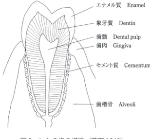
歯の構造は,歯冠・歯頸・歯根からなり,歯冠は
エナメル質の硬い組織に覆われる。その内部に象牙
質があり,さらにその深部に神経や血管が通る歯髄
腔がある(図2)。セメント質は歯根を覆う硬い被膜
である。歯の表記は,他の部位の表記とは異なる(下
顎骨の図を参照)。特に方向を表す際,外側にあたる
頬の側を「頬側」,内側にあたる舌の側を「舌側」と
表記する。ただし切歯と犬歯は,頬側でなく「唇側」
と表記する。そして,切歯などの前方に位置する側
を「近心」,大臼歯などの後方に位置する側を「遠心」
と表記する。また,歯冠のうち歯の噛み合う面を「咬
合面」,頬側を「頬側面」,舌側を「舌側面」といい,
前方を「近心面」,後方を「遠心面」と表記する。
永久歯は,切歯(8本)
・犬歯(4本)
・小臼歯(8本)
・
大臼歯(12本)からなり,合計32本となる。大臼歯
のうち「親知らず」とよばれる第3大臼歯は時代差,
個人差によって終生未萌出の場合もあり,合計28本
の状態で出土することがある。乳歯は成人の歯と異
なり,大臼歯が存在せず,切歯(8本)
・犬歯(4本)
・
臼歯(8本)の合計20本である。歯は,生後6ヶ月
を過ぎた頃から,下顎の中切歯(乳歯)の萌出が始
まり,永久歯は6歳頃に第一大臼歯の萌出が始まる。
そのため,永久歯の第一大臼歯を「6歳臼歯」とも
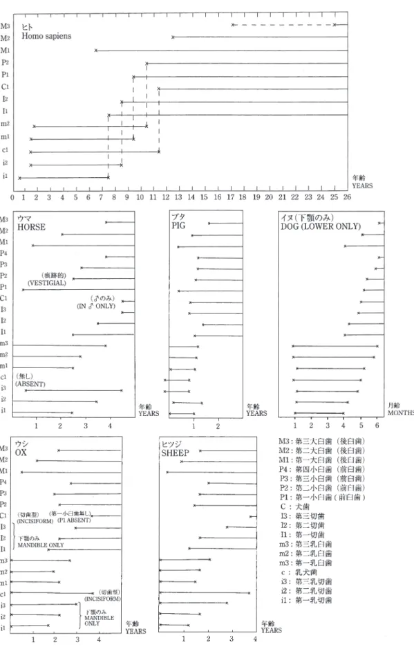
よぶ。「親知らず」とよばれる第三大臼歯は15歳から
TeethTooth enamel is the hardest and the most decay-resistant of the human body tissues. Therefore, among human hard tissue, teeth are the most frequently found through excavation. However, being small in size, they tend to be lost in the soil during excavation; very careful attention is therefore required when retrieving teeth.
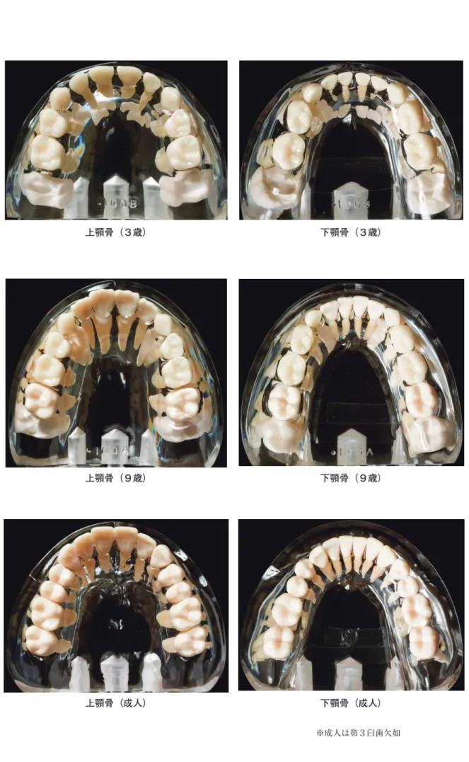
A tooth consists of the crown, the neck, and the root. The crown is covered with hard tissue of enamel, inside which there is dentin (Fig.2). Cementum is a hard membrane covering the root, which has a pulp cavity in which nerves and blood vessels reside. Tooth-related expressions differ from those of other skeletal parts (See the illustration of the mandible). Especially, in directional terms, the positions facing the cheeks are referred to as “buccal”, and those facing the tongue are referred to as “lingual”. However, in the case of the incisors and canines, the outer portions are called "lavial”, instead of buccal. The anterior position in which the incisors are located is called “mesial”, and the posterior positions in which molars are located are called “distal”. Furthermore, in the case of the occlusal surface of tooth too, the buccal is called the “buccal surface”, and the lingual is called the “lingual surface”; meanwhile, the anterior position is called the “mesial surface” and the posterior position is called the “distal surface”. There are 32 permanent teeth in total: 8 incisors, 4 canines, 8 premolars, and 12 molars. Since the third molars (a.k.a. the "wisdom teeth”) do not necessarily erupt, depending on the era and individual, some jaws are found with only 28 teeth. Unlike adult teeth, deciduous teeth include no molars; consequently there is a total of 20 such teeth: 8 incisors, 4 canines, and 8 premolars. Approximately 6 months after birth, the lower central incisors (deciduous teeth) start to erupt. The first permanent molar, the earliest permanent tooth, erupts at about 6 years of age. This is why the first permanent molar is called the 6-year-old molar. The third molars, “wisdom teeth”, usually erupt at ages 15 to 28, varying between individuals, and in some cases they never erupt. The age estimation based on the state of tooth eruption above is the most accurate among some existing age estimation methods (Fig.3).
At six months of age, most deciduous teeth have not yet erupted; however, some are in the preparatory stages in the alveoli. Furthermore, incisors erupt in this phase, followed by the upper deciduous incisors (the central incisors and the lateral incisors, in that order). By 24 to 30 months of age, all the deciduous teeth have completely erupted. The first deciduous molars erupt at 15 to 20 months of age, and the second start at 22 to 26 months. By 18 months, all the deciduous incisors have erupted, and the deciduous canines and deciduous molars begin to erupt. At the same time, the crown of the first permanent molars and incisors come under preparation in the alveoli.
図 2 ヒトの歯の構造(藤田 1949)
18歳頃に萌出することが多いが,個人差があり,萌
出せずに終わる場合もある。このような歯の萌出状
況による年齢推定は,幾つかある推定法の中で,最
も精度が高い(図3)。
生後6ヶ月では多くの乳歯は未萌出だが,歯槽内
に成長段階にある乳歯が存在する。また同じ頃,下
顎の乳中切歯が萌出し,次に上顎の乳切歯が中切歯,
側切歯の順で萌出する。乳歯は生後2年から2年
半くらいで全て生え揃う。第一乳臼歯は15ヶ月から
20ヶ月で,第二乳臼歯は22ヶ月から26ヶ月くらいで
萌出する。そして,乳切歯が完全に萌出し終えるの
は生後18ヶ月で,同時に乳犬歯,乳臼歯が萌出し始
める。この頃,永久歯の第一大臼歯,切歯の歯冠が
歯槽内で形成され始める。
生後3年には上,下顎の全ての乳歯が生え揃うが,
歯根の形成が完成していない乳歯もある。第一大臼
歯の歯冠は,すでに歯槽内で形成が完了している。
また,切歯,犬歯,小臼歯の歯冠も歯槽内で成長を
続ける。生後4年には,全ての乳歯が歯冠,歯根と
も形成が完了する。また,永久歯の第二大臼歯が歯
槽内で形成され始める。生後6年には,乳歯は歯
冠,歯根とも完成する。永久歯の第一大臼歯が萌出
し始め,第二大臼歯は歯槽内で歯冠部の形成が始ま
り,萌出のための準備ができる。切歯,犬歯,小臼
歯の歯冠部の形成は歯槽内で完了する。生後8年に
は,乳切歯が脱落し,永久歯の中切歯,側切歯が萌
出し始める。犬歯,小臼歯,第二大臼歯の歯冠の形
成は歯槽内で完了する。生後10年には,乳犬歯,第
二乳臼歯を除くほとんどの乳歯が脱落する。切歯や
第一小臼歯は萌出しているが,歯根部の形成は依然,
継続する。第三大臼歯の歯冠部の形成が始まり,歯
槽内で萌出のための準備段階になる。生後12年には,
乳歯は全て脱落する。第二大臼歯が萌出し始め,切
歯の歯根が完成する。生後15年には,第三大臼歯を
除いて全ての永久歯が歯冠,歯根とも完成する。第
三大臼歯の歯冠は歯槽内で完成する。
生後21年には,32本全ての永久歯が生え揃う(第
三大臼歯は生えてこない場合もある)。この頃から,
食物の咀嚼による歯の咬耗が目につくようになる。
生後21年以降は,歯の咬耗の進行段階により,おお
よその年齢を推定することが可能である。しかし,
咬耗の進行は,食物の違いや集団差,時代差,生活
環境などの要素に,大きな影響を受ける。
By three years of age, all deciduous teeth on both upper and lower jaws have erupted; however, the roots of some deciduous teeth are incompletely formed. The crown of the first molar is completely formed in the alveoli. Furthermore, the crowns of incisors, canines, and premolars are also growing in the alveoli. At the age of four, the crowns and the roots of all deciduous teeth are completely formed. The formation of the second permanent molars starts in the alveoli. By the age of six, the crowns and the roots of all deciduous teeth are completely formed. The permanent first molars start to erupt; meanwhile, the second molars begin to form the crown in the alveoli in preparation for eruption. The formation of the crowns of the incisors, the canines, and the premolars are completed inside the alveoli. By the age of eight, the deciduous incisors disappear, and both the permanent central incisors and the lateral incisors start to erupt. The formation of the crowns of the canines, premolars, and second molars are completed within the alveoli. By the age of 10, almost all deciduous teeth disappear except the deciduous canines and the second deciduous molar. Although the incisors and the first premolars erupt, the formation of the roots still continues. The third molars begin to form the crowns in preparation for eruption within the alveoli. By the age of 12, all the deciduous teeth have fallen out. The second molars start to erupt, and the incisor roots are complete. By the age of 15, except for the third molars, the crown and the root of all permanent teeth are complete. The crowns of the third molars are completed within the alveoli.
By the age of 21, all 32 permanent teeth have erupted. (In some cases the third molars do not erupt.) From this stage, dental attrition resulting from the mastication of foods begins to be conspicuously identifiable. Age estimation based on dental attrition is possible to some extent for adults of 21 years or older. However, the progress of attrition varies greatly depending on factors including foods, groups, era, living environments, etc.
Types of teeth (Fig.3)
Permanent teeth
1. Incisors
The incisors function to bite off food with their two pairs of teeth (central/frontal) on each of the upper and lower jaws. The crown is wedge-shaped. Unlike the labial surface, the lingual surface is concave to form a lingual fossa. The root is single and generally conical in shape. The mesiodistal width of the upper incisor root is comparatively larger, whereas that of the lower incisor root is narrower to make the root shape flat. In some cases, in the Late Jomon sites, the lower incisors may not be found because the people of those days customarily extracted the teeth.
歯種の分類
(図3)
永久歯(Permanent teeth)
1. 切歯(Incisor)
上下歯列の中央部,つまり最前部にある各二対の
歯で,食物を咬み切る作用を持っている。歯冠部は
楔のような形をしている。舌側面は唇側面とは反対
に凹面で舌側面窩を形成する。歯根は1本で,形は
おおむね円錐形である。上顎切歯の根は近遠心径の
幅が比較的厚く,下顎切歯の根は近遠心径の幅が狭
く扁平な形をしている。縄文時代晩期には下顎切歯
は抜歯により出土しない場合がある。
1-1. 上顎中切歯(Upper central incisor)
正中線の両側にあり,切歯の中で最も大きい。口
を開いたときにすぐ目につくため,容貌にとって重
要な要素をなす。大きな特徴は歯冠の幅が大きいこ
とにある。歯の全形は左右対称的である。舌側面は
辺縁部の隆線が発達して舌側面の中心が深く凹面を
作る。これはシャベル型とよばれ,弥生時代以降の
人骨に多く認められるが,縄文時代の人骨にはあま
り認められない。
1-2. 上顎側切歯(Upper lateral incisor)
中切歯の外側に隣接している1本ずつの歯である。
形は中切歯に似ているが,中切歯に比べると小さく、
歯冠の遠心隅角が鈍円化し、根が細くて長いなどの
特徴がある。舌側面は中切歯と同様に弥生時代以降
の人骨はシャベル型をしていることが多い。歯根部
1-1. Upper central incisor
The upper central incisors are the largest incisors on either side of the median line. This pair of teeth is conspicuous when the mouth is open, being the foremost and important element of appearance. Their distinctive feature lies in the notably large width of the crown. The overall shape is bilaterally symmetric. The lingual surface is very concave at the center with developed marginal bulges. Incisors with this feature are called shovel-type and are often seen in the bones of the Yayoi people or later ones, but rarely found in those of the Jomon people.
1-2. Upper lateral incisor
The upper lateral incisors adjoin on the distal side of the central incisors. Despite their similarities in shape to the central incisors, the upper lateral incisors are distinctive due to their smaller size, more rounded distoincisal angle of the crown, and thin, long root. As with the central incisors, the lingual surface of people of the Yayoi period and later is often shovel-shaped. The roots are quite similar to the central incisors, except for the comparatively longer length and the nearly egg-shaped cross section. In most cases, the apex of the roots is inclined or curved distally.
1-3. Lower central incisor
In the upper jaw, the central incisors are larger than the lateral incisors. In contrast, in the lower jaw, the central incisors are smaller than the lateral incisors. Hence, the lower central incisors are said to be the smallest of all teeth, although their length is no different to other incisors. Without a developed marginal bulge on the lingual surface, which is prominent in the upper jaw, the lower central incisors are not as concave as shovel-type teeth. The root is mesiodistally
図 3 歯
Fig.3. Human Teeth
上顎歯 Upper 下顎歯 Lower 頬側(唇側) buccal(lavial) 大臼歯 Molars Premolars小臼歯 犬歯 Canine Canine 犬歯 切歯 Incisors Incisors 切歯 Premolars 小臼歯 Molars 大臼歯 舌側 lingual
は中切歯とよく似ているが,中切歯よりは比較的長
く,横断面の形が卵形に近い。また,根尖はほとん
どの場合に遠心側に傾斜や弯曲をしている。
1-3. 下顎中切歯(Lower central incisor)
上顎では中切歯の方が側切歯よりも大きいが,下
顎では反対に中切歯の方が側切歯よりも小さい。そ
のため下顎中切歯は歯群の中で最小の歯といわれる
が,長さは他の切歯と変わらない。舌側面は上顎に
認められるような辺縁部の発達がほとんどないため,
シャベル型のような明瞭な凹面は認められない。歯
根は近遠心的につぶれて,水平断面が長楕円形に近
い。歯根の近心面には中央部に縦走する隆線が見ら
れることが多い。反対に遠心面はくぼんでおり浅い
溝を持つことがほとんどである。これは下顎中切歯
の左右を決定するのに重要な標識となる。
1-4. 下顎側切歯(Lower lateral incisor)
この歯は下顎中切歯と非常によく似ている。上顎
の中切歯と側切歯の関係に比べると類似度は非常に
高い。もし,下顎の切歯が1本のみ遺跡から出土し
た場合は,中切歯と側切歯のどちらであるか同定す
るのは非常に困難である。この歯は中切歯に比べる
とやや大きい。次に切縁は中切歯がほぼ水平なのに
対し,側切歯では近心から遠心に向かって傾く。歯
根は中切歯とほとんど同じだが,側切歯の根尖は遠
心側に傾斜や弯曲をしている。歯根の近心面の隆線
と遠心面のくぼみは中切歯に比べると顕著であり,
この点で近心側と遠心側を決定することができ,中
切歯との差と左右の同定を行う重要な標識となる。
2. 犬歯(Canine)
切歯の外側に位置し,上下顎にそれぞれ一対ずつ
合計4本ある。位置は口角部に近く,糸切り歯とい
う別称がある。犬歯は全歯群の中で最も丈の高い歯
で,歯冠は隣の歯よりも高く歯列上に突出し,歯根
の先端もほかの歯に比べると深く顎骨の中にある。
この歯の特徴は歯冠の先端が錐状の尖頭をなしてい
る点である。歯根は1本である。舌側面は菱形をし
ており,唇側に向かって傾斜し,軽度の凹面をして
いる。縄文時代では抜歯されていることが多く,特
に晩期では出土することは少ない。
2-1. 上顎犬歯(Upper canine)
全歯群のうちで最も長い歯である。歯冠の唇側面
は五角形で,切縁は近心辺より遠心辺の方が長い。
squashed and the horizontal cross section is nearly oblong. The mesial surface of root often has a vertical ridge at the center. The distal surface of root, on the other hand, is sunken and commonly has a shallow groove; this provides an important indication of whether a lower central incisor is located on the right or left side of the mouth.
1-4. Lower lateral incisor
These teeth look quite similar to the lower central incisors. The degree of similarity is much higher than that between the upper central incisors and the upper lateral incisors. In the event that a lower incisor is found at an archaeological site, it would be extremely difficult to distinguish it between the central incisor and lateral incisor. These teeth are slightly larger than the central incisors. Furthermore, the incisal edge of the central incisor is almost horizontal, while that of the lateral incisor inclines distally. The root is almost the same as the central incisors, but the apex of the root of the lateral incisors inclines or curves distally. The ridge on the menial surface and the concave distal surface of the root are clearer than in the central incisors. This feature enables determination between the mesial or distal side, serving as an important sign to indicate the difference from the central incisors as well as to distinguish between left and right.
2. Canines
The canines are located on the outer side of the incisors. There is a pair of canines on each of the upper and lower jaws, four in total. They are just at the angle of the mouth, and also known as “eyeteeth”. Tallest among all teeth, canines stand higher than the neighboring teeth, out of the tooth alignment. The point of the root is deeper in the jawbone than the other teeth. The crown is characteristically cuspidate in a conical shape. There is only one root. The lingual surface is rhomboid-shaped and inclines labially with a slight concave. These teeth were often extracted in the Jomon period and are rarely found, especially in the late Jomon sites.
2-1. Upper canines
The longest of all teeth. The labial surface of the crown is pentagonal. The distal incisal edge is longer than the mesial, giving an impression that the distal line angle is projected distally. The roots look similar to those of the upper incisors except that the shape is mesiodistally pressed.
2-2. Lower canines
The lower canines are basically the same in shape as the upper canines, but smaller in size. The size difference is not as great as that between the upper and lower incisors.
3. Premolars
Following the canines, the premolars are two pairs of teeth on each of upper and lower jaws. Many non-human mammals have very large premolars; and, some mammals have bigger
そのため遠心隅角が遠心に向かって突き出ているよ
うに見える。歯根は上顎切歯に似ているが,近遠心
に圧迫されている。
2-2. 下顎犬歯(Lower canine)
基本的な形は上顎犬歯と同様である。しかし,上
顎犬歯に比べると大きさが劣る。しかし,切歯の上
下の差ほどではない。
3. 小臼歯(Premolar)
犬歯の後に続く上下それぞれ二対の歯である。ヒ
ト以外の多くの哺乳動物では極めて大きいものがあ
り,その後方の大臼歯は時に小臼歯よりも小さい場
合があるため,動物骨では小臼歯のことを臼前歯ま
たは,前臼歯という。小臼歯の大きな特徴は頬側と
舌側にそれぞれ咬頭がある点である。そのため双頭
歯とも呼ぶ。
3-1. 上顎第一小臼歯
輪郭のはっきりした丈の高い2つの咬頭を備えて
いること,歯が全体として近遠心的に偏平になって
いることが大きな特徴である。頬舌径は近遠心径よ
りも大きい。歯冠の咬合面は頬側面の咬頭の方が大
きく見える。歯根は頬側と舌側の2根に分かれてい
るものと,単根のものがあり,またその中間型のも
のもある。
3-2. 上顎第二小臼歯
第一小臼歯と形は似ている。そのため,どちらか
一方しか出土しない時は区別のつかないこともある。
歯冠は一般に第一小臼歯よりも小さい。そして第一
大臼歯よりも全体的に丸みを帯びている。頬側と舌
側の咬頭の大きさは第一大臼歯よりも小さい。歯根
は95% 近い確率で単根である。歯根の先端は遠心方
向に傾いている。
3-3. 下顎第一小臼歯
歯冠の頬側面は上顎小臼歯に似ているが,舌側面
は発育が弱い。歯根は単根で,頬舌径に対して近遠
心径が比較的大きい。
3-4. 下顎第二小臼歯
形態は下顎第一小臼歯に似ている。一般に第一小
臼歯よりもやや大きい。この歯の大きな特徴は歯冠
の舌側が第一小臼歯よりもよく発達しているため,
咬合面が大きくなる。歯根の長さは第一小臼歯とほ
とんど同じで単根である。
premolars than the molars located behind them. Therefore, in animal osteology, the premolar is also called the bicuspid In addition, the premolar is outstandingly characteristic of its two cusps on the buccal and lingual sides. Accordingly, this tooth is also referred to as the two-pointed tooth.
3-1. Upper first premolars
The major characteristics are the two chiseled and high cusps and their mesiodistally flat overall shape. The buccal-lingual diameter is larger than the mesiodistal diameter. On the occlusal surface of the crown, the buccal cusp looks larger. Some roots diverge into two (buccal and lingual), some have a single root or an intergrade-type root.
3-2. Upper second premolars
These teeth are similar to the first premolars in shape. When one premolar is unearthed, it may be impossible to identify between them. Generally, the crown is smaller than the first premolars. The overall shape is rounder than the first molars. The buccal and lingual cusp sizes are smaller than the first molars. Nearly 95% of them have a single root. The end of the root is distally inclined.
3-3. Lower first premolars
The buccal surface of the crown is similar to the upper premolars. Howerver, as the lingual surface is less developed, nearly the entire occlusal surface looks buccal surface. They have a single root with a mesiodistal diameter comparatively larger than the buccal-lingual diameter.
3-4. Lower second premolars
The shape is similar to the lower first premolars. Generally, the size is slightly larger than the first premolars. The occlusal surface is larger with the lingual side of the crown characteristically more developed than in the first premolars. The root is single, and the length is almost the same as the first premolars.
4. Molars
Behind the premolars, there are three pairs of molars on each of the upper and lower jaws, 12 in total. The major difference between the upper and lower molars is the number of roots. The first and second molars on the upper jaw have two buccal roots and one lingual root, three roots in total. Those on the lower jaw have one menial root and one distal root, two in total. This does not necessarily apply to the third molars which are highly individual. The crown has four cusps; two on the buccal side and two on the lingual side (i.e., paracone, metacone, protocone and hypocone). The lower molar also has a fifth distal cusp (a.k.a. hypoconulid). The third molars (wisdom teeth) vary widely; a molar that shares no characteristic with the first or second molars is likely to be the third molar.
4. 大臼歯(Molar)
小臼歯の後ろに続く上下それぞれ三対の歯で,全
部で12本ある。しかし最後の歯,第三大臼歯(智歯
または親知らず)は現代人では萌出しないことも多
いため,大臼歯の数は個体によって異なる。上顎大
臼歯と下顎大臼歯の大きな違いは歯根の数である。
第一大臼歯と第二大臼歯の場合,上顎の歯根は頬側
に2本,舌側に1本の合計3本であるのに対し,下
顎は近心と遠心にそれぞれ 1 本ずつの合計2本であ
る。第三大臼歯の場合は歯根が癒合する傾向がある
のでこの限りではない。歯冠部は四咬頭性で,頬側
と舌側にそれぞれ2つの咬頭が並ぶ(近心頬側咬頭,
遠心頬側咬頭,近心舌側咬頭,遠心舌側咬頭という)。
下顎大臼歯には5番目の遠心咬頭がある。第三大臼
歯(親知らず)は個人差が大きいので第一,第二大
臼歯のいずれの特徴にも当てはまらない場合は,第
三大臼歯である可能性が高い。縄文時代には高い比
率で萌出するが,中世以降は退化傾向にあり,現代
では萌出しないケースが多い。
4-1. 上顎第一大臼歯
歯冠の全形は立方体,正方形に近いが,近心頬側
と遠心舌側がわずかに突出した菱形に近い。頬舌径
の方が近遠心径よりもやや大きい。
4-2. 上顎第二大臼歯
第一大臼歯とほとんど同じ形だが,近心頬側と遠
心舌側の突出が第一大臼歯よりも大きくつぶれた印
象を与える。
4-3. 下顎大臼歯
上顎と同様に第一大臼歯が基本形をなし,第二,
第三大臼歯は基本形から少しずつ違っていく。上顎
に比べると,より立方体の形を呈す。下顎大臼歯で
は近遠心径の方が頬舌径よりもやや大きい。咬頭は
5つあり,頬側に3個,舌側に2個となっている。
各咬頭の大きさは,近心頬側咬頭が最大で,遠心咬
頭が最も小さい。5つの咬頭のほかに,遠心咬頭と
遠心舌側咬頭の間に過剰咬頭(第六咬頭)が,舌側
の両咬頭の間に過剰咬頭(第七咬頭)が現れること
がある。これらの出現率から集団差を導き出す研究
がおこなわれている。
乳歯(Primary teeth/Milk teeth)
生まれてから初めに生える歯を乳歯といい,これに
代わって後から生える歯を代生歯(永久歯)という。
Although third molars erupted at a high rate during the Jomon period, they became less frequent in the medieval and later ages, and seldom erupt in modern times.
4-1. Upper first molars
The overall shape of the crown is nearly cubic or square, but somehow rhombic with slight mesial-buccal and distal-lingual projections. The buccal-lingual diameter is slightly larger than the mesiodistal diameter.
4-2. Upper second molars
The shape is almost identical to the first molar, except that the mesial-buccal and distal-lingual projections are larger than the first molars, giving a more flattened impression.
4-3. Lower molars
As with the upper molars, the shapes of the second and the third molars vary little by little from the basic shape of the first molars. The shapes are more cubic than the upper molars. In the lower molars, the mesiodistal diameters are slightly larger than the buccal-lingual diameters. There are five cusps, three on the buccal side and two on the lingual side. Among the cusps, the protoconid is the largest and the hypoconulid is the smallest. In addition to the five cusps, a supernumerary cusp (the sixth cusp) appears occasionally between the hypoconulid and the entoconid. Another supernumerary cusp (the seventh cusp) may appear between the two lingual cusps. Some researchers are attempting to determine appearance ratios of these supernumerary cusp in different groups.
Primary teeth
The first series of teeth to grow after birth are called the primary teeth, and the following teeth are called successional teeth (permanent teeth). Falling out before successional teeth erupt, the primary teeth are also called deciduous teeth. The primary teeth basically have shapes quite similar to the permanent teeth that erupt later.
Compared to permanent teeth, they are characteristically smaller in size, and less likely to be found at archaeological sites.
As the teeth ranging from the primary incisors to the primary canines are shaped quite similarly to the permanent teeth, their descriptions are omitted.
Primary molars
All primary molars have shapes completely different from their successional teeth.
Upper first primary molars
The shape of the upper first primary molars is significantly different from that of the first premolars. They have a large buccal cusp and a small lingual cusp. The buccal and lingual cusps correspond to protoconid and metaconid of the molar respectively. The distal cusp is only vestigial, distally pushed away and reduced. The buccal surface is quadrate-like. The
乳歯が脱落してその後に代生歯が生えることから,
乳歯のことを脱落歯ともいう。乳歯は原則的には後
に萌出する永久歯とよく似た形を備えている。そし
て永久歯よりも小さいという特徴がある。そのため,
遺跡から出土するケースは永久歯に比べると少ない。
乳切歯から乳犬歯までは永久歯の形とよく似ている
ため省略する。
乳臼歯
乳臼歯はすべてその代生歯とは形態が大きく違う。
上顎第一乳臼歯
上顎第一乳臼歯は第一小臼歯とは著しく形が違う。
大きな頬側咬頭と小さい舌側咬頭から成る。この頬
側咬頭と舌側咬頭はそれぞれ大臼歯の近心頬側咬頭
と近心舌側咬頭に相当する。対する遠心の両咬頭は
第一乳臼歯では遠心部に押された形で痕跡が確認で
き,退化的になっている。頬側面は四辺形に近い。
舌側面は頬側面より小さく丸みがあり,膨隆してい
る。歯根は3本で,配列は上顎大臼歯と同様に頬側
に2根,舌側に1根である。
上顎第二乳臼歯
代生歯である第二小臼歯とは似ても似つかない形
態をしている。形は第一大臼歯にきわめてよく似て
いる。歯冠は四咬頭性で,咬合面は菱形,歯根は3
本である。この歯にはしばしばカラベリ結節が認め
られる。
下顎第一乳臼歯
歯冠は咬頭が著しく頬舌的に偏平である。四咬頭
性が一般であるが五咬頭の場合も多く認められる。
歯根は近心と遠心に1本ずつで,下顎大臼歯に似て
いる。この歯の代生歯である第一小臼歯との形態差
は上顎第一乳臼歯と第一小臼歯との差よりもさらに
大きい。
下顎第二乳臼歯
上顎第二乳臼歯が上顎第一大臼歯に似ているのと
同様で,下顎第一大臼歯に似た形をしているが、頬
舌的に押しつぶされた印象である。歯冠は立方形に
近く五咬頭で,歯根は2本,近遠心に並んでいる。
咬合面の浮彫像は第一大臼歯よりも複雑で,六咬頭
性となっているものも少なくない。歯根は偏平の度
合いが大臼歯根よりも著しく,両根が大きく開大し
ている。近心根の方が遠心根よりも大きく,根の末
端が湾曲している。
lingual surface is smaller and rounder than the buccal surface, and convex. There are three roots, two on the buccal side and one on the lingual side as with upper molars.
Upper second primary molars
The shape is completely dissimilar to the second premolars (i.e. successional permanent), but quite similar to the first molars. The crowns have four cusps on the rhombic occlusal surface, and three roots. Carabelli cusps often appear on these teeth.
Lower first primary molars
The crown of this tooth has prominent cusps and is buccal-lingually flat. These teeth commonly have four cusps, and sometimes five cusps. They have a mesial root and a distal root, similar to the lower molars. The difference between the shape of these teeth and their successional first premolars is more acute than that between the upper first primary molars and the first premolars.
Lower second primary molars
As the upper second primary molars are shaped similarly to the upper first molars, the shape of these teeth is similar to that of the lower first molars. The crown is nearly cubic with five cusps, and two roots, mesial and distal. The occlusal surface is a more complex shape than the first molars, often with six cusps. The roots are far flatter than the molar roots, and are widely dilated. The mesial root is larger than the distal root, and is curved at the end.
5.軀幹骨 Axial skeleton
脊柱
脊柱は体幹の中心に位置し、体軸を構成する。32
〜35個の上下に連結する椎骨で構成され、上から頸
椎(7個)、胸椎(12個)、腰椎( 5 個)、仙椎( 5 個:
全体で1個の仙骨となる)、尾椎(3〜6個:全体で
尾骨をつくる)からなる。
頸椎 Cervical vertebrae
頸椎は脊柱の上部,頸の部分に位置する椎骨で計
7個あり,第一頸椎を環椎(Atlas)とよび,第二頸
椎を軸椎(Axis)とよぶ。頸椎は下位に向かうほど
大きくなるが,胸椎,腰椎に比べると椎体は小さく,
厚みも薄い。直立した姿勢では全体のつながりとし
て前湾している。椎骨動静脈が通る横突孔は,左右
に1つずつある。脊髄を通す椎孔は三角形を呈し,
棘突起はほぼ水平に後方へと伸びる。第三頸椎から
第七頸椎の棘突起と横突起の先端は,2つに分かれ
ているが,第七頸椎の棘突起のみ分かれない。第七
頸椎の棘突起は,椎骨の位置を決める基準点に使わ
れ,「隆椎」ともよばれる。椎骨は環椎と軸椎を除き,
椎体と椎弓とに分かれ,成長するにしたがい,椎体
と椎弓とが化骨化して椎骨となる。
第一頸椎は頭蓋の後頭骨と関節し,第二頸椎(軸椎)
の歯突起のまわりを回転する。環椎は,脊椎の一般
的な特徴である椎体がなく,環状の形態をしている。
横突起の上面には後頭骨の後頭顆と関節する上関節
窩とよぶ,大きな長楕円形のくぼみがあり,下面に
は円形に近い下関節窩がある。
第二頸椎は椎体の上面から柱状の歯突起が頭蓋骨
に向かって突出している。この歯突起は,環椎と頭
蓋骨が回転する時に軸となる。歯突起は本来,第一
頸椎の椎体となるべきものが第一頸椎から分離して
第二頸椎と癒合したものといわれている。この歯突
起は火葬の際に「のど仏」として扱
われる。
5.Axial skeleton
Vertebrall column (spinal column)
The vertebrall column is located at the center of the body trunk, forming the body axis. It consists of 32 to 35 vertebrae: (from up to down) 7 cervical, 12 thoracic, and 5 lumbar, followed by the sacral (5 sacral vertebrae form one sacral bone) and the coccygeal (3 – 6 coccygeal vertebrae form one coccyx).
Cervical vertebrae
The cervical vertebrae are 7 neck vertebrae located at the top of the vertebral column. The first cervical vertebra is called the atlas, and the second vertebra is called the axis. Although the cervical vertebrae increase in size from upper to lower in stages, the vertebral body is smaller and thinner than those of the lumbar and the thoracic vertebrae. In standing posture, as an entire connection, these vertebrae curve forward. On each side, there is one transverse foramen to house the vertebral artery and vein. The vertebral foramen through which the spinal cord passes is triangular, and the spinous process projects backwards almost horizontally. The spinous and transverse processes of the third to sixth cervical vertebrae are split in two at the end, whereas the spinous process of the seventh cervical vertebrae is not split. The spinous process of the seventh cervical vertebrae is also known as the "vertebra prominence" and is used as the reference point for the positions of the vertebrae. With the exception of the atlas and axis vertebrae, the vertebrae consist of a vertebral body and the vertebral arch. With growth, the vertebral body and the vertebral arch fuse together to form the vertebra.
First cervical vertebra
The first cervical vertebra (atlas) articulates with the cranium occipital, and rotates upon the dens of the second cervical vertebra (axis). Unlike general vertebrae, the atlas has no vertebral body, and is ring-shaped. On the upper face of the transverse process, there is a large, oblong groove called the superior articular facet for articulation with the occipital condyle, whereas on the lower face there is the circular inferior articular facet.
Second cervical vertebra
The second cervical vertebra (axis) has a column-like process projecting toward the cranium from the upper portion of the vertebral body. The odontoid process forms a pivot for the rotation of the atlas and cranium. It is said that the odontoid process is the parts, which were supposed to become the body of vertebra; but were separated from the first cervical, and fused with the second cervical. Incidentally, after a cremation in Japan, this odontoid process, called “Nodo-botoke (lit. Buddha in the throat),” is picked up and treated carefully.
横突孔 Foramen transversarium
環椎(第一頚椎)
Atlas
軸椎(第二頚椎)
Axis
Lower cervical vertebra( 7 th)
隆椎(第七頚椎)
横突起 Transverse process 椎体 body 棘突起 Spinous process 歯突起 Dens 椎孔 Vertebral foramen 上面 upper surface 前面 front view 下面 lower surface 左側面 left side
胸椎 Thoracic vertebrae
胸椎は12個あり,それぞれ肋骨と関節するための
関節面を持つ。直立した姿勢では全体のつながりと
して後湾している。第一胸椎から第九胸椎までは,
肋骨と関節する上肋骨窩と下肋骨窩の両方を持つ。
しかし,第十胸椎には上肋骨窩のみで,第十一胸椎
と第十二胸椎には,椎体側面の中央部に肋骨窩があ
る。椎体は下位のものほど大きくなる。上位の椎体
は横長の頸椎に似た楕円形を呈し,中位では前後径
が長く前後に長いハート形を呈し,下位では左右径
が長くなり腰椎と似た楕円形になる。棘突起は長く
下方に強く傾斜する。横突起はよく発達し,上方の
胸椎から下方に行くにしたがい大きくなり,第八胸
椎で最も大きくなり,下に向かい再び小さく短くな
る。
腰椎 Lumbar vertebrae
腰椎は 5 個あり,大きく頑丈である。椎体は横楕
円形で,椎骨の中でも最も大きく,幅は下位ほど広い。
椎弓は厚く強大で,上椎切痕と下椎切痕は,ともに
顕著である。直立した姿勢では前湾している。棘突
起は胸椎のものより強大であるが短く,ほぼ水平に
後方へとのびる。
仙骨 Sacrum
仙骨は1個であるが,元来,5個の仙椎が癒合し
たもので,骨盤の後壁を形成する。形は逆三角形で,
厚さが上方から下方になるにしたがい薄くなり,前
方に向かい湾曲する。前面は平滑で,後面は不平坦
な凸面で,5本の長い高まりが縦に走る。いずれも
各仙椎の突起が連なったものである。仙骨の形状は,
性差が強く現れ,女性のそれは幅が広くて短く,湾
曲の度合いが小さいのに対し,男性のそれは幅が狭
くて長く,湾曲が強い。仙骨の下方には,3個ない
し6個の尾椎が化骨化した尾骨が
続く。
Thoracic vertebraeThe thoracic vertebrae consist of 12 vertebrae in the chest region, each of which has articular surfaces joined to ribs. In a standing posture, as an entire connection, these vertebrae curve backward. The first to the ninth thoracic vertebrae have both superior and inferior costal facets for articulation with the ribs. However, the tenth thoracic vertebra has superior costal facets only. The eleventh and twelfth thoracic vertebrae have costal facets at the center of the lateral surface of the vertebral body. The vertebral body increases in size from upper to lower. The body of the upper thoracic vertebrae is oval in the lateral direction, similar to the body of the cervical vertebrae. The body of the mid thoracic vertebrae is heart-shaped and oval in the anterior-posterior direction. The body of the lower thoracic vertebrae is long laterally and oval-shaped similar to the body of the lumbar vertebrae. The spinous process is long, projecting downward steeply. The lateral processes are well developed, increasing in size from superior to inferior, attain the maximum size at the eighth thoracic vertebra, and then decrease toward the furthest inferior.
Lumbar vertebrae
The lumbar vertebrae consist of five large and firm vertebrae. The vertebral body is oval-shaped, long laterally, and the largest of all vertebrae. The lower the location is, the larger the width increases. The vertebral arch is thick and solid with a prominent superior vertebral notch and an inferior vertebral notch. In a standing posture, these vertebrae curve forwards. The spinous processes are larger but shorter than those of the thoracic vertebrae, and project almost flatly backward.
Sacrum
The sacrum is a single bone. However, it is formed by the fusion of five sacral vertebrae. The sacrum also forms the posterior wall of the pelvis. The shape is an inverted triangle. The lower the location is, the smaller the thickness becomes. The sacrum curves forwards. The anterior surface is smooth and the posterior surface is an uneven convex with five long vertical ridges, which are the remains of the piled sacral processes. The configurations of the sacrum differ noticeably between the sexes. The female sacrum is wider, shorter, and less curved. The male sacrum, in contrast, is narrower, longer, and more fully curved. Below the sacrum is the coccyx, comprising three or six coccygeal vertebrae fused together.
耳状面 Auricular surface
仙骨
Sacrum
上面 upper surface 岬骨 Promontory 上面 upper surface 上面 upper surface胸椎
Thoracic Vertebra
前面 front view 前面 front view 前仙骨孔 Anterior sacral foramina 後面 back view 前面 front view 左側面 left side 左側面 left side腰椎
Lumbar Vertebra
左側面 left side6.上肢骨
肩甲骨 Scapula
肩甲骨は,ほぼ三角形の扁平骨で胸郭の背側上外
部で,第二から第八肋骨の間に位置する。上肢帯を
形成する上で重要な骨であり,体幹と上腕骨を連結
する。外側上方には,上腕骨と関節する関節窩があり,
前面には烏口突起,後面には肩甲棘がある。特徴の
ある形態をしているが,薄くて幅広いため土圧など
の影響によって,完形でとり上げられることは稀で
ある。
鎖骨 Clavicle
S字状に緩く捻れた長骨で,左右の胸郭上方に1
個ずつある。内側端は胸骨端とも呼ばれており,そ
の断面は円形を呈している。鎖骨下面には,鎖骨下
筋溝などの筋肉粗面があり,上面に比べて粗くなる。
胸骨端は丸い断面を持つが,外側の肩峰端は,薄く
広がる。
胸骨・肋骨 Sternum, Ribs
胸骨は胸郭の前部に位置し、上部で鎖骨と関節す
る。また、その両側面で上位七対の肋骨と肋軟骨を
介して関節する。胸骨は上から柄、体、剣状突起の
3 部からなる。柄と体の連結部はやや前方に突出し、
胸骨角と呼ばれる。
肋骨は胸郭の側面を構成し、十二対の弓状の骨か
らなる。上位 七 対の肋骨は後方では胸椎と、前方で
は肋軟骨を介して胸骨と関節する。第一肋骨から第
七肋骨にかけては次第に長さを増し、第八肋骨以降
は次第に短くなっている。
6.Bones of upper limbs
Scapula
The scapula is a triangular, flat bone in the region of the upper dorsal thorax between the second and eighth ribs. It is an important bone forming the pectoral girdle and connecting the humerus with the axial skeleton. On the upper external border is the glenoid cavity where the humerus articulates. The coracoid process is on the anterior surface and the spine of the scapula is on the posterior surface. Although it is uniquely shaped, this element is thin and flat, and thus is rarely retrieved without damage due to factors such as earth pressure.
Clavicle
The clavicles are long bones gently twisted in an S-shape located on both sides of the upper chest. The medial (sternal) end is circular in cross section. With the subclavian groove and other muscular tuberosities, the inferior surface is rougher than the superior. Unlike the round sternal end, the acromial (lateral) end expands flatly.
Sternum, Ribs
The sternum is a flattened bone, forming the anterior wall of the thorax. Its upper end supports the clavicles, and its margins articulate with the cartilages of the first seven pairs of ribs. It consists of three parts, named from above downward, the manubrium, the body or gladiolus, and the xiphoid process. The junction of the manubrium and the body forming the sternal angle.
The ribs are elastic arches of bone, which form a large part of the thoracic skeleton. They are twelve in number on either side. The first seven are connected behind with the vertebral column, and in front, through the intervention of the costal cartilages, with the sternum. The ribs increase in length from the first to the seventh, below which they diminish to the twelfth.
烏口突起 Coracoid process 肩峰 Acromion 肩甲棘 Spine 関節窩 Glenoid cavity