Study on the efficient selection of porcine in vitro produced
embryos and their expression of pluripotency-associated genes
The United Graduate School of Veterinary Science
Yamaguchi University
NGUYEN THI HIEP
September 2020
TABLE OF CONTENTS
LIST OF ABBREVIATIONS... 4
SUMMARY ... 6
CHAPTER 1 GENERAL INTRODUCTION ... 9
1.1 IVEPIN PIGS... 9
1.1.1 IVM in pigs ... 9
1.1.2 IVF in pigs ... 13
1.1.3 IVC in pigs... 17
1.2 SELECTION OF GOOD QUALITY EMBRYOS BASED ON MORPHOLOGY AT EARLY STAGE IN IVEPSYSTEMS... 19
1.3 GENE EXPRESSION IN MAMMALIAN EMBRYOS... 20
1.3.1 Genome activation... 20
1.3.2 Housekeeping genes ... 22
1.3.3 Pluripotency-associated genes and their expression in mammalian embryos and embryonic stem (ES) cells... 23
1.3.4 Allelic gene expression... 25
1.3.5 Gene positioning and expression ... 26
1.3.6 Fluorescent in situ hybridization (FISH) as a tool to study allelic gene expression and gene positioning in single cells... 27
AIMS OF RESEARCH ... 29 CHAPTER 2 SELECTION BASED ON MORPHOLOGICAL FEATURES OF PORCINE EMBRYOS PRODUCED BY IN VITRO FERTILIZATION: TIMING
OF EARLY CLEAVAGES AND THE EFFECT OF POLYSPERMY... 30
2.1 ABSTRACT ... 30
2.2 INTRODUCTION ... 31
2.3 MATERIALS AND METHODS ... 33
2.3.1 Reagents ... 33
2.3.2 Oocyte collection and IVM... 34
2.3.3 Preparation of epididymal spermatozoa ... 35
2.3.4 IVF and IVC ... 35
2.3.5 Polyspermy evaluation ... 36
2.3.6 Embryo evaluation... 38
2.3.7 Nuclear status evaluation ... 38
2.3.8 Chromosome analysis... 39 2.3.9 Experimental design ... 41 2.3.9.1 Experiment 1 ... 41 2.3.9.2 Experiment 2 ... 42 2.3.9.3 Experiment 3 ... 42 2.3.9.4 Experiment 4 ... 43 2.3.9.5 Experiment 5 ... 43 2.3.10 Statistical analysis ... 43 2.4 RESULTS ... 44 2.4.1 Experiment 1... 44 2.4.2 Experiment 2... 45 2.4.3. Experiment 3... 46 2.4.4. Experiment 4... 49
2.4.5. Experiment 5... 49
2.5 DISCUSSION... 52
CHAPTER 3 PLURIPOTENCY-ASSOCIATED GENES REPOSITION DURING EARLY EMBRYONIC DEVELOPMENTAL STAGES IN PIGS... 56
3. 1 ABSTRACT ... 56
3.2 INTRODUCTION... 56
3.3 MATERIALS AND METHODS ... 58
3.3.1 Oocyte collection and in vitro maturation... 58
3.3.2 In vitro fertilization and in vitro culture ... 59
3.3.3 RNA and DNA FISH ... 60
3.3.4 Statistical analysis ... 62
3.4 RESULTS ... 63
3.4.1 Allelic gene expression during early development ... 63
3.4.2 Gene repositioning during early development ... 65
3.5 DISCUSSION ... 68
CHAPTER 4 GENERAL DISCUSSION... 73
ACKNOWLEDGEMENTS ... 76
LIST OF ABBREVIATIONS
AD-MSC adipose-tissue-derived mesenchymal stem cells BM-MSC bone-marrow-derived mesenchymal stem cells
BSA bovine serum albumin
cAMP cyclic adenosine monophosphate
DAPI -diamidino-2-phenylindole
dbcAMP dibutyryl cAMP
DNA deoxyribonucleic acid
eCG equine chorionic gonadotropin
EGA embryonic genome activation
EGF epidermal growth factor
ES embryonic stem
FCS fetal calf serum
FISH fluorescent in situ hybridization
FSH follicle-stimulating hormone
GFAP glial fibrillary acidic protein
GJ gap junction
GSH glutathione
GV germinal vesicle
hCG human chorionic gonadotropin
HEPES hydroxyethyl-piperazineethane-sulfonic acid buffer Hoechst 33342 bisBenzimide H33342 trihydrochloride
heavy chain
IVC in vitro culture
IVEP in vitro embryo production
IVF in vitro fertilization
IVM in vitro maturation
IVP in vitro production
LH luteinizing hormone
MET maternal to embryonic transition
MII metaphase II
MPN male pronucleus
mRNA messenger RNA
NCSU North Carolina State University
POM porcine Oocyte medium
PPARG peroxisome proliferator-activated
PSCs pluripotent stem cells
PZM Porcine zygotic medium
RNA ribonucleic acid
SCNT somatic cell nuclear transfer
ZGA zygotic genome activation
SUMMARY
In vitro embryo production (IVEP) of porcine embryos is an important tool not only for porcine gene banking but also for human biomedical research. However, in pigs, in vitro fertilization (IVF) is characteristic with a high frequency of polyspermy, causing chromosomal abnormalities in embryos. Although various approaches have been tried for reduction of polyspermy in pigs, it still remains as major obstacle. Therefore, a reliable selection system for high quality embryos under polyspermy condition is essential for reproduction studies in pigs of which high polyspermy is often unavoidable.
Study on gene expression especially pluripotency-associated genes and their repositioning during embryonic genome activation (EGA) is important for embryonic development and stem cell research. However, the information about allelic expression patterns, location and repositioning of pluripotency-associated genes in mammalian embryos other than mice is little known. Although DNA/RNA fluorescence in situ hybridization (FISH) have been established in mouse embryos, they have not been set up in porcine embryos yet. Besides, the efficiency of FISH assays can be affected by frequency of abnormalities in embryos.
Therefore, the first study (Chapter 2) was aimed to examine the efficiency of embryo selection based on morphological features and timing of early cleavage in order to select good quality embryos under polyspermy condition. The embryos were produced by IVF under moderate and high polyspermy conditions. The 4-cell embryos were selected at 48 hr after IVF (single selection) and 8-cell embryos were selected at 79 hr after IVF from the collected 4-cell embryos (double selection). Both of single and double
selection embryos showed high developmental competence to blastocyst under both moderate and high polyspermy conditions. However, blastocysts derived under high polyspermy condition had significantly fewer cells than those produced under moderate polyspermy condition. Moreover, the frequency of nuclear and chromosomal abnormalities in 4- and 8-cell embryos produced under high polyspermy condition were significantly higher in comparison to those under moderate polyspermy condition. These findings suggest that although high polyspermy affects the frequency of anomalies in nucleus and number of chromosome in porcine embryos produced by IVF, subsequent selection based on morphological features of 4- and 8-cell embryos even under high polyspermy condition, could be an alternative option for selecting porcine embryos with high developmental ability. Furthermore, the 4- and 8-cell embryos produced under moderate polyspermy condition showed low rate of abnormalities. It would be associated improving the efficiency of DNA/RNA FISH assays which were utilized to examine allelic gene expression.
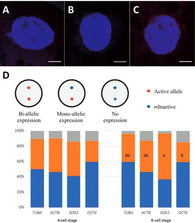
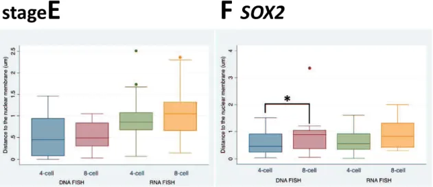
The second study (Chapter 3) was designed to evaluate the allelic expression and positioning of two pluripotency-associated genes, OCT4 and SOX2, and two housekeeping genes, ACTB and TUBA, in 4- and 8-cell porcine embryos which coincide with embryonic genome activation (EGA), utilizing RNA and DNA FISH assays. The expression of SOX2 in bi-allelism increased from 45% at the 4-cell stage to 60% at the 8-cell stage. Moreover, SOX2 was expressed bi-allelically in 8-8-cell embryos in significantly more blastomeres than those of OCT4. Also, this was associated with a tendency that SOX2 alleles move toward the nuclear interior during 4- to 8-cell transition. However, OCT4 alleles did not change significantly radial location during this transition. The locations of active and inactive alleles also were measured based on DNA/RNA FISH
assays. Active OCT4 alleles were more centrally disposed in the nucleus meanwhile inactive OCT4 alleles located in very close to the nuclear membrane. Nevertheless, active and inactive SOX2 alleles did not change location in the nucleus in either 4- or 8-cell blastomeres. The present results provide novel information on the allelic expression patterns and positioning of pluripotency-associated genes, OCT4 and SOX2, during EGA in pigs.
In conclusion, the first study demonstrated that although polyspermy affects the frequency of abnormalities in embryos, subsequent selection of 4- and 8-cell embryos based on morphological features and timing of early cleavage would be effective. This knowledge will contribute as an alternative option for selection of high developmental competence embryos produced by IVF even under high polyspermy condition. The second study revealed that the repositioning of SOX2 alleles coincided with an increase in the percentage of blastomeres with bi-allelic expression during these stages of EGA, and the expression of OCT4 correlated with its nuclear location. To my knowledge, it is the first study on allelic expression in mammalian embryos other than mice. The information should be useful for improvement of embryonic development as well as stem cell research.
Key words: abnormalities, allelic expression, chromosome, gene positioning,
CHAPTER 1 GENERAL INTRODUCTION
1.1 IVEP IN PIGS
IVEP is the technology of production of embryos from gametes under laboratory conditions. In pigs, IVEP is an important technology for the utilization of frozen sperm kept in gene banks (Kikuchi et al., 2016). Also, IVEP is a basic technology necessary for the production of genetically modified pigs, which have great importance for human biomedical research (Prather et al., 2003). The first successful piglet production by the transfer of in vitro produced blastocyst-stage embryos was reported in 2001 by Marchal et al. The most common source of oocytes for IVEP is ovaries of slaughtered pigs from commercial slaughterhouses; however, harvesting immature oocytes from live pigs by ovum pick up is also possible (Yoshioka et al., 2020). Although fresh sperm can be utilized to generate embryos, it is more common to use frozen sperm, both from ejaculation and epididymis. IVEP consists of three steps; 1) in vitro maturation (IVM) of immature oocytes; 2) in vitro fertilization (IVF) of matured oocytes and; 3) the subsequent in vitro culture (IVC) of fertilized oocytes to embryos, usually to the blastocyst stage. Aspects of IVM, IVF and IVC will be discussed in this chapter.
1.1.1 IVM in pigs
The basis of IVM is the phenomenon of “spontaneous maturation” which was first described by Pincus and Enzmann (1935). In antral follicles, mammalian oocytes stay arrested at the first meiotic prophase (also known as germinal vesicle (GV) stage) until the gonadotropins resume meiosis (Motlik and Fulka, 1976). However, Pincus and
Enzmann observed that once removed from the follicles (and hence from the meiosis-suppressing factors from the ovary) and cultured in isotonic solutions, rabbit oocytes resumed meiosis spontaneously. Based on this phenomenon, mammalian oocytes have been successfully matured to the Metaphase-II (MII) stage and embryos/offspring have been produced from such oocytes in rabbit (Chang, 1959), mice (Mukherjee and Cohen, 1970), sheep (Cheng et al., 1986), pig (Mattioli, 1989), and cattle (Bracket et al., 1982; Fukuda et al., 1990).
During IVM, the oocytes must acquire the ability to be fertilized and to develop subsequently to healthy embryos and offspring which requires both nuclear and cytoplasmic maturation. Nuclear maturation is a term that refers to the resumption of meiosis and the progression of the oocyte to the MII stage at which it remains temporarily arrested until fertilization. The failure of oocyte nuclear maturation normally leads to abnormal development after fertilization which results in the formation of embryos with abnormal chromosome numbers. Such abnormal embryos are very unlikely to develop to offspring (Kikuchi et al., 2009). In another word, the success of nuclear maturation has key importance for the normality of chromosome numbers in the developing embryo. Nuclear maturation of oocyte can be evaluated by morphology based on the presence of the first polar body or by nuclear staining such as with orcein or DAPI. (reviewed by Dang-Nguyen et al., 2011).
Cytoplasmic maturation is a broad term that refers to all cytoplasmic events occurring during maturation that prepare the oocyte for fertilization and preimplantation development. It includes the accumulation of cytoplasmic glutathione (GSH) (Yoshida et al., 1993a,b), the redistribution of organelles (Ferreira et al., 2009), and after reaching the MII stage, the adjustment of levels of cytoplasmic protein kinases to a level that will allow
oocyte activation by the fertilizing sperm (Kikuchi et al., 1995). GSH compounds major non-protein sulfhydryl which exists in mammalian cells (Abeydeera et al., 1998a) and it is very important for cytoplasmic maturation (Eppig, 1996). GSH protects cells against the effects of reactive oxygen species (Meister, 1983) as well as supports the male pronucleus (MPN) formation after fertilization (Yoshida, 1993a, b). Also, GSH extends the transportation of amino acids, and stimulates DNA and protein synthesis (Grupen et al., 1995). Therefore, the cytoplasmic maturation can be improved by increasing the concentration of GSH in the oocyte such as supplementation composition of maturation medium with cystein (Yoshida et al., 1993a), cysteamine (Yamauchi and Nagai, 1999), -mercaptoethanol, glutamine (Abeydeera et al., 1998a, Jeong and Yang, 2001) and epidermal growth factor (EGF) (Abeydeera et al., 2000). The cytoplasmic maturation could not be analyzed directly, but indirectly by examination of GSH content, the formation of MPN after fertilization or the competence to develop to blastocyst and cell number in blastocysts (reviewed by Dang-Nguyen et al., 2011).
The cumulus cells surround and connect with the mammalian oocyte via gap junctions (GJ) in mammals (Anderson and Albertini, 1976). During IVM, cumulus cells are essential since they play important roles in the initiation of nuclear maturation and the process of cytoplasmic maturation of the oocyte in cattle and pigs (Yamauchi and Nagai, 1999; reviewed by Nagai, 2001; Tanghe et al., 2002) and also for the process of fertilization (Kikuchi et al., 1993). Cumulus cells synthesize and transport GSH into the oocytes through GJ (Maedomari et al., 2007).
The success of IVM to provide oocytes with the competence to embryonic development depends on many factors such as 1) the initial capacity of oocytes to mature and develop at the time of oocyte collection (i.e. “oocyte quality”) (Mermillod et al.,
2008) and 2) the culture conditions during IVM (Nagai, 2001). In slaughterhouse-derived ovaries from commercial pig breeds, fully grown oocytes obtained from 3-6 mm follicles with at least 2 layers of cumulus cells are known to have acquired the ability to mature and develop (Marchal et al., 2002). The morphology and behavior of cumulus cells during IVM affects nuclear and cytoplasmic of the oocytes and thus affect IVF results (Somfai et al., 2004). Besides, age of pigs also has effects on blastocyst development since young gilts oocytes have lower competence than mature sows (Bagg et al., 2006).
Among the culture conditions, the medium has a great importance for the success of maturation. Immature porcine oocytes were traditionally cultured in media such as Whitten’s, Waymouth MB 752/1, Tissue Culture Medium 199, North Carolina State University (NCSU)-23 or NCSU-37, usually enriched with a non-defined protein supplementation such as fetal calf serum (FCS) or follicle fluid (FF) and other supplements, such as gonadotropins and growth factors (reviewed by Abeydeera, 2002; Grupen, 2014). Later, a chemically defined medium called Porcine Oocyte Medium (POM) was developed (Yoshioka et al., 2008). Since a chemically defined medium eliminates undefined factors present in biological materials, its application for IVM of oocytes (and later embryo culture) has various great advantages, especially for studying the effects of chemicals on embryonic development (reviewed by Dang-Nguyen et al., 2011).
Porcine IVM media are generally supplemented with gonadotropins, follicle-stimulating hormone (FSH), luteinizing hormone (LH); or its analogues such as equine chorionic gonadotropin (eCG) and human chorionic gonadotropin (hCG). The gonadotropins improve nuclear and cytoplasmic maturation of porcine oocytes (Mattioli et al., 1991; Funahashi et al., 1994). Evidence shows that gonadotrophins contribute to
the synthesis of GSH in cumulus cells, which is transferred to the oocytes via GJs (Ozawa et al., 2010). The supplementation of gonadotropins together with EGF improve MPN formation (Ding et al., 1994), indicating the beneficial effects of EGF on cytoplasmic maturation (Abeydeera, 2002). The treatment improves the subsequent synchrony of oocyte nuclear and cytoplasmic maturation and increases the subsequent blastocyst development rates (Funahashi et al., 1997). Also, elevating cAMP levels during the first half of IVM have other various positive effects on the oocyte developmental ability (reviewed by Appeltant et al., 2016). IVM of porcine oocytes is normally performed under 38.5-39oC in the humified atmosphere of 5% CO
2 (Kikuchi et al., 2002a).
1.1.2 IVF in pigs
In IVEP systems, IVF is achieved by the co-incubation of matured cumulus-enclosed oocytes with spermatozoa. Boar spermatozoa can be frozen-thawed ejaculated (Wang et al., 1991) or epididymal (Kikuchi et al., 1998; Kikuchi et al., 1999b). Fertilization medium of in vitro systems is designed to mimic the in vivo systems. Several different media have been used for IVF in pigs such as TCM199 (Nagai and Moor, 1990; Yoshida et al., 1990; Funahashi et al., 2000), Brackett and Oliphant (BO) medium (Kikuchi et al., 1993; Wang et al., 1995), modified tris-buffered medium (Abeydeera and Day, 1997) and Pig fertilization medium (PigFM) (Suzuki et al., 2002). It was found that mammalian spermatozoa need extracellular calcium for capacitation and maximal acrosomal exocytosis (Fraser, 1995). Bovine serum albumin (BSA) and caffeine are also crucial modulators of sperm penetration. The addition of BSA into IVF medium increased the number of sperm penetrating the oocyte (Abeydeera and Day, 1997) whereas caffeine
promoted the capacitation of boar spermatozoa and fertilization (Wang et al., 1991; Nagai et al., 1993).
The efficiency of IVF is affected by sperm concentration and interval of sperm and oocyte co-incubation (Nagai, 1996). In traditional porcine IVF systems, it was found that sperm penetration takes places around 3 hr after insemination and is completed by 6 hr. Therefore, numerous laboratories incubate gametes for about 6 hr (reviewed by Dang-Nguyen et al., 2011). However, the incidence of polyspermy increases as the co-culture duration is extended (Funahashi et al., 2000). The incubation with sperm for 6 hr doubles the average number of sperm per oocyte in comparison with 3 hr thus increased polyspermy whereas did not improve the MPN formation (Kikuchi et al., 2006). The optimum sperm concentration for frozen-thawed epididymal sperm, was 1 x 10⁵sperm/ml,
and co-incubation with oocytes for 3 hr gave the best results (Kikuchi et al., 2002a). Furthermore, when shorter co-culture periods were applied, the penetration and polyspermy rates were not affected (Gil et al., 2004). Since then 3 hr gametes incubation has been suggested.
However, the effectiveness of a brief gamete co-incubation in decreasing polyspermy also dependent on the sperm:oocyte ratio used (Gil et al., 2007), which must be optimized based on boar and/or storage on sperm fertilizing capacity (Gil et al., 2008; Alminana et al., 2005). The optimal duration and sperm concentration for successful penetration with low incidences of polyspermy may differ among samples from different sources (ejaculate or epididymal), preservation (fresh, stored or frozen/thawed), boar or even the lot (i.e. ejaculate) even when taken from the same boar. Therefore, it is recommended to set up the optimal concentration and duration of gametes incubation specifically for each frozen lot (reviewed by Dang-Nguyen et al., 2011).
Polyspermy occurs as a result of simultaneous penetration of an oocyte by two or more spermatozoa (Kikuchi et al., 2009). It is one of the major obstacles of porcine IVF systems (reviewed by Nagai et al., 2006; Dang-Nguyen et al., 2011; Grupen, 2014), which significantly interfere embryonic development (reviewed by Kikuchi et al., 2009). Therefore, multiple of studies have been carried out to improve IVF conditions in order to reduce polyspermy in pigs (reviewed by Nagai et al., 2006; Dang-Nguyen et al., 2011; Grupen, 2014). The degree of polyspermy was closely linked with the number of sperm per oocyte at fertilization (Rath, 1992). However, simply reducing the sperm concentration within a fertilization droplet containing oocytes also results in a decrease in the overall penetration rates. Also, various methods have been examined to restrict the number of sperm reaching the oocytes during fertilization such as the climbing-over-a-wall method (Funahashi and Nagai, 2000); straw IVF (Li et al., 2003); biomimetic microchannel IVF system (Clark et al., 2005); modified swim up method (Park et al., 2009) or microfluidic sperm sorter (Sano et al., 2010) to be ensured that only highly motile sperm are able to make contact with the oocytes. Each of systems attempts to mimic the in vivo selection of the best sperm by forcing sperm to overcome some sort of ratification obstacle. Although this approach can reduce the incidence of polyspermy, it does not eliminate the problem completely.
Another method to reduce polyspermy in pigs is ‘zona hardening’ in which the ZP is hardened by pre-treating oocytes with an amine-reactive cross-linker (Coy et al., 2008). By this method, the oocytes were pre-treated with the cross-linker. As the results, the monospermy rate increased and the number of penetrated sperm per oocyte reduced significantly, and the IVF efficiency improved 45% (Coy et al., 2008). Besides, the modified IVF method established by Grupen and Nottle (2000) also supports reducing
polyspermy in pigs. In this method, the oocytes are briefly co-incubated with sperm before transfer to fresh insemination droplets. It was based on the report that the sperm which binds to the zona pellucida (ZP) within the first 10 minutes of insemination effectively penetrate a high proportion of oocytes (Alminana et al., 2008; Gil et al., 2004; Grupen and Nottle, 2000). Removing the oocytes and bound sperm from the excess sperm also increased the sperm penetration rate and the subsequent embryonic development (Grupen and Nottle, 2000). Similarly, a brief gamete co-incubation in the presence of caffeine, followed by insemination culture in the absence of caffeine, reduced the rate of polyspermic penetration (Funahashi and Romar, 2004).
Another question is the necessity and essential of cumulus cells for the successful of IVF in pigs. In cattle IVF system, cumulus cells play an important role during fertilization since removal cumulus cells from oocytes reduced efficiency of IVF systems (Cox et al., 1993). The role of cumulus cells on IVF of in vitro-matured oocytes was contradicting. Early research has reported the importance of follicle cells on MPN formation and efficiency of IVF in vitro-matured porcine oocyte (Kikuchi et al., 1993). However, later research showed that removal of cumulus cells before IVF did not reduce the penetration rate when using certain lots of frozen sperm (reviewed by Nagai et al., 2006; Dang-Nguyen et al., 2011; Grupen, 2014).
An issue that confounds the problem of polyspermic fertilization in porcine embryos is that polyspermic embryos are still able to develop to the blastocyst stage (Han et al., 1999a, Somfai et al., 2008). However, only mixoploid embryos could develop to term and most of them fail to develop to offspring (Han et al., 1999b). Therefore, it is important to select normal and good quality embryos for the successful production of piglets.
1.1.3 IVC in pigs
Early attempts to culture porcine embryos in vitro throughout the pre-implantation stages were unsuccessful. In vivo embryos could develop to the blastocyst stage in various media only when cultured from the four-cell stage, but the development was arrested the four-cell stage when cultured from the one-cell stage (Davis, 1985). To solve this problem, porcine embryos after IVF were transferred into the oviducts of recipient pigs (reviewed by Grupen, 2014). Early stage porcine embryos transferred to the ligated oviducts of other species could develop to the morula, blastocyst stages, and give rise to offspring after subsequent transfer to recipient pigs (Prather et al., 1991, Kikuchi et al., 1999a). Therefore, embryo culture media had to be modified to more closely mimic the composition of oviductal fluid. The approaches such as supplementing medium with oviductal fluid, co-culturing with oviductal epithelial cells improve in vitro porcine embryonic development (reviewed by Petters and Wells, 1993).
Until now many culture systems have been developed for in vitro produced porcine embryos (reviewed by Grupen et al., 2014). In early studies, embryos were cultured in media such as Whitten’s medium (Menino and Wright, 1982), modified Kreb’s Ringer bicarbonate medium (Krisher et al., 1989), NCSU-23 medium (Petters and Wells, 1993) modified NCSU-23 (Abeydeera and Day, 1997) or NCSU-37 (Kikuchi et al., 2002a) media. Differences in the presence and abundance of glucose, pyruvate, and lactate were considered to be the primary cause for the observed variance in results between media. It was suggested that porcine embryos did not require pyruvate or lactate, because NCSU-23 medium only contained high levels of glucose, and that lactate
inhibited the development of porcine embryos in the presence of glucose (reviewed by Grupen, 2014). However, the concentration of glucose in porcine oviductal fluid was found to decrease markedly from the pre- to post-ovulatory period via an unidentified systemic mechanism (Nichol et al., 1998; Nichol et al., 1992). Furthermore, analysis of IVEP porcine embryo metabolism revealed that glucose utilization increases from the one-cell to the blastocyst stage (Gandhi et al., 2001), as it occurs in the embryos of other species (reviewed by Gardner, 1998). Therefore, changing the culture medium composition after 2 days to simulate the changing in vivo conditions seems a valid rationale (reviewed by Grupen, 2014). Kikuchi et al. (2002) found that culture of porcine embryos in NCSU-37 medium lacking glucose and containing low concentrations of pyruvate (0.17mm/L) and lactate (2.73 mm/L) for the first 48 hours, followed by NCSU-37 with glucose (5.55 mm/L), improved blastocyst development. This is because the by-product of glucose metabolism is hydrogen peroxide (H2O2) which is very toxic at high
levels to pig embryos (Karja et al., 2006). However, embryos at later stages, from Day 2 (the day of IVF is Day 0) need glucose. Therefore, embryo culture is normally performed in glucose-free medium for the first 2 days after IVF, then in glucose-containing medium on Day 2.
However, a more recently described single-step culture medium, porcine zygote medium (PZM) was also effective (Yoshioka et al., 2002). Its composition is based on the concentration of inorganic elements and energy substrates in porcine oviducts (Nichol et al., 1992; Iritani et al., 1974). Wang et al. (2009) found that PZM-3 medium supported porcine early embryonic development more efficiently than NCSU-23 medium. Also, the later developed PZM-4 and PZM-5 media supported the in vitro development of in vivo-derived zygotes and IVP embryos for 5 days and the embryos could develop to term
(Yoshioka et al., 2002; Yoshioka et al., 2012; Yoshioka et al., 2003). Besides, the supplementation of glutamine and hypotaurine to PZM media improved the blastocyst formation (Suzuki and Yoshioka, 2006).
Despite these advances, porcine embryo culture media will continue to be considered suboptimal while the blastocyst development of porcine zygotes cultured in vitro remains poorer than that obtained in vivo (Kikuchi, 2004). The development of fully defined cultured media has been essential to future progress in this area.
1.2 SELECTION OF GOOD QUALITY EMBRYOS BASED ON MORPHOLOGY AT EARLY STAGE IN IVEP SYSTEMS
The quality of embryo affects the embryonic developmental ability in human and mammals. The good embryos had high competence to develop to blastocyst and to induce pregnancy (Hardarson et al., 2001; Magli et al., 2007; Dang-Nguyen et al., 2010; Sugimura et al., 2012; Ochota and Nizanski, 2016). It was found that timing of developmental stages and morphology of early embryos were linked with the quality of embryos in human and other mammalian species (Edirisinghe et al., 1992; McKiernan and Bavister, 1994; Lonergan et al., 1999; Alikani et al., 2000; Hardarson et al., 2001; Magli et al., 2007; Ulloa et al., 2008a,b; Dang-Nguyen et al., 2010; Sugimura et al., 2012; Ochota and Nizanski, 2016). The time for selection of good quality embryos is different between species and normally early cleaved embryos are good (Lundin et al., 2001; Dang-Nguyen et al., 2010). For example, bovine embryos were recommended to be selected on Day 2 (Day 0 = IVF), at the 5- to 8-cell stage (Ulloa Ulloa et al., 2008b). Meanwhile, porcine embryos were good at the 3- to 4-cell stage and 5- to 8-cell stage selected 52 h
after IVF (Ulloa Ulloa et al., 2008a), or 2-cell stage embryos selected 30 h after IVF (Dang-Nguyen et al., 2010). Those embryos showed lower incidences of chromosomal abnormalities and high developmental competence (Dang-Nguyen et al., 2010).
Beside timing of cleavage and morphology of embryos, evenness of division and degree of fragmentation could also be useful criteria to predict blastocyst formation ability of early embryos in pigs (Mateusen et al., 2005; Booth et al., 2007; Dang-Nguyen et al., 2010). Blastocyst from evenly cleaved embryos had higher cell number in blastocyst than those from unevenly cleaved embryos (Dang-Nguyen et al., 2010). Similarly, in human, the evenly cleaved embryos showed higher implantation and pregnancy rate compare with unevenly cleaved embryos (Hardarson et al., 2001). It can partly be explained by a higher rate of chromosomal abnormalities in unevenly cleaved embryos (Hardarson et al., 2001). However, it has not been examined if such morphological evaluation also effective for selecting high quality embryos under high polyspermy condition.
For this reason, one of the objectives of my study was to find out, whether such embryonic morphology selection method at specific time points, a simple and non-invasive method, is still effective under high polyspermy condition (Chapter 2). A reliable selection system for high quality embryos under polyspermy condition is essential for reproduction studies in pigs since dealing with high polyspermy rate is often unavoidable.
1.3 GENE EXPRESSION IN MAMMALIAN EMBRYOS
1.3.1 Genome activation
An oocyte and sperm fuse during fertilization to form a zygote. Embryonic development starts after syngamy, when the genome of embryo is formed. Interestingly,
in all animal embryos, early stages of preimplantation development occur in the absence of transcription (Bogliotti and Ross, 2015). During this period, development relies on maternal proteins and mRNAs stored in cytoplasm of oocyte during oogenesis (Tadros and Lipshitz, 2009). The transition from maternal to embryonic control of development happens in successive waves of increasing intensity until the major activation of the embryonic genome occurs. This transition has been known with different names through the literature such as maternal to embryonic transition (MET), zygotic genome activation (ZGA), or embryonic genome activation (EGA). It was found that EGA starts at different time point depending on species such as between the 1- and 2-cell stage in mice (Flach et al., 1982; Schultz, 1993); 4-cell stage in pigs (Jarrell et al., 1991); by 4- to 8-cell stages in humans (Braude et al., 1998); by 8- to 16-cell stage in bovine (Memili and First, 2000). Despite the differences in timing of EGA across species, its major features, such as the degradation of maternal mRNA and proteins and the massive transcriptional activation of the embryonic genome are conserved in all metazoan (Schultz, 2002; Schier, 2007; Walser and Lipshitz, 2011).
In cattle, the largest proportion of up-regulated genes was found at the 8-cell stage, coinciding with EGA. Among the first embryonic genes to be expressed was heterogeneous nuclear ribonucleoprotein A2/B1 (HNRNPA2B1), which is known to interact with SOX2, a key transcription factor for embryonic stem cell pluripotency (Masui et al., 2007), as well as KLF17, which can activate/suppress transcription (van Vliet et al., 2006), and the Nanog homeobox (NANOG) coinciding with previous reports (Khan et al., 2012). In pigs, EGA occurred at 8-cell-stage in somatic cell nuclear transfer (SCNT) embryos, one cycle delayed compared to in vivo fertilized embryos (Cao et al., 2014). Hierarchical clustering of DNA binding protein transcripts, characteristic of EGA,
appeared at the 4-cell stage for in vivo embryos, and at the 8-cell stage for SCNT derived embryos (Cao et al., 2014). Maternal genes are largely conserved among the divergent species, while the earliest embryonic genes are not, suggesting species-specific functions of embryonic genes during EGA and central conserved maternal program across species (Heyn et al., 2014).
EGA is a key event during preimplantation development, as demonstrated by the development block when transcription is inhibited from the embryonic genome (Bogliotti and Ross, 2015); however, the specific mechanisms involved in this process are still poor understood. A better understanding about the activities of genes during EGA may lead to a clear conception of the achievement of totipotency and later differentiation. The next parts will be discussed about housekeeping genes, pluripotency-associated genes and their expression in mammalian embryos and stem cells.
1.3.2 Housekeeping genes
In molecular biology, housekeeping genes are typically constitutive genes that are required for the maintenance of basic cellular function. They are considered to produce the essential transcripts necessary for normal cellular physiology (Butte et al., 2001). Among housekeeping genes, beta ( -actin (ACTB) and -tubulin (TUBA) are good candidates for control genes in gene expression research.
ACTB gene provides instructions for making a protein beta ( -actin which is a part of the actin protein family. These proteins are organized into a network of actin cytoskeleton which makes up the structural framework inside cells. ( -actin play important roles in determining cell shape and controlling cell movement. It was suggested that ( -actin may also be involved in relaying chemical signals within cells (according
to United State, National library of medicine).
Whereas, TUBA gene provides instruction for making alpha-tubulin ( -tubulin) protein. This protein is part of tubulin family that form and organize structures named microtubules. Microtubules are rigid, hollow fibers that make up the cell’s structural framework (the cytoskeletal). Microtubules are necessary for cell division and movement (according to United State, National library of medicine).
1.3.3 Pluripotency-associated genes and their expression in mammalian embryos and embryonic stem (ES) cells
The pluripotency-associated genes have important roles in maintenance of pluripotency in early embryos and ES cells. In human and mouse, Octamer-binding transcription (OCT4), Sex-determining region Y-box 2 (SOX2) and NANOG were considered as core transcription factors in maintenance ES cells state. ES cells are isolated from the ICM of blastocyst embryos. They have capacity for self-renewal and pluripotency. They can remain undifferentiated in long term culture and can differentiated into cells of all three germ layers (Ivan et al., 2010). Therefore, ES cells are used extensively not only in biomedical research but also as a model to study early mammalian development (Hou et al., 2016).
Likewise, early mammalian embryogenesis is controlled by mechanisms governing the balance between pluripotency and differentiation. The dynamic expressions of core pluripotency-associated genes such as Oct4, Nanog, and Sox2 were considered to be associated with large-scale changes of embryonic genome activation and essential to maintain totipotency of the ICM (Boyer et al., 2005).
Sox2, a member of the SoxB1 transcription factor family, is essential in the formation of pluripotent cells in early embryos. It is also a critical factor for embryonic development, differentiation of pluripotent stem cells (PSCs)as well as in somatic cell reprogramming(Keramari et al., 2010; Pan and Schultz, 2011; Zhang and Cui, 2014; Liu et al., 2015). Sox2 is together with octamer-binding transcription factor 4 (Oct4) and Nanog are the core factors for regulating pluripotency, control gene expression PSCs
(Boyer et al., 2005; Chen et al., 2008) as well as maintain self-renewal in human and mouse ES cells (Wang et al., 2012).
OCT4 expressed specifically in the ICM of blastocyst and ES cells of mice and humans (Kirchhof et al., 2000).Moreover, Oct4, Sox2, Kruppel-like factor 4 (Klf4) and c-Myc are common factors used to generate induced pluripotent stem cells (iPSCs) from somatic cell (Takahashi et al., 2007). Besides, Oct4 and Sox2 protein maintains ES cells identity and arranges germ layer fate selection in mouse (Thomson et al., 2011). In addition, Oct4 and Sox2 co-occupy a large number of enhancers/promoters and regulate the expression levels of their target genes (reviewed by Zhang and Cui, 2014). However, the expression pattern and regulation mechanism of OCT4 in porcine embryos are quite different from those in mice (reviewed by Han et al. 2019).
The ES cells have been established successfully in mouse (Evans and Kaufman 1981), human (Thomson et al., 1998) and rat (Li et al., 2008). However, in pigs, although porcine embryonic stem-like cells have established from 1990, until now, they are still not satisfied for the real ES cells because germline transmission is not successful yet (Hou et al. 2016). Therefore, more information about gene expression of such key transcription factors OCT4, SOX2 and NANOG would be helpful for establishment of ES cells in pigs in the future.
1.3.4 Allelic gene expression
Allelic gene expression is considered as one of approaches to study gene expression. It was revealed that during differentiation of mouse ES cells to neural progenitor cells, mono-allelic expression increased 5.6-fold, from 67 genes expressed in mono-allelic in mouse ES cells to 376 genes expressed in mono-allelic in neural progenitor cell (Eckersley-Maslin et al., 2014). Also, Nanog can control the ground-state pluripotency by allelic regulation (Miyanari and Torres-Padilla, 2012). It was revealed that Nanog expressed in mono-allelic in 4- and 8-cell stage embryos in mice. Nanog then undergoes a progressive switch to bi-allelic expression during the transition towards ground-state pluripotency in the naive epiblast of the late blastocyst. Similarly, Nanog expressed in mono-allelic in undifferentiated ES cells and switches to bi-allelic expression when ES cells become differentiated. Besides, Nanog-heterozygous blastocysts have fewer ICM and delayed primitive endoderm formation, indicating a role for the bi-allelic expression of Nanog in the timely maturation of the ICM into a fully reprogrammed pluripotent epiblast. Therefore, allelic regulation of Nanog can control the ground state pluripotency in both in vivo and in vitro in mice (Miyanari and Torres-Padilla, 2012). However, allelic expression of pluripotency-associated genes in human and porcine embryos are not known!
Allelic expression of genes has another advantage for the investigation of the position of those genes of interest. Genes expressed in mono-allelism can be used as models to study the relationship between gene activity and its localization in the nucleus (Takizawa et al., 2008). In mouse, the astrocyte marker glial fibrillary acidic protein
(GFAP) is of mono-allelic expression and the functionally distinct alleles occupy differential radial positions within the cell nucleus and differentially associate with intranuclear compartments (Takizawa et al., 2008).
Allelic gene expression also links with its movement during differentiation (Stachecka et al., 2019). In pigs, proper expression of the peroxisome proliferator activated (PPARG) gene, which encodes a key transcription factor of adipogenesis, is important for the formation of mature adipocytes. The adipocytes were established via adipose-tissue-derived mesenchymal stem cells (AD-MSC) or bone-marrow-derived mesenchymal stem cells (BM-MSC). PPARG moved from nuclear periphery to the interior during differentiation of adipocytes. Using DNA/RNA fluorescent in situ hybridization, it was found that transcription of PPARG begins with one allele, but both alleles are active in later stages of differentiation.
Allelic gene expression has been studied in humans and mice, but not yet in other mammalian embryos, especially with pluripotency-associated genes that affect the establishment and maintenance of pluripotency in early embryonic development as well as in stem cells.
1.3.5 Gene positioning and expression
The genome of high eukaryotes is organized nonrandomly in the nucleus of a cell (Misteli, 2007; Schneider and Grosschedl, 2007). The chromatin of eukaryotes contains euchromatin and heterochromatin. Heterochromatin is a tightly packed form of DNA or condensed DNA and associated with the di- and tri-methylation of H3K9 and regarded as inactive. Whereas, euchromatin is an uncoiled packed form of chromatin and are genetically active. The degree of chromatin condensation is thought to be linked to
transcriptional activity (reviewed by Fedorova and Zink., 2008). It has been suggested that nuclear position can affect expression of heterochromatic regions (Jachowicz et al., 2013). Also, the repositioning occurs during physiological processes such as differentiation (reviewed by Tanizawa et al., 2008). In certain genes, position of a gene in the nucleus changes when they become highly expressed. Several genes move from the peripheral area into interior part of the nucleus upon their activation during differentiation such as IgH and c-maf of B cells and T cells, respectively (Kosak et al., 2002; Hewitt et al., 2004); -globin and Mash1 of erythroid cells and neurons, respectively (Ragoczy et al., 2006; William et al., 2006) as well as NANOG and OCT4 in human ES cells compared with their location in differentiated lymphoblastoid cells (Wiblin, 2005).
The change of location toward a more internal area of the nucleus also happens with GFAP in murine astrocytes or HoxB1 and HoxB9 in mouse embryos upon activation. Similarly, the PPARG gene moved from the nuclear periphery to the nuclear center as its transcriptional activity increased during the differentiation from mesenchymal stem cells to matured adipocytes in pigs (Stachecka et al., 2019). Active alleles preferentially occupy the central part of the nucleus, while the inactive alleles are found on the nuclear periphery. Study on genes position and their activities may be useful in gene controlling and stem cell research.
DNA/RNA FISH is a useful tool for studying allelic gene expression and the relationship between their activities and position in the nucleus. Consequently, FISH technique will be discussed in the following part of the chapter.
and gene positioning in single cells
FISH is a molecular cytogenetic technique that uses fluorescent probes that bind to only those parts of a nucleic acid sequence with a high degree of sequence complementarity. DNA FISH can detect and localize the presence or absence of specific DNA sequences on chromosomes whereas RNA FISH can be used to detect and localize specific RNA targets such as mRNA. The RNA/DNA FISH method involves a hybridization reaction between a labeled nucleotide probe and complementary target RNA or DNA sequences. These probes are labeled with fluorescent-labeled bases (Jensen, 2014). The binding of complementary probes to the target of interest enables its detection and can be visualized by fluorescent microscopy (Reviewed by Jin and Lloyd, 1997; Jensen, 2014, Huber et al., 2018).
The procedure of RNA FISH requires cells to be fixed rapidly to prevent deterioration of the RNA, which is then hybridized to a hapten-labelled probe. The type of probe will determine whether only nascent transcript is detected (intronic probe) or whether later forms of the mRNA are also visualized (genomic or exonic probes). Such hybridizations require careful controls to exclude background signal, non-specific hybridization of the probe. The probes are then annealed to matching sequences in fixed cells or tissue (Reviewed by Jin and Lloyd, 1997; Jensen, 2014, Huber et al., 2018).
RNA-FISH permits the sensitive detection of specific transcripts within individual cells while preserving the cellular morphology (Brown and Buckle, 2010). Also, RNA FISH technique can enable the maximum use of a tissue that is difficult to obtain such as embryos and clinical biopsies (reviewed by Jensen, 2014). Recently, RNA FISH becomes an extremely powerful tool when used in combination with the detection of specific regions of DNA (RNA-DNA-FISH) which can be used to investigate gene activity and
its position in nuclear organization simultaneously (Takizawa et al., 2008; reviewed by Jensen, 2014). Therefore, DNA and RNA FISH are useful tools to study allelic expression and gene positioning in single cells.
AIMS OF RESEARCH
As mentioned above, IVF in pigs is associated with a high frequency of polyspermy which causes abnormalities in embryos (Abeydeera & Day, 1997; McCauley et al., 2003). Those embryos with abnormalities normally have low developmental competence and fail to develop to terms (Han et al., 1999b). To date, polyspermy still remains as difficult obstacle in pigs (reviewed by Nagai et al., 2006; Grupen, 2014). Therefore, the establishment of reliable selection system for high quality embryos under polyspermy condition is necessary for reproduction studies in pigs.
Pluripotency-associated genes have important roles in maintenance of pluripotency in stem cells and early embryonic development, and mono-allelic expression might be one possible mechanism for this in mouse (Miyanari and Torres-Padilla, 2012). Moreover, mono-allelic gene expression increased 5.6-fold during differentiation from ES cells to neural progenitor cells in mouse. Also, it was reported that some genes can change position when gene activities change especially during embryonic genome activation (reviewed in Takizawa et al., 2008). However, until now, there is no information reported about allelic expression patterns, location and repositioning of pluripotency-associated genes in other mammalian embryos other than mice. In mouse embryo, DNA/RNA FISH have been established successfully. However, they have not set up in porcine embryos yet and the frequency of abnormalities in embryos can affect the
efficiency of FISH assays.
As a consequence, the first study (Chapter 2) was aimed to clarify whether selection of 4- and 8-cell stage embryos as single- and double-selected embryos at specific time points might be effective for selection of good quality embryos, and how degree of polyspermy might affect the developmental competence, nuclear status and karyotype of those selected embryos. The data from this study would lead to an alternative option for selection of good embryos based on their morphological features at fixed time points under high polyspermy conditions. It would be essential for reproduction studies in pigs where a high polyspermy rate is often unavoidable.
The second study (Chapter 3) was designed to examine the allelic expression and positioning of two pluripotency-associated genes, OCT4 and SOX2, and two housekeeping genes, ACTB and TUBA, in 4- and 8-cell porcine embryos, utilizing RNA and DNA FISH in single blastomeres. The expected results would give better understanding on allelic expression patterns, location and repositioning of such important genes. It would be potentially useful for embryonic development and stem cell research in pigs.
CHAPTER 2 SELECTION BASED ON MORPHOLOGICAL FEATURES OF PORCINE EMBRYOS PRODUCED BY IN VITRO FERTILIZATION: TIMING OF EARLY CLEAVAGES AND THE EFFECT OF POLYSPERMY
2.1 ABSTRACT
The aim of this study was to examine whether a morphological approach is efficient for selecting high-quality porcine embryos produced by IVF under high
polyspermy condition. Frozen-thawed Meishan epididymal spermatozoa showing moderate and high polyspermy were subjected to IVF (1 × 10⁵ sperms/ml). Under
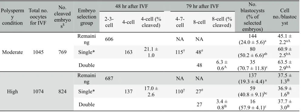
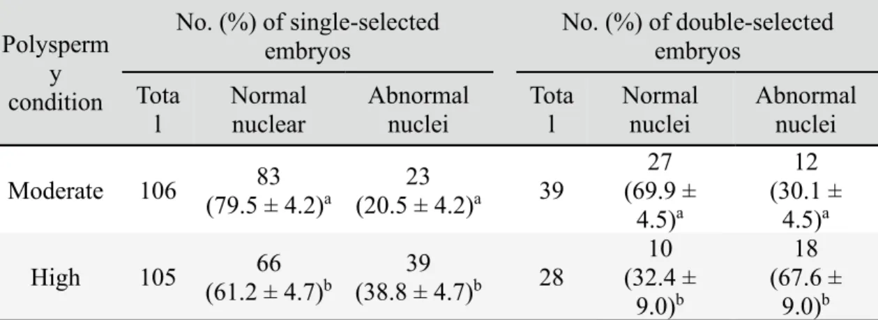
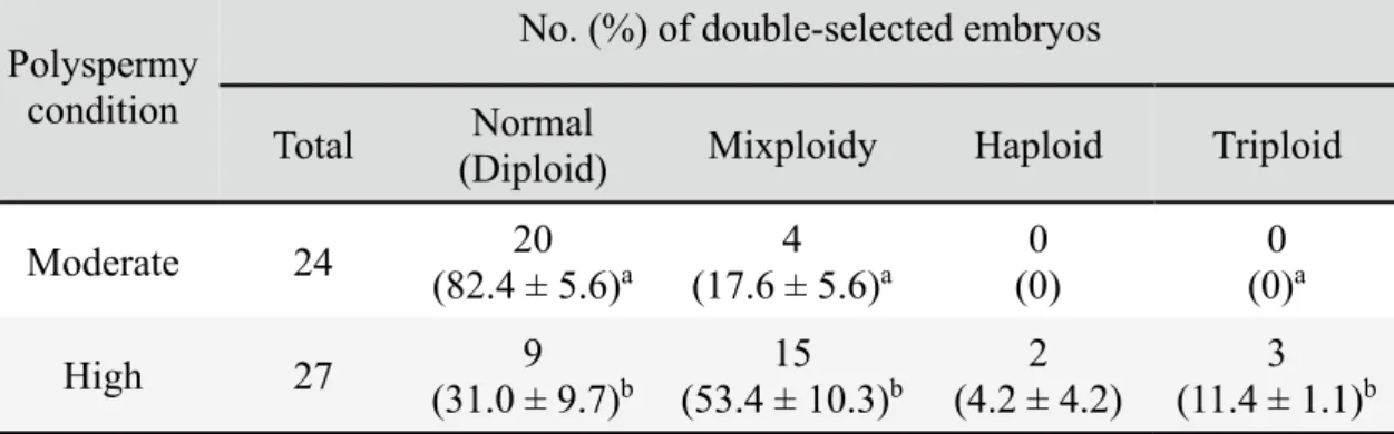
condition of moderate polyspermy, 4-cell embryos were selected at 48 hr after IVF (single selection) and 8-cell embryos were selected at 79 hr after IVF from the collected 4-cell embryos (double selection) showed high developmental competence. Likewise, 4- and 8-cell embryos produced by IVF under high polyspermy condition also showed high competence for development to blastocysts. However, blastocysts derived from high polyspermy condition had significantly fewer cells than those produced under moderate polyspermy condition. Furthermore, the frequency of nuclear and chromosomal abnormalities in 4- and 8-cell embryos produced under condition of high polyspermy was significantly (p < 0.05) higher in comparison to moderate polyspermy condition. These findings suggest that although high polyspermy affects the frequency of nuclear and chromosomal anomalies in porcine IVF embryos, subsequent selection based on morphological features of 4- and 8-cell embryos even under high polyspermy condition, could be an alternative option for selecting porcine IVF embryos with high development ability.
2.2 INTRODUCTION
In vitro production of porcine embryos is an important tool for porcine gene banking (Kikuchi et al., 2016) as well as for human biomedical research (Niemann & Rath, 2001). However, IVF in pigs is associated with a high frequency of polyspermy, causing chromosomal abnormalities in embryos (Abeydeera & Day, 1997; Herrick et al., 2003; Koo et al., 2005; Nagai et al., 2006). Polyspermy also occurs during IVF in humans (Rudak et al., 1984; Kola et al., 1987) and cattle (Iwasaki et al., 1989; Iwasaki et al., 1992)
but at lower frequency. Although various approaches have been tried for reduction of polyspermy in pigs, production of normal porcine embryos in vitro remains a challenge (Nagai et al., 2006; Grupen, 2014).
Although IVP embryos are able to develop to term after embryo transfer (ET) (Yoshida et al., 1993; Kikuchi et al., 2002), their quality is still lower than those of embryos produced in vivo (Han et al., 1999a; Kikuchi et al., 1999b; McCauley et al., 2003). A higher incidence of chromosomal abnormalities has been reported in IVP embryos relative to their in vivo counterparts, especially in pigs (van der Hoeven et al., 1985; McCauley et al., 2003; Ulloa Ulloa et al., 2008a). Although some polyspermic zygotes have been reported to develop to piglets (Han et al., 1999b), most embryos with chromosomal abnormalities fail to develop to term (Plachot, 1989; Causio et al., 2002, reviewed in Yoshizawa, 2003). Therefore, it is important to select embryos with a low frequency of chromosomal abnormalities utilizing simple and reliable procedures.
In humans and many other mammalian species it has been shown that the developmental competence of embryos is linked with the timing of early cleavages after IVF (Edirisingle et al., 1992; McKiernan & Bavister, 1994; Lonergan et al., 1999; Alikani et al., 2000; Magli et al., 2007; Ulloa Ulloa et al., 2008a, 2008b; Dang-Nguyen et al., 2010). Those previous studies showed that good quality embryos could be selected based on their morphological features and the timing of early cleavages. Bovine embryos at the 5- to 8-cell stage selected on Day 2 (Day 0 = IVF) had lower incidences of chromosomal abnormalities (Ulloa Ulloa et al., 2008b). Similarly, porcine embryos at the 3- to 4-cell stage and the 5- to 8-cell stage selected 52 hr after IVF (Ulloa Ulloa et al., 2008a), or 2-cell stage embryos selected 30 hr after IVF (Dang-Nguyen et al., 2010) were proven to have lower incidences of chromosomal abnormalities and high developmental
competence. Other studies have also shown that the timing of cleavage, evenness of division, and the degree of fragmentation can also be useful criteria for predicting the blastocyst formation ability of early porcine embryos (Mateusen et al., 2005; Booth et al., 2007; Dang-Nguyen et al., 2010). However, whether or not such morphological evaluation could also be effective for selecting high-quality embryos under high polyspermy condition has not been investigated.
In a preliminary experiment using the current IVP system in our laboratory, with the support of time lapse cinematography, It was found that single selection of 4-cell embryos and double selection of 8-cell embryos showed a high potential for rapid development to good quality blastocysts. Consequently, the present study was conducted to clarify whether selection of 4- and 8-cell stage embryos as single- and double-selected embryos at specific time points might be effective for ensuring good quality embryos, and the degree to which a high rate of polyspermy might affect the developmental competence, nuclear status and karyotype of the selected embryos. It was considered that the data from this study would lead to an alternative option for selection of embryos at fixed time points based on their morphological features, and furthermore yield insight into whether a selection approach based on embryonic morphology would still be effective under high polyspermy condition, an aspect that is essential for porcine reproduction studies where a high polyspermy is oftennot able to be avoided.
2.3 MATERIALS AND METHODS
2.3.1 Reagents
USA) unless otherwise stated.
2.3.2 Oocyte collection and IVM
The ovaries were collected from prepubertal cross-bred gilts (Landrace × Large White × Duroc) at a local slaughterhouse, and carried to the laboratory in Dulbecco’s phosphate-buffered saline (PBS) (Nissui Pharmaceutical Co. Ltd., Tokyo, Japan) at 35–
-oocyte complexes (COCs) were collected from follicles 3–6 mm in diameter in collection medium consisting of Medium 199 (with Hanks’ salts) supplemented with 10% fetal bovine serum (Gibco; Thermo Scientific, Carlsbad, CA, USA), 20 mM HEPES (Dojindo Laboratories, Kumamoto, Japan), and antibiotics (100 units/ml penicillin G potassium and 0.1 mg/ml streptomycin sulfate). IVM of oocytes was carried out as reported previously (Kikuchi et al., 1999b). In brief, about 50 COCs were cultured in each 500 µl of maturation medium, a modified NCSU-37 solution (Petters & Wells, 1993) containing 10% (v/v) porcine follicular fluid, 0.6 mM cysteine, 50 mM
-,5 -monophosphate (dbcAMP), 10 IU/ml equine chorionic gonadotropin (Serotropin; ASKA Pharmaceutical Co. Ltd., Tokyo, Japan), and 10 IU/ml human chorionic gonadotropin (Gonatropin; ASKA) in four-well dishes (Nunclon Multidishes, Nunc; Thermo Fisher Scientific) for 22 hr in an atmosphere of 5% CO2, 5% O2, and 90% N2
maturation medium without dbcAMP and hormones for an additional 24 hr under the same atmosphere.
2.3.3 Preparation of epididymal spermatozoa
Epididymal spermatozoa were collected and frozen according to Kikuchi et al. (1998, 1999a). Briefly, epididymides from four Meishan boars were brought to the laboratory just after the slaughter at Institute of Livestock and Grassland Science, NARO (NILGS) at room temperature. Luminal fluid containing spermatozoa was extruded from the distal portion of the cauda epididymidis by air pressure using a syringe. The fluid was diluted with 30 ml of collection solution at room temperature. The sperm suspension was then cooled to 15oC over about 3 hr. The solution containing the spermatozoa was
centrifuged at 1,200 × g in for 10 min at 4oC and the supernatant was discarded. The
precipitated spermatozoa were gently resuspended with Niwa and Sasaki Freezing (NSF)-I extender at 4oC (Niwa 1989), then diluted with the same volume of NSF-II at the same
temperature. The concentrations of sperm were diluted to 1 × 10⁹ (Boar A) or 5 × 10⁸
sperm/ml (Boars B–D) before freezing. Whereas, motility after collection was higher than 80% in all the four examined boars. The sperm suspension was then transferred into 0.25-ml plastic straws (IMV, L’Aigle, France), which were placed in liquid nitrogen vapor for 10 min and finally stored in liquid nitrogen.
2.3.4 IVF and IVC
COCs were treated with 0.1% (w/v) hyaluronidase to remove part of the cumulus using a glass pipet. In vitro fertilization was performed according to the 2-step IVF method of Grupen and Nottle (2000) with some modifications. The medium used for IVF was a modified Pig-FM medium (Suzuki et al., 2002) containing 10 mM HEPES, 2 mM
caffeine and 5 mg/ml bovine serum albumin (BSA). The oocytes were washed 3 times in IVF medium. They were then transferred into 90-µl IVF droplets (approximately 20 oocytes in each droplet) covered with paraffin oil (Paraffin Liquid; Nacalai Tesque, Kyoto, Japan). Frozen-thawed epididymal spermatozoa from each of Meishan boars were preincubated at 38.5°C in Medium 199 (with Earle’s salts, Gibco, pH adjusted to 7.8) for 15 min (Kikuchi et al., 1998; Ikeda et al., 2002). Matured oocytes were co-incubated with preincubated sperm at 1 × 10⁵sperms/ml (Kikuchi et al, 2002; Ikeda et al., 2002) for 30
min at 38.5°C under 5% CO2, 5% O2 and 90% N2. The oocytes with the zona-bound
sperm were then transferred to other fresh droplets of the IVF medium and subsequently incubated for 2.5 hr. At the end of IVF, spermatozoa were removed from the surface of the zona pellucida by gentle pipetting with a fine glass pipette. The day of IVF was defined as Day 0. The basic IVC medium was NCSU-37 medium containing 4 mg/ml BSA and 50 mM -mercaptoethanol. The putative zygotes were cultured in 500 µl drops of IVC-PyrLac, basic medium supplemented with 0.17 mM sodium pyruvate and 2.73 mM sodium lactate, for Days 0–2 and in IVC-Glu, basic medium with 5.55 mM D-glucose (Wako Pure Chemical Industries, Ltd., Osaka, Japan) for Days 2–6 (Kikuchi et al., 2002) in four-well dishes in an atmosphere of 5% CO2, 5% O2and 90% N2at 39°C.
2.3.5 Polyspermy evaluation
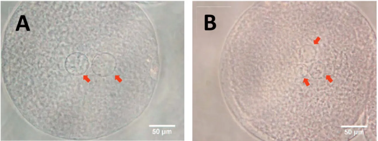
Putative zygotes were fixed at 10 hr after IVF in a mixture of acetic acid and absolute ethanol (1:3) for at least 3 days, stained with 1% aceto-orcein and examined for sperm penetration and male pronucleus formation (MPN) under a phase-contrast microscope. Zygotes with one female pronucleus and a single sperm head or MPN in the
cytoplasm were considered to be monospermic (Fig 1A). Zygotes with more than one sperm and/or MPNs were considered to be polyspermic (Fig 1B). In the present study, polyspermy rates of approximately 60% or lower and 90% or higher were considered as "moderate" and "high" polyspermy conditions, respectively.
Figure 2.1. Pronucleus(ei) in zygotes at 10 hr after IVF. A monopsermic (A) and a
polyspermic (B) zygote. Arrows show pronucleus(ei).
2.3.6 Embryo evaluation
An embryo with more than 10 cells and a clear blastocoel was defined as a blastocyst. The rate of blastocyst formation and the total number of cells per blastocyst were examined on Day 6 (Day 0 = IVF). The blastocysts were fixed and stained in ethanol containing 25 g/ml Hoechst 33342 (Calbiochem; EMD Biosciences Inc., San Diego, CA, USA) and examined under an epifluorescence microscope (Olympus, Tokyo, Japan). The total number of cells per blastocyst was evaluated as an indicator of embryo quality.
2.3.7 Nuclear status evaluation
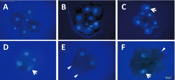
The zona pellucida was removed from single- and double-selected embryos using 1% pronase (protease, P8811). They were then fixed in 4% paraformaldehyde (PFA) for -diamidino-2-phenylindole (DAPI) in a mounting medium (Vectashield; Vector Laboratories Inc., Burlingame, CA, USA) and visualized using an epifluorescence microscope (Olympus). Embryos in which all blastomeres contained exactly one nucleus were considered normal (Fig 2A, B). Embryos with one or several blastomere(s) carrying no or more than one nucleus(ei) were considered abnormal (Fig 2C–F).
Figure 2.2. Nuclear staining of 4- and 8-cell stage embryos. Normal embryos at the
4-cell stage (A) and 8-cell stage (B). Nuclear abnormalities in 4- and 8-cell stage embryos: an 8-cell embryo carrying a binuclear blastomere (C), a 4-cell embryo with a binuclear blastomere (D), a 4-cell embryo containing two anuclear blastomeres (E) and 4-cell embryos with a binuclear and an anuclear blastomeres (F). Arrows show binuclear blastomeres and arrowheads show anuclear blastomeres.
2.3.8 Chromosome analysis
Chromosome samples from single- and double-selected embryos, respectively, were prepared according Yoshizawa et al. (1998) with some modifications. Briefly, the single- and double-selected embryos were treated with 20 ng/ml colcemid in IVC Glu for 17–20 hr. The embryos were then washed and incubated in 0.4 ml of 1% (w/v) hypotonic sodium citrate solution for 15 min and fixed mildly by adding 0.02 ml of acetic acid: methanol (1:1) to the sodium citrate solution for 2 min. A single embryo was placed on a glass slide with a minimal volume of hypotonic solution, immediately covered with a very small droplet of acetic acid to separate the cells, and then re-fixed with several drops of acetic acid: ethanol (1:3). After being allowed to dry completely, chromosome samples were stained with DAPI in Vectashield and then visualized under an epifluorescence microscope (Olympus). Only embryos containing at least two well-spread metaphases
A
B
C
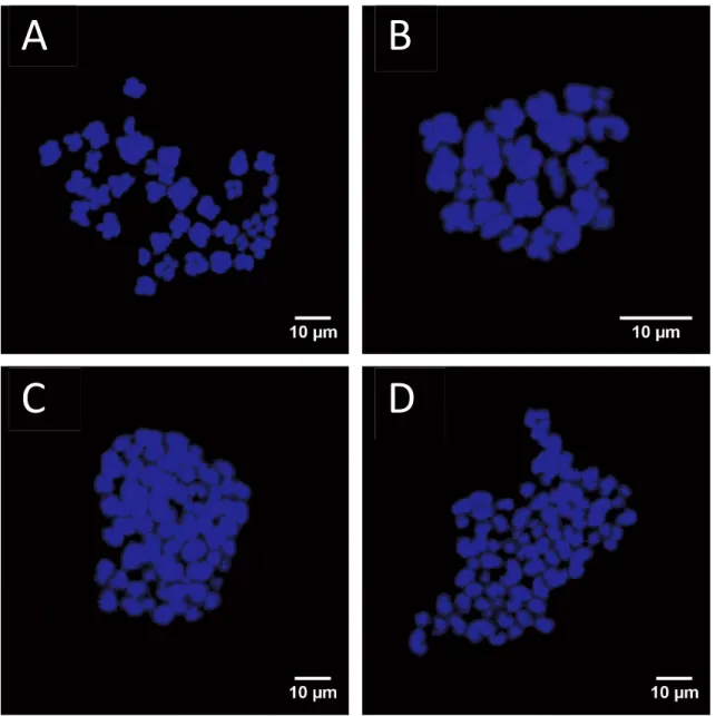
plates (intact and non-overlapping) were analyzed. Embryos that had two sets of chromosomes (2n = 38) in all analyzable metaphase plates were defined as being diploid (Fig. 3A), whereas those with only one set of chromosomes (n = 19) in all analyzable metaphase plates were defined as being haploid (Fig. 3B). Embryos containing more than two sets of chromosomes in all countable metaphase plates were defined as being polyploid (3n, 4n, etc.) (Fig. 3C, D). Embryos containing a mixture of diploid and haploid cells (n/2n), triploid (2n/3n), tetraploid (n/4n) or other types of polyploid cells were defined as being mixoploid.
Figure 2.3. Blastomere ploidy in porcine embryos. Metaphase spread from a diploid
(2n) cell with two chromosome sets (A). Ploidy abnormalities: haploid (n) (B), triploid (3n) (C) and tetraploid (4n) (D).
2.3.9 Experimental design
2.3.9.1 Experiment 1
I first aimed to establish moderate and high polyspermy conditions. Firstly, four Meishan boars (A-D) have been examined to select "moderate" and "high" polyspermy
B
C
D
rate for the purpose of the present study. In the system of our laboratory, sperm acceptable for reasonable in vitro embryo production show polyspermy rate is 50%-100% and the penetration rate is 80% to 100%. The low polyspermy rate could be obtained when reduce sperm concentration. However, it also means low penetration rate, and therefore resulting in low embryonic development. In the present study, the polyspermy rates at 50-70% and >80% to be consider as "moderate" and "high" polyspermy rate, respectively.
2.3.9.2 Experiment 2
In order to compare the developmental competence of embryos produced under moderate and high polyspermy conditions (evaluated and selected in Experiment 1), in vitro matured oocytes were fertilized with each type of sperm. Blastocyst formation rates and the numbers of cells per blastocyst were recorded after 6 days of IVC. The experiment was replicated six times.
2.3.9.3 Experiment 3
In order to compare the effect of selection on developmental competence, three embryo groups were set. I have separated 4-cell embryos and the other "remaining" cleaved (2-3-cell) embryos at 48 hr after in vitro fertilization under moderate and high polyspermy conditions (as evaluated in Experiment 2). Both groups of embryos were cultured separately. Subsequently, from a group of the 4-cell embryos, 8-cell embryos as "double selected" embryos and the other cleaved (4–7-cell) embryos were re-separated at 79 hr after IVF. Those embryos, as well as "remaining" embryos, were cultured until Day 6 after IVF. In this experiment, data of double-selected and the other cleaved embryos are
pooled and analyzed as "single-selected" embryos. Blastocyst formation rates calculated from the selected embryos and cell numbers per blastocyst in the respective groups (single-selected, double-selected and remaining embryos) were analyzed. Three to six replications with each sperm line were carried out.
2.3.9.4 Experiment 4
In order to compare nuclear abnormalities of single- and double-selected embryos produced under moderate and high polyspermy conditions (evaluated in Experiment 3), in vitro-matured oocytes were fertilized with each type of sperm. The single- and double-selected embryos were subjected to nuclear staining to evaluate nuclear abnormalities as described above. Three to five replications with each sperm line were carried out.
2.3.9.5 Experiment 5
In order to compare chromosomal abnormalities in single- and double-selected embryos produced under moderate and high polyspermy conditions (evaluated in Experiment 3), in vitro-matured oocytes were fertilized with each type of sperm. The single- and double-selected embryos were then used for chromosome spreads as described above. Three to six replications with each sperm line were carried out.
2.3.10 Statistical analysis
All data were expressed as mean ± SEM values. The data were analyzed by one-way ANOVA followed by Bonferroni correction by using the Stata/SE 15.0 software
package (StataCorp., College Station, TX, USA). Differences at p < 0.05 were considered statistically significant when it is not identified in the text.
2.4 RESULTS
2.4.1 Experiment 1
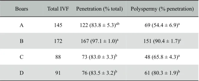
All four Meishan boars (A–D) were informed at the slaughter that they were over 1.5 years of age and used for this study. Penetration and polyspermy rates of these boar sperm lines were presented in Table 1. Zygotes derived from oocytes fertilized by Boar B sperm (90.4 ± 1.7%) had a significantly higher (p < 0.05) polyspermy rate than zygotes derived from oocytes fertilized by Boars D, C, and A sperm (80.3 ± 1.9%; 65.8 ± 4.3% and 54.4 ± 6.9%, respectively) (Table 1). In addition, the percentage of oocytes penetrated by Boar B sperm (97.1 ± 1.0%) was equal to that of Boar A sperm (83.8 ± 5.3%). As a result, sperm from Boars A and B were used for IVF to assess in the following experiments. Boar A was defined as “moderate” polyspermy rate, since the proportion of polyspermy produced from Boar A was lowest in the all samples. Boar B was selected to produce high polyspermy conditions.
Table 1. Penetration and polyspermy rates of different boar sperm lines
Boars Total IVF Penetration (% total) Polyspermy (% penetration)
A 145 122 (83.8 ± 5.3)ab 69 (54.4 ± 6.9)a
B 172 167 (97.1 ± 1.0)a 151 (90.4 ± 1.7)c
C 88 73 (83.0 ± 3.3)b 48 (65.8 ± 4.3)a
D 91 76 (83.5 ± 3.2)b 61 (80.3 ± 1.9)b
Epididymal sperm were collected from Boars A-D.
a-cValues with different superscripts in the same column are significantly different (P <
0.05).
Three to ten replications of each sperm line were carried out. Results are presented as mean percentage ± SEM.
2.4.2 Experiment 2
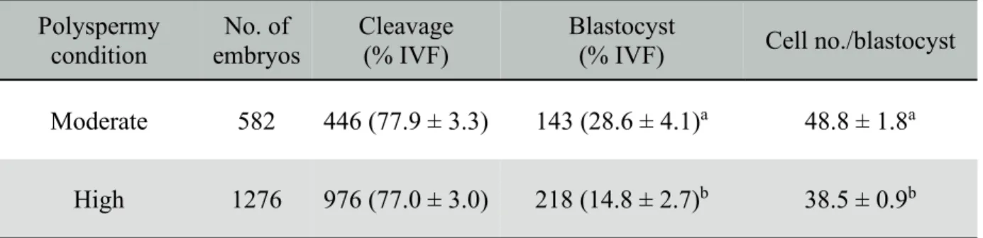
The data for development of embryos produced under moderate and high polyspermy conditions are shown in Table 2. There were no differences in cleavage rate between embryos produced under moderate (77.9 ± 3.3%) and high (77.0 ± 3.0%) polyspermy conditions. However, the proportion of embryos that formed blastocysts and the total number of cells in blastocysts formed under moderate polyspermy condition (28.6 ± 4.1% and 48.8 ± 1.8, respectively) were significantly higher (p < 0.05) than the corresponding figures for high polyspermy conditions (14.8 ± 2.7% and 38.5 ± 0.9, respectively).