A combination of desmopressin and docetaxel inhibit cell proliferation and invasion mediated by urokinase-type plasminogen activator (uPA) in human prostate cancer cells
Hiroshi Sasaki a , Laurence H. Klotz a , Linda M. Sugar b , Alexander Kiss c , Vasundara Venkateswaran a , *
aDivision of Urology, Sunnybrook Health Sciences Center, Toronto, ON, Canada
bDepartment of Pathology, Sunnybrook Health Sciences Center, Toronto, ON, Canada
cDepartment of Research Design and Biostatistics, Institute for Clinical Evaluative Sciences, Sunnybrook Health Sciences Center, Toronto, ON, Canada
a r t i c l e i n f o
Article history:
Received 5 July 2015 Accepted 9 July 2015 Available online 13 July 2015
Keywords:
Prostate cancer Desmopressin Docetaxel uPA
Matrix metalloproteinase
a b s t r a c t
Background:
This study was designed to assess the effectiveness of a combination treatment using both desmopressin and docetaxel in prostate cancer treatment. Desmopressin is a well-known synthetic analogue of the antidiuretic hormone vasopressin. It has recently been demonstrated to inhibit tumor progression and metastasis in
in vivomodels. Docetaxel is widely used for the treatment of castration resistant prostate cancer (CRPC) patients. However, durable responses have been uncommon to date. In this study, we investigated the anti-tumor effect of desmopressin in combination with docetaxel
in vitroand
in vivo.Methods:
Two prostate cancer cells (PC3, LNCaP) were treated with different concentrations of desmo- pressin alone, docetaxel alone, and a combination of desmopressin and docetaxel. Cell proliferation was determined by MTS assay. The anti-invasive and anti-migration potential of desmopressin and in com- bination with docetaxel were examined by wound healing assay, migration chamber assay, and matrigel invasion assay.
Results:
The combination of desmopressin and docetaxel resulted in a significant inhibition of PC3 and LNCaP cell proliferation (p
<0.01). Additionally, cell migration and invasion were also inhibited by the combination when compared to that of either treatment alone in PC3 cells (p
<0.01). The anti-tumor effect of this combination treatment was associated with down-regulation of both urokinase-type plasminogen activator (uPA) and matrix metalloproteinase (MMP-2 and MMP-9) in PC3 cells.
Conclusions:
We are the
first to elucidate the anti-tumor and anti-metastatic potential of desmopressinin combination with docetaxel in a prostate cancer model via the uPA-MMP pathway. Our
finding couldpotentially contribute to the therapeutic profile of desmopressin and enhance the efficacy of docetaxel based treatment for CRPC.
©
2015 Elsevier Inc. All rights reserved.
1. Introduction
Docetaxel is the fi rst cytotoxic drug of choice for management of CRPC [1,2]. Although docetaxel-based combination chemotherapy has signi fi cantly improved survival of CRPC patients, durable re- sponses are uncommon [2,3]. Furthermore, docetaxel causes
adverse events such as grade 3 or 4 neutropenia, fatigue, alopecia, and nausea. High doses of docetaxel induce signi fi cant toxicity [2,3]. A non-toxic agent that enhances the ef fi cacy of docetaxel without increasing toxicity would enhance the treatment of CRPC.
Therefore, a lower dose of docetaxel could be achieved in a com- bination treatment, and potentially provide us with a better outcome. Desmopressin is a synthetic derivative of the antidiuretic hormone vasopressin. Desmopressin is a safe and effective hemo- static agent in patients with von Willebrand disease, hemophilia A, and other bleeding disorders [4,5]. Recent reports suggest that desmopressin inhibits tumor progression and metastasis in in vivo
*Corresponding author. Department of Surgery, University of Toronto Division of Urology, Sunnybrook Health Sciences Centre, 2075 Bayview Avenue, Toronto, Ontario, M4N 3M5, Canada.
E-mail address:[email protected](V. Venkateswaran).
Contents lists available at ScienceDirect
Biochemical and Biophysical Research Communications
j o u r n a l h o m e p a g e : w w w . e l s e v ie r . c o m / l o c a t e / y b b rc
http://dx.doi.org/10.1016/j.bbrc.2015.07.050 0006-291X/©2015 Elsevier Inc. All rights reserved.
Biochemical and Biophysical Research Communications 464 (2015) 848e854
models [6 e 8]. Although these studies suggest that desmopressin has both anti-tumor and anti-metastatic effects on cancer cells, the mechanism of action has not been determined.
The aim of this study was to investigate the anti-tumor effects of desmopressin in combination with the chemotherapeutic agent docetaxel in vitro and in vivo. We demonstrate that desmopressin monotherapy in cultured prostate cancer cells resulted in impeding proliferation. Furthermore, we have delineated a potential mech- anism of action of desmopressin on human prostate cancer cells, whereby treatment signi fi cantly decreased uPA, MMP-2 and MMP- 9 expression levels in PC3 cells.
2. Materials and methods 2.1. Cell culture
Two human prostate cancer cell lines (PC3 and LNCaP) were obtained from the American Type Culture Collection (Rockville, MD, USA). All cells were maintained as previously described [9,10].
2.2. Chemicals
Docetaxel was purchased from Santa Cruz Biotechnology (CA, USA). Docetaxel was prepared in dimethyl sulfoxide (DMSO; Sig- ma e Aldrich, MO, USA) and diluted with a cell culture medium at a fi nal concentration of 0.01% DMSO. Desmopressin was provided by Ferring Inc (ON, Canada).
2.3. Cell proliferation assay
Cell proliferation was determined by MTS assay as previously described [9,10]. Cells (5 10
3/well) were plated using 96 well plates. After 24 h of cultivation, a range of concentrations of des- mopressin (0 nM e 1 m M) and/or docetaxel (0 nM e 100 nM) were added and the culture was allowed to continue for up to 72 h.
2.4. Western blot analysis
Protein lysates from the desmopressin monotherapy dose response study (1 nM-1uM), combination study and concentrated media employed for Western blot analysis, were prepared as pre- viously described [9 e 11]. The membranes were incubated with primary antibodies against Bax, bcl-2, p21, p27, cdk2, uPA, uPAR, MMP-2 and MMP-9 (1:100 e 200, Santacruz Biotechnology, CA, USA).
2.5. Flow cytometry
To analyze cell cycle pro fi les, cells were plated at a density of 1 10
6per 10 cm dish. Asynchronously growing cells were pulse labeled with 10 mM bromodeoxyuridine (BrdU) for 2 h with or without prior treatment of the antioxidants at the end of 24 h. Cell cycle analysis was carried out on the FACS Calibur fl ow cytometer using the Cell Quest Pro software package (Becton e Dickinson, CA, USA). Ten thousand events were counted for each experiment.
2.6. Wound healing assay
Cell motility was assessed using a wound healing assay, per- formed according to a protocol described by Liang et al. [12]. Cells (2 10
4/well) were plated in 6-well plates. The cells were treated with desmopressin and/or docetaxel and allowed to migrate for 24 h. A computer-based microscopy imaging system with a mi- croscope at 200X magni fi cation was used to determine wound healing after scratching the plate.
2.7. Cell migration and invasion assay
The migration of cells was measured using transwell insert plates (BD Biosciences, MA, USA) according to the manufacture's protocol. Cells were subjected to 24 h of serum deprivation in DMEM/F12 supplemented with FBS. PC3 (5 10
4) cells were plated onto fi lters in 8.0 m m transwell insert plates and treated with these compounds - desmopressin (1 nM, 1 m M) and 5 nM docetaxel, in serum-free medium. Bottom wells were fi lled with complete me- dium. Cells were allowed to migrate for 24 h. After the treatment, cells remaining on the top surface of the fi lter were removed using cotton swabs. The migrated cells from random fi elds were chosen then counted, using the computer-based microscopy imaging sys- tem. For the invasion assay, the same procedures were performed as described in the migration assay, except that the cells were plated onto 24-well matrigel-coated transwell plates (BD Bio- sciences, MA, USA).
2.8. Elisa activity assay
PC3 cells were seeded in a six-well plate (1.0 10
5per well) and incubated at 37
C. After 24 h, the complete medium was removed and cells were washed with serum-free medium. Cells were incu- bated in serum-free medium supplemented with desmopressin (1 nM and 1 m M) and/or 5 nM docetaxel for 24 h. MMP-9, MMP-2 and uPA activity in the cell lysate was quanti fi ed using human MMP-9, MMP-2 and uPA assay kit (R & D Systems, MN, USA) ac- cording to the manufacturer's instruction.
2.9. In vivo studies using xenografts
The mice were housed and maintained in laminar fl ow cabinets, under speci fi c pathogen-free conditions, in facilities approved by the University of Toronto Animal Research Ethics Board, and in accordance with their regulations and standards by the Canadian Council on Animal Care (CCAC). Cells (1 10
6PC3 cells with 100 m l martrigel solution (BD Biosciences, CA, USA)) were inoculated subcutaneously (sc) into 6-8 week-old male nude mice (Charles river, QC, CANADA). After 14 days, the developing tumors were measured and mice randomly assigned to different treatment groups. Tumor volumes were determined by measurement of tu- mor length (L) and width (W) with a caliper and calculated twice a week according to the following formula: V ¼ (L W
2) ( p /6). Mice were randomized into four groups; control (n ¼ 15), Desmopressin alone (n ¼ 15), docetaxel alone (n ¼ 10) and desmopressin in combination with docetaxel (n ¼ 10). Mice received desmopressin intravenously in the saline at a fi nal dose of 2 m g/ml/body weight (50 ng/0.3 ml saline dose). Mice were administrated docetaxel at a dose of 5 mg/kg intravenously as every 3 weeks. Desmopresin was administered in 2 doses, 30 min prior to and 24 h after the administration of docetaxel. The animals that were administered docetaxel or desmopressin in combination with docetaxel were sacri fi ced 35 days after cell inoculation.
2.10. Statistical analyses
Each experiment was repeated 2 e 3 times and the correspond- ing statistical analysis was performed. The data represented mean
± the standard error of the mean. Statistical analysis was done by
Student's t test at a signi fi cance level of p < 0.05 using SAS Version
9.3 (SAS Institute, Cary, NC, USA). Analyses of the in vivo results
were performed using either the Student's t-test, or repeated
measures of one-way ANOVA techniques.
3. Results
3.1. Docetaxel and/or desmopressin inhibit cell proliferation in prostate cancer cells
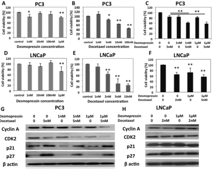
The MTS cell proliferation assay was carried out at 72 h with the cells treated with a range of doses, 0 nM e 1 m M desmopressin and 0 nM e 100 nM docetaxel. PC3 cells, treated with 1 nM and 1 m M of desmopresssin, resulted in an 18% and 20% reduction in cell proliferation (Fig. 1 , A, * p < 0.05 and
**p < 0.01). Treatment of PC3 cells with docetaxel for 72 h decreased cell viability in a dose dependent manner (Fig. 1, B). In PC3 cells, combination therapy of desmopressin (1 nM, 1 m M) and 5 nM docetaxel resulted in a signi fi cant decrease in cell proliferation when compared to treatment with desmopressin as monotherapy (Fig. 1, C,
**
p < 0.01). LNCaP cells treated with 1 m M of desmopresssin resulted in a 32% reduction in cell proliferation (Fig. 1, D,
**
p < 0.01). Also, treatment of LNCaP cells with docetaxel for 72 h decreased cell viability in a dose dependent manner (Fig. 1, E). In LNCaP cells, combination therapy of desmopressin (1 m M) and 2 nM docetaxel resulted in a signi fi cant decrease in cell prolifer- ation when compared to treatment with desmopressin as mono- therapy (Fig. 1, F,
**p < 0.01).
3.2. Desmopressin enhances the apoptotic effect of docetaxel as determined by western blot analysis, and desmopressin in combination with docetaxel induces cell cycle arrest
Docetaxel has the ability to alter key regulatory molecules, including the suppression of microtubules with consequent mitotic spindle disruption, leading to G2/M phase cell cycle arrest and in- duction of bcl-2 phosphorylation ultimately leading to apoptosis [13,14]. In PC3 cells, the expression of cyclin A and CDK2 were reduced in the cells treated with desmopressin and docetaxel
Fig. 1.Cell proliferation assay after 72 h. (A) MTS assay with PC3 cells treated with a dose range of 0e1mM desmopressin for 72 h. (B) MTS assay with PC3 cells treated with a dose range of 0e100 nM docetaxel for 72 h. Significant reductions were seen after 5 nM. (C) PC3 cells were incubated in the presence of the indicated concentration of each drug for 72 h.
Each combination therapy resulted in a significant reduction in proliferation when compared to control and corresponding docetaxel alone as well as desmopressin alone groups (*p<0.05 and **p<0.01, Student's t-test). (D) MTS assay with LNCaP cells treated with a dose range of 0e1mM desmopressin for 72 h. (E) MTS assay with LNCaP cells treated with a dose range of 0e10 nM docetaxel for 72 h. Significant reductions were seen after 2 nM. (F) LNCaP cells were incubated in the presence of the indicated concentration of each drug for 72 h. The combination therapy resulted in a significant reduction in proliferation when compared to the control and corresponding docetaxel alone, as well as desmopressin alone group (**p<0.01, Student's t-test). (G) Western blot shows the effect of a combination treatment of desmopressin and docetaxel on cell cycle regulatory proteins in PC3 cells.
(H) Western blot shows the effect of a combination treatment of desmopressin and docetaxel on cell cycle regulatory proteins in LNCaP cells.
(Fig. 1, G). In addition, the expression of CDK inhibitory proteins, p21(waf1/cip1) and p27(kip1), were elevated under the same conditions (Fig. 1, G). These data suggest that desmopressin in combination with docetaxel therapy inhibit the molecules associ- ated with cell cycle progression, and at the same time induce cell cycle arrest in PC3 cells. However, in LNCaP cells p21 and p27 were not signi fi cant elevated under the treatment with desmopressin and desmopressin (Fig. 1, H). In terms of the apoptotic pathway, docetaxel monotherapy signi fi cantly inhibited bcl-2 expression in both cell lines (Supplementary Fig. 1, A, B). The ratio of Bax/Bcl-2 revealed a 4-fold and 8-fold increased expression in cells treated with 1 nM desmopressin in combination with docetaxel and 1 m M desmopressin in combination with docetaxel respectively in PC3 cells (Supplementary Fig. 1, C). However in LNCaP cells, the ratio of Bax/Bcl-2 revealed a one-third decreased expression in cells treated with 1 m M desmopressin in combination with docetaxel. This result indicates that desmopressin may in fl uence the apoptotic effect of docetaxel in PC3 cells.
3.3. Desmopressin does not alter cell cycle distribution on PC3 cells
We investigated alterations in cell cycle pro fi les by fl ow cyto- metric analysis using BrdU labeling on PC3 cells treated with des- mopressin and docetaxel alone and in combination. Treatment with 5 nM docetaxel or a combination with 1 nM/1 m M desmo- pressin showed a signi fi cant increase in the proportion of cells in G2 phase, consistent with a G2M cell cycle arrest (Supplementary Fig. 1, D). It should be noted that there was no change in cell cy- cle distribution following treatment of cells with desmopressin monotherapy.
3.4. Differential in fl uences of desmopressin on migration of PC3 cells in a dose dependent manner
Since desmopressin and docetaxel treatment resulted in an anti- proliferative effect on PC3 cells and LNCaP cells in culture, we examined the effect of desmopressin alone and in combination with docetaxel treatment on cell migration with the wound- healing assay. As shown in Fig. 2, the difference in distance migrated by the control between 0 h and 24 h was measured and compared with that of the treated PC3 cells. At the end of 24 h, there was signi fi cant decreased cell migration with desmopressin monotherapy compared to the control (p < 0.05). We noticed a signi fi cant decrease in cell migration with a combination of 1 nM and 1 m M desmopressin and 5 nM docetaxel than with either treatment alone on PC3 (Fig. 2, A). However, we could not demonstrate anti-migrative effect during 72hr on LNCaP (data not shown). The migratory capability of PC3 cells was further quanti fi ed using the migration chamber transwell plates. Different doses of desmopressin (1 nM and 1 m M) or docetaxel monotherapy treat- ment signi fi cantly inhibited cell migration (Fig. 2). Moreover, each combination treatment with desmopressin (1 nM and 1 m M) and 5 nM docetaxel resulted in a further signi fi cant reduction in cell migration compared to controls and either monotherapy (Fig. 2, B and C,
**p < 0.01).
3.5. Desmopressin exhibits anti-invasive potential evidenced by its effect on PC3 cells
To assess the anti-metastatic ability of desmopressin, we per- formed a matrigel invasion assay. As shown in Fig. 3, 5 nM doce- taxel alone and the two different doses of desmopressin (1 nM and 1uM) treatment induced signi fi cant inhibition of cell invasion.
Furthermore, each combination treatment of 5 nM docetaxel and desmopressin (1 nM and 1 m M) resulted in a signi fi cant reduction in
cell invasion when compared to that of the control and either monotherapy (Fig. 2, D and E,
**p < 0.01), indicative of the anti- invasive ability of this combination.
3.6. uPA-MMP pathway mediates the desmopressin-induced migration and invasion of prostate cancer cells
The two important molecules MMP and uPA are involved in cancer cell invasion, motility, and tumor dormancy [15 e 17]. Based on the results of both the migration assay and invasion assay in PC3 cells, we anticipated that desmopressin used as a monotherapy may have anti-metastatic potential. Dose standardization study was carried out with desmopressin. Desmopressin monotherapy altered the expression of precursor pro-uPA, active uPA, MMP-2, and MMP-9, all of which were reduced in a dose dependent manner (Supplementary Fig. 2). Desmopressin monotherapy reduced zymogen type uPA expression, thus attenuating uPA ac- tivity on the cell surface. These results demonstrate that desmo- pressin has the ability to inhibit tumor cell migration and invasion via the uPA-MMP pathway. We have looked at the expression (Western blotting) as well as the levels of activity of uPA, MMP-2 and MMP-9 (ELISA) following treatment with desmopressin and docetaxel. As shown in Fig. 3, combination treatment resulted in a more signi fi cant reduction in the expression of uPA when compared to the control (Fig. 3, A,
**p < 0.01), and desmopressin monotherapy (Fig. 3, A,
*p < 0.05). In addition, treatment with 5 nM docetaxel in combination with 1 m M desmopressin showed a sig- ni fi cant decrease in uPAR expression (Fig. 3, B * p < 0.05). The expression of MMP-2 was also signi fi cantly reduced in the cells treated with each combination therapy when compared to the control (Fig. 3, C,
**p < 0.01), but not signi fi cantly different to each monotherapy. The expression of MMP-9 was signi fi cantly reduced in the cells treated with each combination therapy when compared to control and either of the monotherapy (Fig. 3, D,
**p < 0.01). We found that decompression alone signi fi cantly inhibited the activity of uPA, MMP-2, and MMP-9 as determined by ELISA. Also, combi- nation therapy resulted in decreased uPA, MMP-2, and MMP-9 expressions (Fig. 3, E, F and G). Consequently, these results clearly suggest that desmopressin in combination with docetaxel in- fl uences the uPA-MMP activity.
3.7. Desmopressin in combination with docetaxel inhibits PC-3 cell growth in a xenograft model
Tumors in control animals grew rapidly, measuring a volume of
923 mm
3on day 35 post tumor inoculation. In contrast, tumor
growth in the desmopressin-treated mice had a signi fi cantly slower
rate of tumor development reaching a mean volume of 642 mm
3on
day 35, resulting in a 30.4% reduction when compared to the
control (Fig. 4, Student's t-test p < 0.01). The tumor volumes be-
tween the control and desmopressin treatment were signi fi cantly
different (ANOVA, p < 0.01). We went on to further investigate
whether desmopressin was able to enhance the sensitivity of PC3
cells to docetaxel in vivo. As shown in Fig. 4, the tumors in the
combination group were signi fi cantly smaller than either the
control group, desmopressin treated group (ANOVA, p < 0.01), or
the docetaxel treated group (ANOVA, p < 0.05). Statistical analysis
con fi rmed that the combination treatment resulted in a signi fi cant
inhibitory effect on tumor growth in the PC3 xenografts. Treatment
with intravenous injection of docetaxel and/or desmopressin was
well tolerated. All mice consistently maintained their body weight
during each study.
4. Discussion
In this study we found desmopressin to have anti-proliferative, anti-migration, and anti-invasive effects on prostate cancer cells in vitro and in vivo. We are the fi rst to report the mechanism of action of desmopressin in combination with docetaxel in a pros- tate cancer model. Our results have shown that in PC3 cells, both a low dose desmopressin (1 nM) as well as a higher dose (1 m M), have a signi fi cant anti-proliferative effect in in vitro. Desmo- pressin has different pharmacologic effects including anti- hemostatic and anti-diuretic effects, with. higher doses having an anti-hemostatic effect [18]. Our results suggest that a high dose of desmopressin inhibited cell proliferation via the uPA-MMP pathway. The growth of prostatic tumor cells related to the acti- vation of the plasminogen activator system is derived from the demonstration that growth rates and uPA production in tumor cells cultured at a low density are higher than those observed in cells grown at a higher cell density. This modulation may result in altered tumor cell proliferation [19]. Alteration in cellular prolif- eration was not dose dependent in comparison to the expression of uPA activity; which was decreased with increasing doses of desmopressin. It is possible that many cytokines and growth factors such as TGF b 1, IGF-1, FGF, EGF and bombesin induce the expression of components of the uPA system [15,20 e 22]. These factors may also be associated with uPA expression and tumor cell proliferation. Further studies are needed to elucidate the mech- anism that causes the anti-proliferative effect for low-dose des- mopressin in PC3 cells. Desmopressin monotherapy did not alter cell cycle distribution and the expression levels of apoptosis related proteins in both cell lines. Nonetheless, in PC3 cells, we observed that desmopressin, in combination with docetaxel, enhanced cell cycle arrest and the apoptotic effect. Our study
demonstrated that desmopressin functions as a docetaxel che- mosensitizer for androgen independent cells. Further analysis could be needed to elucidate the mechanism that leads to the anti-tumor effect of desmopressin for androgen dependent cells.
We have shown that the anti-metastatic activity of desmo- pressin is mediated through the uPA pathway. The uPA and its re- ceptor uPAR are expressed in most solid and invasive cancers including PC3 cells. However, LNCaP cell does not express the uPA and uPAR [19]. The uPA protein is involved in the degradation of the extracellular matrix, facilitating invasiveness and growth [23]. Our present study demonstrated desmopressin as monotherapy and in combination with docetaxel signi fi cantly inhibited the expression of uPA, MMP-2, and MMP-9. uPA activates MMP-2 and MMP-9 during the migration and invasion of prostate cancer [24]. The concept of the key role of the binding of uPA to uPAR derives from several studies demonstrating that the ability of tumor cells to invade and metastasize is downregulated by uPA inhibitors. Our results suggest that desmopressin monotherapy reduced pro-uPA expression as well as active uPA, thus attenuating uPA activity on the cell surface. Accordingly, desmopressin may be acting to inhibit uPA activity in prostate cancer.
One of the limitations associated with this study was the animal model. We have not performed vivo experiments to explain the anti-metastatic effects. Future studies could be conducted in transgenic animals to elucidate the potential anti-metastatic effects in an in vivo setting. In conclusion, this is the fi rst study revealing the anti-tumor effect of desmopressin in combination with doce- taxel for prostate cancer cells working activity. Our fi nding could potentially contribute to the therapeutic pro fi le of desmopressin and enhance the ef fi cacy of docetaxel based treatment for castrate resistant prostate cancer. Clinical studies of this innocuous com- bination are warranted.
Fig. 2.Desmopressin in combination with docetaxel treatment inhibits the migration and the invasion of PC3. (A) Wound healing assay, after 24 h, the numbers of cells were confluent in control group. Whereas, decreased cell numbers in docetaxel monotherapy, each desmopressin treatment groups and combination groups. (B) The migration of PC3 cells through a transwellfilter, were assessed with or without 5 nM docetaxel in the presence of 1 nM or 1mM of desmopressin. (C) Each combination therapy resulted in a significant reduction in cell migration compared to control and corresponding docetaxel alone as well as desmopressin alone group in the migration chamber assay (**p<0.01, Student's t-test). (D) The invasion of PC3 cells through a matrigel-coated transwellfilter, were assessed with or without 5 nM docetaxel in the presence of 1 nM or 1mM of desmopressin. (E) Invasion assay showed that each combination treatment of desmopressin and docetaxel resulted in a significant reduction in cell invasion when compared to the control and corresponding docetaxel alone as well as desmopressin alone group (**p<0.01, Student's t-test).
Con fl ict of interest
The authors declare no con fl ict of interest.
Acknowledgment
This work was supported by funds provided by Ferring Inc.
(Canada). The funders had no role in this study design, data collection, analysis, and decision to publish. We would also like to thank the staff in the comparative research department, at the Sunnybrook Research Institute, for their technical support.
Appendix A. Supplementary data
Supplementary data related to this article can be found at http://
dx.doi.org/10.1016/j.bbrc.2015.07.050.
Transparency document
Transparency document related to this article can be found online at http://dx.doi.org/10.1016/j.bbrc.2015.07.050.
Fig. 3.Western blot analysis was used to demonstrate the anti-metastatic effect of desmopressin used in combination with docetaxel study on PC3 cells. Blots showing levels of expression of (A) uPA, (B) uPAR, (C) MMP-2, and (D) MMP-9. Both combination therapies decreased uPA, MMP-2 and MMP-9 expressions (*p<0.05 and **p<0.01, Student's t-test).
Also activity assays were used to demonstrate the anti-metastatic effect of desmopressin used in combination with docetaxel study on PC3 cells. Assays showing levels of activity of (E) uPA, (F) MMP-2, and (G) MMP-9. Both combination therapies decreased uPA, MMP-2 and MMP-9 activities. (*p<0.05 and **p<0.01, Student's t-test).
Fig. 4.Effects of desmopressin and/or docetaxel treatments on growth of PC3 xeno- graft model. Nude mice were subcutaneously inoculated with 1106PC3 cells per mouse. The tumor volumes were measured twice per week for 35 days (*p<0.05 and
**p<0.01, Student's t-test or ANOVA).
References
[1] D.P. Petrylak, C.M. Tangen, M.H. Hussain, et al., Docetaxel and estramustine compared with mitoxantrone and prednisone for advanced refractory pros- tate cancer, N. Engl. J. Med. 351 (2004) 1513e1520.
[2] I.F. Tannock, R. de Wit, W.R. Berry, et al., Docetaxel plus prednisone or mitoxantrone plus prednisone for advanced prostate cancer, N. Engl. J. Med.
351 (2004) 1502e1512.
[3] D. Friedland, J. Cohen, R. Miller Jr., et al., A phase II trial of docetaxel (Taxotere) in hormone-refractory prostate cancer: correlation of antitumor effect to phosphorylation of Bcl-2, Semin. Oncol. 26 (1999) 19e23.
[4] D.W. Richardson, A.G. Robinson, Desmopressin, Ann. Intern Med. 103 (1985) 228e239.
[5] P.M. Mannucci, M. Aberg, I.M. Nilsson, et al., Mechanism of plasminogen activator and factor VIII increase after vasoactive drugs, Br. J. Haematol. 30 (1975) 81e93.
[6] D.F. Alonso, G. Skilton, E.F. Farías, et al., Antimetastatic effect of desmopressin in a mouse mammary tumor model, Breast Cancer Res. Treat. 57 (1999) 271e275.
[7] S. Giron, A.M. Tejera, G.V. Ripoll, et al., Desmopressin inhibits lung and lymph node metastasis in a mouse mammary carcinoma model of surgical manip- ulation, J. Surg. Oncol. 81 (2002) 38e44.
[8] G.V. Ripoll, S. Giron, M.J. Krzymuski, et al., Antitumor effects of desmopressin in combination with chemotherapeutic agents in a mouse model of breast cancer, Anticancer Res. 28 (2008) 2607e2611.
[9] V. Venkateswaran, L.H. Klotz, N.E. Fleshner, Selenium modulation of cell proliferation and cell cycle biomarkers in human prostate carcinoma cell lines selenium modulation of cell proliferation and cell cycle biomarkers in human prostate carcinoma cell lines, Cancer Res. 62 (2002) 2540e2545.
[10] N.A. Venier, A.J. Colquhoun, H. Sasaki, et al., Capsaicin: a novel radio- sensitizing agent for prostate cancer, Prostate 75 (2015) 113e125.
[11] A.J. Colquhoun, N.A. Venier, A.D. Vandersluis, et al., Metformin enhances the antiproliferative and apoptotic effect of bicalutamide in prostate cancer, Prostate Cancer Prostatic. Dis. 15 (2012) 346e352.
[12] C.-C. Liang, A.Y. Park, J.-L. Guan, In vitro scratch assay: a convenient and inexpensive method for analysis of cell migration in vitro, Nat. Protoc. 2 (2007) 329e333.
[13] M.L. Immordino, P. Brusa, S. Arpicco, et al., Preparation, characterization, cytotoxicity and pharmacokinetics of liposomes containing docetaxel, J. Control Release 91 (2003) 417e429.
[14] K.J. Pienta, Preclinical mechanisms of action of docetaxel and docetaxel combinations in prostate cancer, Semin. Oncol. 28 (2001) 3e7.
[15] K. Forbes, K. Gillette, L.A. Kelley, et al., Increased levels of urokinase plas- minogen activator receptor in prostate cancer cells derived from repeated metastasis, World J. Urol. 22 (2004) 67e71.
[16] A. Moroz, F.K. Delella, L.M. Lacorte, et al., Fibronectin induces MMP2 expres- sion in human prostate cancer cells, Biochem. Biophys. Res. Commun. 430 (2013) 1319e1321.
[17] A.D. Kacsinta, C.S. Rubenstein, I.C. Sroka, et al., Intracellular modifiers of integrin alpha 6p production in aggressive prostate and breast cancer cell lines, Biochem. Biophys. Res. Commun. 454 (2014) 335e340.
[18] J.E. Kaufmann, U.M. Vischer, Cellular mechanisms of the hemostatic effects of desmopressin (DDAVP), J. Thromb. Haemost. 1 (2003) 682e689.
[19] C. Festuccia, V. Dolo, F. Guerra, et al., Plasminogen activator system modulates invasive capacity and proliferation in prostatic tumor cells, Clin. Exp. Metas- tasis 16 (1998) 513e528.
[20] M. Bologna, C. Festuccia, P. Muzi, et al., Bombesin stimulates growth of human prostatic cancer cells in vitro, Cancer 63 (1989) 1714e1720.
[21] D.F. Jarrard, B.F. Blitz, R.C. Smith, et al., Effect of epidermal growth factor on prostate cancer cell line PC3 growth and invasion, Prostate 24 (1994) 46e53.
[22] K. Danø, P.A. Andreasen, J. Grøndahl-Hansen, P. Kristensen, et al., Plasminogen activators, tissue degradation, and cancer, Adv. Cancer Res. 44 (1985) 139e266.
[23] P.H. Quax, A.C. de Bart, J.A. Schalken, et al., Plasminogen activator and matrix metalloproteinase production and extracellular matrix degradation by rat prostate cancer cells in vitro: correlation with metastatic behavior in vivo, Prostate 32 (1997) 196e204.
[24] C. Festuccia, D. Giunciuglio, F. Guerra, et al., Osteoblasts modulate secretion of urokinase-type plasminogen activator (uPA) and matrix metalloproteinase-9 (MMP-9) in human prostate cancer cells promoting migration and matrigel invasion, Oncol. Res. 11 (1999) 17e31.