Acta Med. Nagasaki 40: 18-21
Pancreatic Pseudocysts - Proposal for Diagnosis and Surgical Management -
Takatoshi SHIMOYAMA, M.D.1), Hiroshi ISHIKAWA, M.D.2), Teruhisa SHIMIZU, M.D.2), Hiroshi HISANO,M.D.1), Seiji HONJO, M.D.1), Satoshi MATSUO, M.D.1), Yorihisa SUMIDA, M.D.'), Seiji MATSUO, M.D.2),
Kiyoomi NISHIKAWA, M.D.2), Ryusuke TERADA, M.D.2), Hiroyuki KUSANO, M.D.2), Tohru NAKAGOE, M.D.2), Toshio MIURA, M.D.2), Masao TOMITA, M.D.2), Akio KAWAGUCHI, M.D.3), and Kazuto ASHIZAWA, M.D.4)
1) Department of Surgery, National Ureshino Hospital, Ureshinocho Fujitsugun, Saga 843-03, Japan
2) First Department of Surgery, Nagasaki University School of Medicine, 1-7-1 sakamoto, Nagasaki 852, Japan 3) Department of Surgery, Inoue Hospital, 8-9 Takaramachi, Nagasaki 852, Japan
4) Department of Radiology, National Ureshino Hospital, Ureshinocho Fujitsugun, Saga 843-03, Japan
To determine the clinical features and problems presented by a pancreatic pseudocyst, eighteen patients managed at our hospitals since 1980 were reviewed. Males predominated with a ratio of 16: 2, and the average age was 36.2 years. Pseu- docyst was located in the pancreatic head in 6, in the body in 6, in the tail in 5 and multiple pseudocysts in one case. The cause of pancreatic pseudocyst was chronic pancreatitis in 9 (including 8 patients with histories of alcoholism), acute pancreatitis in 2 and trauma in 7 patients, respectively. Three patients were associated with pancreatic calculi. Rupture of pseudocyst occurred in one patient with multiple cysts.
Autopsy showed acute pancreatic necrosis and penetration to the stomach and duodenum. The indications for surgery were principally cyst-related symptoms(infection, hemorrhage, enlargement and compression of adjacent organs) and differential diagnosis from malignancy. Most of the patients underwent internal drainage ; 9 cystojejunostomy, 2 cys- togastrostomy). Three patients underwent cystojejunostomy with longitudinal pancreaticojejunostomy for chronic pancreatitis. Massive hemorrhage after cystogastrostomy occurred in one patient with infected pseudocysts, which required removal of the cysts. Postoperative outcome of the 17 surgical patients was excellent in 16 patients. Only one patient died of pancreatitis associated with alcohol abuse, resulting in a 5.9% mortality. This study suggests that adequate internal drainage by pancreaticojejunostomy to treat the underlying pancreatic disease is most important to obtain an excellent prognosis in patients with pseudocysts.
Introduction
Pancreatic psuedocysts are a common complication of acute pancreatitis, occurring in up to 10% of patients'.
Recently, ultrasonographic studies demonstrated an incidence as high as 11 to 53% in acute pancreatitis due to alcoholism'-'). Pseudocysts are also associated with chronic pancreatitis and pancreatic trauma, either external or
surgical'). Although recent progress in imaging techniques has resulted in increased detection of pancreatic pseudocysts, there is still an inadequate appreciation of the diagnostic and therapeutic approaches as well as the pitfalls involved. This paper reviews our experiences in diagnosing and managing pancreatic pseudocysts.
Patients and Materials
Since 1980, a total of 18 patients were diagnosed with pancreatic pseudocysts at our hospitals. There were 16 males and 2 females and the mean age of the patients was 36.2 years (range 3.5 to 61 years). The cause of pancreatic peudocyst was chronic pancreatitis in 9 including 8 associ- ated with alcoholism, acute pancreatitis associated with gallstone in 2 and trauma in seven (Table 1). Three pa- tients were associated with calculi in the dilated pancreatic duct. Pseudocysts were located in the pancreatic head in 5, body in 5, tail in 2 and were multiple in one patient.
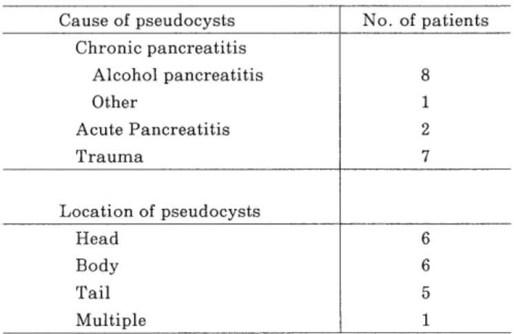
Table 1. Cause and Location of Pancreatic Pseudocysts.
Cause of pseudocysts No. of patients
Chronic pancreatitis
Alcohol pancreatitis 8
Other 1
Acute Pancreatitis 2
Trauma 7
Location of pseudocysts
Head 6
Body 6
Tail 5
Multiple 1
Table 2. Indications for Surgical Management of Pancreatic Pseudocyst.
No. of patients
Infection 3
Abdominal pain associated with a palpable mass 7
Expanding mass 3
Esophageal obstruction 1
Pseudocyst associated with biliary obstruction 1
Differential diagnosis (malignancy) 2
Total 17
Table 3. Surgical Procedures of Pancreatic Pseudocysts.
No. of patients
Cystojejunostomy 6
Cystojejunostomy with Peustow procedure 3
Cystogastrostomy 2
Resection of pseudocyst 3
Pancreatoduodenectomy 1
External drainnage 2
Total 17
Results
Clinical Feutures
Pain was chief symptoms on admission in all patients.
Palpable mass was noted in 7, fever in 4, jaundice in 2 and dysphagia due to compression of the esophagogastric portion by the enlarged pseudocyst was noted in two patients. Three patients had active pancreatitis on admis- sion. Diagnosis was based primarily on ultrasound in 4 patients, physical examination in 3, upper gastrointestinal tract series in 2, CT scanning in 7 and ERCP in 7 patients.
Among 13 patients, ultrasound was parformed in 9 with 7 correct (78%) with two false-negative (22%) results. In those false-negative cases, US images could not adequately demonstrate the pseudocyst due to the coexistence of pancreatic calculi. ERCP was performed in 9 patients, but there was no evidence of fistula between the pancreatic duct and the pseudocyst.
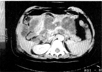
Pseudocyst rupture occurred in a patient with multiple pseudocysts. This patient was a 56-year-old female who
Fig. 1. CT scan of a patient with multiple pseudocysts around the head and body of the pancreas. She died of pseudocysts rupture.
was referred to Nagasaki University Hospital because sudden onset of abdominal pain. She had been diagnosed with Parkinson disease 10 years earliers. CT scan revealed multiple pseudocysts around the pancreatic head (Fig. 1).
She was conservatively managed for active pancreatitis as the underling pancreatic disease in association with subsequent multiple pseudocysts. However, she died of ruptured pseudocysts 32 days after admission. Autopsy revealed acute pancreatic necrosis and penetration of the pseudocysts to the stomach and duodenum.
Surgical Management and Prognosis
Indication for surgical treatment in 17 patients are shown in Table 2. In two other patients with hemorrhage, the pseudocyst could not be distinguished from malignant cystic tumor, preoperatively.
Surgical procedures in the 17 patients who required surgery are summarized in Table 3. Selection of cyst- ogastrostomy or cystojejunostomy was based on cyst location. However, three patients required cystojejuno- stomy with side-to-side longitudinal pancreaticojejuno- stomy for underlying chronic pancreatitis. Two patients showed unilocular cyst with wall thickness which could not be differentiated from malignant cystic tumor. One patient underwent pancreaticoduodenectomy for cyst in the pancreatic head, and another patient developed pseudocyst hemorrhage requiring the removal of the cyst along with splenectomy (Fig. 2).
Subsequent surgeries were performed in 2 patients. One patient with cystogastrostomy developed massive pseudocyst hemorrhage associated with infection, which was managed by resection of the pseudocyst 32 days after the initial surgery. Another patient developed a parsistent external pancreatic fistula due to external operative drainage of the pseudocyst. He subsequently underwent fistulectomy.
The postoperative outcomes of the 17 surgical patients were excellent in 16 patients. One patient died during the follow-up study resulting in a 5.9 percent mortality. This
Fig. 2. A : CT scan showing irregular thickening wall of pseudocyst. It was difficult to distinguish the cyst from cystadenocarcinoma.
B : Resected specimen measured 8 X 6 cm.
patient was a 33-year-old male with pseudocyst due to alcohol-related pancreatitis (Fig. 3A). He was successfully treated by cystojejunostomy with longitudinal pancreati- cojejunostomy, but he died of acute pancreatitis due to massive intake of ethanol one month after discharge.
Autopsy showed acute hemorrhagic infarction of the stomach and intestine due to multiple organ failure (MOF) (Fig. 3B).
Discussion
It is generally agreed that patients with pancreatic pseudocyst may be asymptomatic or demonstrate nonspe- cific symptoms even if the cyst enlarged",'). In the present series, unusual presentations, such as a palpable abdomi- nal mass, intractable pain, fever, jaundice, dysphagia and periodic follow-up examination for underlying pancreas disease led to cyst detection. The detection rates of
Fig. 3. A : CT scan of a patient with alcohol-related pancreatitis associated with a pseudocyst measured 13cm.
B : Autopsy showing acute homorrahgic infarction of the intestine due to MOF.
pancreatic pseudocyst by US, CT and ERCP were described as approximately 90% for each modality"'. However, the differential diagnosis of a cystic lesion in the pancreas must include a variety of neoplasms, particularly in the absence of antecedent factors or events that could generate a pseudocysta). Irregularity or loculation in the cyst or a solid component noted on CT scan seems to indicate a tumor, but the possibility of a malignant component should still be considered even in absence of such findings on scan"'). ERCP might be an especially valuable diagnos- tic adjunct when the biliary tract or pancreatic duct are suspected to communicate with the pseudocyst.
The chief indication in the present series was cyst-related symptoms and this agrees with the surgical indications described in other studies',"). However, indications for surgical treatment remain controversial. Regarding the natural history of pancreatic pseudocysts, spontaneous regression in 8 to 60% of the instances has been reported"). However, a 33 to 40% incidence of serious
complications in patients with untreated pancreatic pseudocysts has been reported',"'. one of our patients, spontaneous rupture of the pseudocysts occurred during medical treatment for acute pancreatitis. Spontaneous
rupture of pseudocysts have been reported in 2 to 20% of patients, almost all of these draining directly into the peritoneal cavity or the gastrointestinal tract resulting in a high mortality rate of 50 to 70 percent"). Several factors contributing to rupture of the pancreatic pseudocysts have been noted ; infection, hemorrhage and increasing pressure on the cyst. In our series, rupture occurred in a patient with active pancreatitis. It was considered that a cyst growing expansively in coexisting active pancreatitis was particularly prone to rupture. These physiological factors would seem to play important roles in the pathogenesis of the spontaneous rupture. Rupture of a pseudocyst into the esophagus, portal vein, common bile duct or pleural space has also been reported'). Therefore, it is difficult to obtain a balance between careful monitoring and early surgical intervention in patients with pancreatic pseudocysts.
Recently, it was reported that the size of the pseudocyst (> 4 em') or > 6 crn41 in diameter) was a significant indicator for surgical drainage, while asymptomatic pseudocysts less than 4 cm or 6 cm in diameter can often be followed with the expectation of complete resolution in most cases and with a low incidence of complications4'9)However, it is generally accepted that a six week period should elapse between an episode of acute pancreatitis and elective internal drainage41.
The selection of surgical procedures for pancreatic pseudocysts also remains controversial. Internal drainage seems to be superior to resection or external drainage in the elective management of pseudocysts with regard to the morbidity and mortality rates",'). In the present series, most of the patients were successfully treated with cystoenterostomy. However, massive hemorrhage after cystogastrostomy occurred in one patient with an infected pseudocyst. This suggests that if the patient has severe infections on admission, the internal drainage should be preceded by conservative management until the pseudocyst infection improves. Regarding patients with chronic pancreatitis and pseudocysts, there are three main surgical options - internal drainage of the cyst or bilioenteric bypass combined with pancreaticojejunostomy or resection procedures, such as pancreaticoduodenectomy. Longitudi- nal pancreatic ojejunostomy combined with cystojejuno- stomy to treat chronic pancreatitis with marked ductal
dilatation seems to provide better long term results than other drainage or resection procedures""'. We employed these procedures in three patients, expecting to improve the underlying chronic pancreatitis with good results.
Recently, percutaneous catheter drainage (PCD) has become a well established and often preferred alternative to surgical treatment in the management of pancreatic pseudocysts. However, the current data suggest that
despite early success with percutaneous drainage, the rate of recurrent pseudocysts remains high"-"). Therefore, the long term success of PCD in the treatment of pancreatic pseudocysts requires further investigation.
References
1) O'Malley VP, Cannon JP and Posteier RG. Pancreatic pseudocysts : Cause, therapy, and results. Am J Surg 150: 680-682, 1985.
2) Czaja AJ, Fisher M and Marin GA. Spontaneous resolution of pancreatic masses (pseudocysts ?). Arch Intern Med 135 : 558-562, 1975.
3) Agha EP. Spontaneous resolution of acute pancreatic pseudocysts.
Surg Gynec Obstet 158: 22-27, 1984.
4) Yeo CJ, Bastidas JA, Lynch-Nyhan A, Fishman E and Zinner MJ. The natural history of pancreatic pseudocysts documented by computed
tomography. Surg Gynec Obstet 170: 411-417, 1990.
5) Bradley III EL, Clements JL and Gonzales AC. The natural history of pancreatic pseudocysts : A unified concept of management. Am J Surg
137: 135-141, 1979.
6) Eilliford ME, Foster WL, Halvorsen RE, et al. Pancreatic pseudocyst : comparative evaluation by sonography and computed tomography. A.
J. R. 140: 53-57, 1983.
7) Sugawa C and Walt A. Endoscopic retrograde pancreatography in the surgery of pancreatic pseudocysts. Surgery 86: 639-647, 1979.
8) Warshaw AL, Compton CC, Lewandrowski K, et al. Cystic tumors of the pancreas. New clinical, radiologic, and pathologic observations in
67 patients. Ann Surg 212: 432-445, 1990.
9) Beebe DS, Bubrick MP, Onstad GR and Hitchcock CR. Management of pancreatic pseudocysts. Surg Gynec Obstet 159: 562-564, 1984.
10) Sankaran S and Walt AJ. The natural and unnatural history of pancreatic pseudocysts. Br J Surg 62 : 37-44, 1975.
11) Grace RR and Jordan PH Jr. Unresolved problems of pancreatic pseudocysts. Ann Surg 184: 16-21, 1984.
12) Kuroda A, Konishi T, Kimura W et al. Cystopancreaticostomy and longitudinal pancreaticojejunostomy as a simpler technique of com-
bined drainage operation for chronic pancreatitis with pancreatic
pseudocyst causing persistent cholestasis. Surg Gynec Obstet 177:
183-185, 1993.
13) Nealon W and Thompson J. Progressive loss of pancreatic function in chronic pancreatitis is delayed by main pancreatic duct decompression.
A longitudinal prospective analysis of the modified Puestow procedure.
Ann Surg 217: 458-468, 1993.
14) Adams D and Anderson M. Percutaneous catheter drainage compared with internal drainage in the management of pancreatic pseudocyst.
Ann Surg 215: 571-578, 1992.
15) Criado E, Stefano AAD, Weiner TM and Jaques PJ. long term results of percutaneous catheter drainage of pancreatic pseudocysts. Surg
Gynec Obstet 175: 293-298, 1992.