Acta Med. Nagasaki 41: 38-44
Relationship between the Immunohistochemical Expressions of Cathepsin B, Laminin and Tenascin and Clinicopathologic Features in Gallbladder Carcinomas
Atsushi NANASHIMA, Hiroyuki YAMAGUCHI, Sinichi SHIBASAKI, Juan-Eiki NISHIZAWA-TAKANO, Terumitsu SAWAI, Toru YASUTAKE, Hiroyuki KUSANO, Tohru NAKAGOE and Hiroyoshi AYABE
First Department of Surgery, Nagasaki University School of Medicine 1-7-1 Sakamoto, Nagasaki 852, Japan
In the present study, the expression of protease indicated by cathepsin B (CB) and the expression of extracellular matrix
indicated by laminin (LN) and tenascin (TN) was immuno- histochemically examined in 25 gallbladder carcinomas. The incidence of expression of .CB and TN in normal epithelium
was 1/25 (4%) and 0/25 (0%), respectively, and significantly increased in carcinomas (14/25 : 56% and 21/25 : 84%, respec- tively) (p<0.01). The LN expression was detected in the basement membrane of all normal epithelium, and the incidence of LN expression was significantly decreased in the carcinomas (7/25: 28%) (p<0.01). The incidence of CB expression in poorly differentiated adenocarcinomas (1/8:
13%) was significantly lower than that in papillary, well- and moderately differentiated adenocarcinomas (11/15:
74%) (p<0.01). However, these responses were not signifi- cantly related with other histologic features or nuclear DNA ploidy pattern. The LN expression of the hepatic metastasis group (4/6 :67%) was significantly greater than that in the non-metastatic group (3/19 :16%) (p<0.05). The expressions of CB, LN and TN were not associated with the postoperative prognosis. In conclusion, the increased expressions of CB and TN, and the decreased expression of LN were cancer- associated alterations. The expression of CB was correlated with the histological grade of differentiation, and the expres- sion of LN was correlated with the hepatic metastasis.
Key words : gallbladder carcinoma, cathepsin B, laminin, tenascin
Introduction
Gallbladder carcinoma is one of the frequent carcinomas in Japan.') Despite improvements in the preoperative diagnosis and surgical procedure, the prognosis of gall- bladder carcinoma is still poor among carcinomas of the digestive tract.") In gallbladder carcinomas, the high frequency of direct invasion to surrounding organs, lymph node metastasis and liver metastasis through the lym- phatic route') are characteristic. Such characteristics in gall bladder cancer are influenced by tumor-related factors
(e. g., the biological grade of the malignancy) and host- related factors (e. g., the anatomy and structure of the gallbladder wall).''
In the present study, we have investigated tumor-related factors such as protease secreted from cancer cells and the extracellular matrix around the cancer. Cathepsin B (CB),') a cysteine endopeptidase contained in the lysosome, is implicated in the invasion of cancer cells, and the expression of CB is closely associated with the staging or the prognosis of gastrointestinal") and pulmonary can- cers. 9) Laminin (LN)10' is a glycoprotein and one of the major structural components of basement membrane. In gastric and colorectal carcinomas,",") LN was found to be closely linked to hepatic metastasis. Tenascin (TN)') is also an extracellular matrix glycoprotein and is involved in epithelial proliferation, differentiation, cell adhesion, epithelial cell shedding from the surface, demarcation of tissue boundaries, hemagglutination and cell migration during embryonic development. An increased serum level of TN has been observed in various cancer stromas, increasing further with the advancement of the malig- nancy.",")
The aim of the present study was to clarify the biologi- cal behavior of gallbladder carcinomas by examining the relationship between the immunohistochemical expressions of CB, LN and TN, and the clinicopathologic features or the prognosis in resected gallbladder carcinomas.
Materials and Methods
Materials
Twenty-five specimens of gallbladder cancer including
normal epithelium which had been surgically resected at
the First Department of Surgery of the Nagasaki
University School of Medicine between 1982 and 1993 were
examined. Samples were fixed with 10% formalin and
embedded in paraffin blocks.
Fig. 1. The immunohistochemical responses of cathepsin B, laminin and tenascin (X 100). a. Cathepsin B expression was observed in the cytoplasm of cancer cells. b. Laminin was observed on the basement membrane of surrounding vessels but not on that of cancer cell nests. c. Laminin strongly expressed on the basement membrane of cancer cell nests in this case. d.
Tenascin expression was observed in the stroma around the cancer cell nests.
DNA flow cytometry
The 50-Prn sections of paraffin-embedded tissue were pretreated according to the method reported by Schutte et al.,') and the nuclear DNA content was measured by a FACScan flow cytometer (Becton-Dickinson, San Jose, CA). Ten thousand nuclei were examined in each specimen.
The heterogeneity of DNA content was also evaluated and was determined when at least one different pattern of DNA content in five sections per specimen was detected .
Immunohistochemical staining
For immunohistochemical staining, 3-am sections were cut. Sections were deparaffinized by xylene twice and rehydrated in an ethanol series (100%, 90%, 80%), fol- lowed by a rinse in distilled water.
Cathepsin B stain : The avidin-biotin complex peroxidase
method was applied. Using 1 % hydrogen peroxide, the
inhibition of endogeneous peroxidase activity was per-
formed. After incubation with normal rabbit serum (1 :
50 ; Vector Lab, Burlingame, CA), anti-cathepsin B
antibody (1 : 100 ; sheep anti-sera purchased from the
Binding Site Ltd., Birmingham, England) was applied at
room temperature for 60 minutes. Each section was
reacted with biotinylated rabbit anti-sheep gamma-
globulin (1 : 200 ; Vector Lab.) and treated with avidin-
biotin peroxydase complex solution. The peroxidase
reaction was visualized with 0.05% of 3,3'-diamino-
benzidine (DAB) containing 0.01% of H202 in Tris-HCI
buffered solution (pH = 7.6). The expression of CB was
seen in the cytoplasm of the carcinoma cells (Fig. la).
Laminin stain : Initially, sections were incubated with 0.4% pepsin in 0.01N HC1 for 90 minutes at 37C.") °The avidin-biotin complex-alkaline phosphatase method was then performed for the detection of laminin. After treat- ment with normal goat serum (1 : 50 ; Nichirei, Tokyo), anti-laminin antibody (1 : 300 ; rabbit anti-sera purchased from Chemicon International, West Temecula, CA) was applied for 60 minutes at room temperature. Each section was reacted with biotinylated goat anti-rabbit gamma globulin (1 : 200 ; Nichirei) and treated with alkaline phosphatase-labelled streptoavidin solution (Nichirei).
The alkaline phosphatase reaction was detected by a solution containing phosphate ester of 6-bromo-2-hydoxy- 3-naphthoic acid as buffered substrate solution, and diazotised 4'-amino-2',5'-diethoxy-benzanilide as phthalo- blue solution and activator solution (HistoMark Blue ; Kirkegaard & Perry Lab., Gaithersburg, MD). Figures lb and c show the negative and positive expression of LN in cancer tissues, respectively. The positive expressions of LN in cancer tissue was seen on the basement membrane of carcinoma cell nests (Fig. lc).
Tenascin stain : First, sections were incubated with 0.4%
pepsin in 0.01N HC1 for 120 minutes at 37°C14) and treated with 1 % hydrogen peroxide to inhibit endogeneous peroxi- dase activity. Anti-tenascin antibody (1 : 100 ; Life Technologies, Grand Island, NY) was applied for 24 hours at 4t. The avidin-biotin complex peroxidase method was performed in the same manner as for the cathepsin B stain, and visualized with DAB. The expression of TN was seen in the stroma around the carcinoma cell nests (Fig. ld).
Evaluation of stains
Each staining was determined in the superficial and the deepest layer of cancer tissues. The evaluation of CB was determined as the percentage of CB-positive cancer cells in the cancer tissue : stained less than 20% ( - ), 20-80%
(+), more than 80% (++); both (+) and (++) were considered positive. When more than 20% of LN immuno- reactivity on the basement membrane of cancer nest was observed in a cancer tissue, the tissue was considered positive for LN. The evaluation of TN was determined as the percentage of TN positive around cancer tissues : stained less than 20% (-), 20-80% (+), more than 80%
(+ +) ; both (+) and (+ +) were considered positive.
Statistics
The results of the immunohistochemical study were analyzed using the X 2 test, and a p value less than 0.05 was considered significant. The survival rate was calculated by the Kaplan-Meier method and the differences were tested by the generalized-Wilcoxon test.
Results
Clinico-histological features
Of the 25 patients, 6 were male and 19 were female, and the age ranged from 46 to 78 years old (the mean age was 61.5 y. o.). Their gallbladder carcinomas consisted of 3 carcinomas invading the muscularis propria, 7 carcinomas invading the subserosa, 8 carcinomas that exposed the serosa, and 6 carcinomas infiltrating organs. Lymph node metastasis was detected in 8 cases (44 %) , and lymphatic involvement was detected in 18 cases (72%). Venous involvement was detected in 13 cases (52 %) , and perineu- ral involvement was detected in 13 cases (52%). According to the General Rules for Surgical and Pathological Studies on Cancer of the Biliary Tract, 5 cases were in stage 1, 4 were in stage 2, 3 were in stage 3 and 14 were in stage 4.
Histologically, 3 cases were papillary-, 5 were well-, 7 were moderately- and 8 were poorly-differentiated adeno- carcinomas. Metachronous hepatic metastasis after operation was confirmed in 6 cases (24%). The mean survival time was 4.1 years, and both the three- and five-year survival rates were 34%.
DNA ploidy pattern
DNA diploidy and aneuploidy were detected in 2 cases (8.7%) and 21 cases (91.3%), respectively. Two of 22 specimens showed multiploidy. The heterogeneity of DNA ploidy was detected in 9 cases (39.1%). DNA ploidy and its heterogeneity were not associated with the histological features or prognosis of these carcinomas.
Expression in normal epithelium and gallbladder carcino- mas (Table 1)
In the normal epithelium of the gallbladder, the expres- sion of LN was detected in all specimens, and the expres- sions of CB and TN were hardly detected. In contrast, in the carcinomas, the incidence of LN expression was signif i- cantly decreased and those of CB and TN expression were significantly increased (p<0.01, respectively).
Table 1. Expression of cathepsin B, laminin and tenascin in normal epithelium and gallbladder carcinomas
Immunohistochemical response cathepsin B laminin tenascin
Normal epithlium 1/25 25/25 0/25
(4%) (100%) (0%)
t** t** $**
Carcinomas 14/25 7/25 21/25
(56%) (28%) (84%)
**: p<0 .01 X 2 test
Inter-layer difference of expression in carcinomas (Table 2)
There was no significant difference of these immuno- histochemical expressions between the superficial and the deeper layer of carcinomas.
Relationship with histological features and DNA content (Table 3)
The positive expression of CB was significantly lower in poorly differentiated adenocarcinomas compared with papillary-, well- and moderately-differentiated adeno- carcinomas (p<0.01). However, the expressions of CB, LN and TN were not associated with other parameters at all.
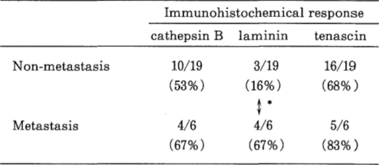
Relationship with the hepatic metastasis after operation (Table 4)
Table 2. Inter-layer difference of expression in carcinomas Immunohistochemical response
cathepsin B laminin tenascin Superficial layer 10/25 11/25 17/25
(40%) (44%) (68%)
t#
Deepest layer 14/25 7/25 21/25
(56%) (28%) (84%)
# : p<0.08 X 2 test
Table 4. Relationship with the hepatic metastasis Immunohistochemical response
cathepsin B laminin tenascin
Non-metastasis 10/19 3/19 16/19
(53%) (16%) (68%)
Metastasis 4/6 4/6 5/6
(67%) (67%) (83%)
*: p<0 .05 X 2 test
Table 3. Relationship with histologic features and DNA ploidy pattern
cathepsin B laminin tenascin
negative positive negative positive negative positive depth
m, PM, ss 5 5 7 3 2 8
se, si 5 9 10 4 3 11
lymphnode
meta (-) 4 4 5 3 1 7
meta (+) 3 7 6 4 3 7
lymphatic
inv. (-) 4 3 5 2 1 6
inv. (+) 7 11 13 5 4 14
venous