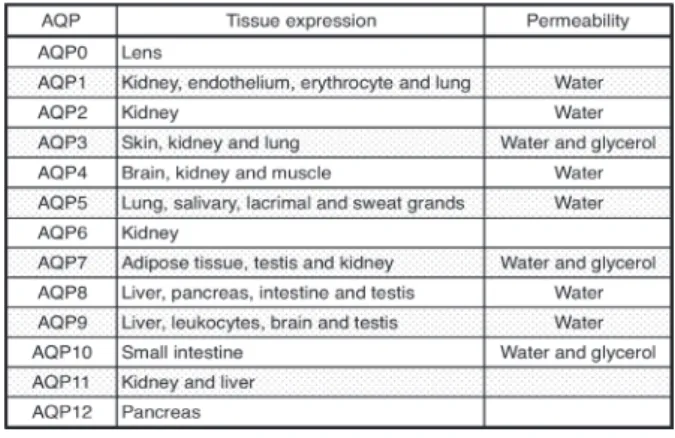
Table 1. Tissue distribution and permeability of human aquaporins.
The Roles of Aquaporins in Inflammatory and Ischemic Diseases
Ichiro H
ORIE, Yusuke G
OTOH, Satomi K
ITA, Makoto F
UJII, Takahiro I
WAMOTODepartment of Pharmacology, Faculty of Medicine, Fukuoka University
Abstract
:Aquaporins
(AQPs)are membrane proteins that function as a water channel and, in some cases, also transport small solutes such as glycerol and CO
2. AQPs are expressed in various tissues such as the kidney, lung, brain, skin, and glandular epithelia. Abnormalities of water metabolism are one of characteristics of inflammatory and ischemic diseases. However, their molecular mechanisms are poorly understood. Pathophysiological stimuli modulate the AQP expression level in inflammatory and ischemic tissues. This short review summarizes the pathophysiological roles of AQPs in abnormal water metabolism of lung, skin, and kidney, based on studies with AQPs-deficient mice.
Key words
:Aquaporin, Gene Expression, Inflammation, Ischemia/Reperfusion, Water Metabolism
Introduction
Water constitutes about 70
% of our body mass and theappropriate regulation of water transport is therefore necessary to maintain a sufficient water balance in various organs. Aquaporins
(AQPs)are a family of small
(about30 kDa) , hydrophobic, integral membrane proteins that dominantly function as water channels
1). AQPs facilitate trans-epithelial water transport in response to osmotic gradients such as fluid absorption by kidney proximal tubule and fluid secretion in the salivary gland. Some AQPs also transport small solutes such as glycerol and CO
2across the plasma membrane. Phenotype studies of mice lacking each AQP suggest that the physiological functions of AQPs are involved in urinary concentration, glandular fluid secretion, and maintenance of water balance in the brain and skin. Therefore, AQPs may be therapeutic targets for abnormalities of water metabolism.
The dysregulation of the water metabolism is observed in inflammatory and ischemic diseases. For example, inflammation in the lung induces lung edema which is an end-stage feature in alveolus, and mucus retention
(low water content in mucus)
in the airway. In addition, brain edema, which is most frequently associated with the destruction of water homeostasis, is caused by a brain
stroke. Therefore, inflammation and ischemia are the main causes of an abnormal water metabolism, although their molecular mechanisms are still poorly understood.
This review focuses on the pathophysiological roles of AQPs in lung, skin, and kidney, based on studies with AQPs-deficient mice.
Physiological functions of AQPs
AQPs are constitutively expressed in the plasma membranes of various organs
(Table 1). Thirteen AQPs
(AQP0-12)
have been identified in mammals and divided into two groups on the basis of their permeability
1)–4).
Correspondence to : Takahiro Iwamoto, Ph.D. Department of Pharmacology, Faculty of Medicine, Fukuoka University, 7-45-1 Nanakuma, Jonan-ku, Fukuoka 814-0180, Japan
TEL : +81-92-801-1011 FAX : +81-92-865-4384 E-mail : [email protected]
Figure. 1 Schematic model of down-regulation of AQP1 and AQP5 in lung inflammation.
I n f la m mator y st imul i (adenov ir us in fect ion, LPS, and inflammatory cytokines) decrease the AQP1 and AQP5 expression level in the alveolus.
Inflammation-induced decreases in AQP1 and AQP5 reduce alveolar-capillary water permeability and subsequently cause pulmonary edema.
AQP1, 2, 4, 5 and 8 are selectively permeable to water.
Computer simulations based on the AQP1 crystal structure suggest the single-file passage of water through a narrow pore within the molecule
5). On the other hand, AQP3, 7, 9 and 10 transport not only water but also glycerol, therefore, they are called aquaglyceroporins .
AQPs have no gating system in water permeable pores.
Therefore, functions of AQPs depend on their expression level in the plasma membrane. For example, AQP2, which normally exists in intracellular storage vesicles of the collecting duct cells, translocates to the apical plasma membrane in response to vasopressin stimulation, leading to water reabsorption for urine concentration
6). On the other hand, the expression level of other AQPs is regulated by changing the transcriptional-level. NF-κB, a pro-inflammatory transcription factor, decreases AQP5 gene transcription in lung epithelial cells
7).
Phenotype studies of AQPs-deficient mice suggest multiple physiological roles of AQPs. Deficiency of AQP1 and AQP2, which are expressed in the kidney, caused polyuria in mice due to abnormal urinary concentration
8),9). In addition, AQP2 mutations in humans cause congenital nephrogenic diabetes insipidus
(NDI). NDI is characterized by the inability to concentrate urine despite a normal level of vasopressin in plasma, resulting in a massive loss of water from the kidney
10)–12). A study with AQP5-deficient mice revealed that AQP5, which is expressed in salivary glandular epithelial cells, is thought to play an important role in the secretion of saliva
13).
Roles of AQP1 and AQP5 in the lung
AQP1 and AQP5 are main subtypes in lung. AQP1 is expressed in vascular endothelial cells and AQP5 is expressed in type 1 alveolar epithelial cells
14),15). Double knockout mice for AQP1 and AQP5 show decreased alveolar-capillary water permeability
16). Therefore, AQP1 and AQP5 play critical roles in the regulation of water content in the alveolus. Lung inflammation, such as adenoviral infection and acute lung injury is a respiratory dysfunction, which finally induces pulmonary edema.
Pulmonary edema is destruction of water homeostasis in the alveolus. Intratracheal infection with adenovirus, which causes airway inflammation and pulmonary edema, decreases the expression levels of AQP1 and AQP5 in the lung
17). In addition, pro-inflammatory cytokines such as a tumor necrosis factor-α
(TNF-α)also decrease AQP5 expression in lung epithelial cells
13). These findings suggest that decreases in AQP1 and AQP5 expression in
the lung induce low water excretion from the alveolus and finally cause pulmonary edema in lung inflammation
(Fig.1) . Interestingly, AQP1 is also permeable to CO
2gas
18),19). Therefore, the AQP1 and AQP5 expression levels may contribute not only to the dysregulation of water homeostasis in the alveolus, but also to an impairment of gas exchange in lung inflammation.
AQP5 is also expressed in air way epithelial cells.
The airway has a lot of water, so-called airway-surface liquid
(ASL). ASL is a first defense system whichremoves pathogens and viruses and maintains airway humidity. However, a decrease in ASL induced by airway inflammation such as cystic fibrosis
(CF)causes mucus retention
(low water content in mucus). Decreases in AQP5 expression in the air way may reduce water secretion to the airway lumen and decrease ASL volume.
Therefore, AQP5 may also play an important role in the pathophysiology of airway inflammation.
Role of AQP3 in the skin
Impaired hydration of the stratum corneum, so-called
dry skin , is a characteristic feature of inflammatory skin
diseases, such as atopic dermatitis, eczema, psoriasis,
senile xerosis, and hereditar y ichthyosis
20)–24). Low
hydration of the stratum corneum is frequently correlated
to disruption of the skin barrier function
25)and probably
Figure. 2 Decrease of AQP3 expression induced by inflammatory cytokines in the skin.
A) TNF-α decreased AQP3 mRNA expression in human keratinocytes cell lines.
B) TNF-α decreased water permeability of plasma membrane in human keratinocytes cell lines.
C) A schematic model of down-regulation of AQP3 in skin inflammation.
influences epidermal differentiation and wound repair.
AQP3 is the major subtype in skin and is specifically expressed in the basal layer of keratinocytes
26). Impaired stratum corneum hydration, reduced skin elasticity, and delayed recovery of barrier function after removal of the stratum corneum are observed AQP3-deficient mice
27),28). However, the molecular mechanisms that control the AQP3 expression level in inflammator y skin are poorly understood. TNF-α decreases AQP3 expression in keratinocytes and this reduction affects water permeability in the plasma membrane
(Fig. 2)29). AQP3 down-regulation may also affect glycerol transport.
Keratinocytes-mediated water and glycerol transport are important for maintaining hydration of the stratum corneum. Therefore, impaired stratum corneum hydration due to skin inflammation may be caused by a decrease in AQP3 expression in keratinocytes.
Skin inflammation causes a delay of wound-healing.
Interestingly, delayed wound-healing is observed in AQP3- deficient mice
28)and keratinocytes derived from AQP3- deficient mice have low migration activity, which causes delayed wound-healing
30),31). Inflammation-induced decrease in AQP3 expression in keratinocytes may affect the wound-healing activity in skin. Therefore, the AQP3 expression level is important for the skin barrier function via water homeostasis and epidermis regeneration and AQP3 plays a role in the pathophysiology of skin inflammation.
Role of AQPs in the kidney
AQP1, AQP2, and AQP3 are the main subtypes in the
kidney, although other AQPs
(AQP4, 6, 7, and 8)12)are
also expressed
(Table 2). AQP1 is highly abundant in the
proximal and descending thin limb and plays a critical
role in the constitutive re-absorption of water in these
segments and their role in urinary concentration
32). In
Table 2. Subcellular localization and regulation of renal aquaporins.
addition, AQP2, AQP3, and AQP4 are expressed in the collecting duct. AQP2 is found in the apical membrane in the principal cells of the collecting duct. On the other hand, AQP3 is found in the in basolateral membrane.
Therefore, AQP2 and AQP3 may be involved in water re- absorption from the collecting ducts.
Acute kidney injur y
(AKI)caused by ischemia/
reperfusion
(I/R)injury is a common clinical problem associated with a high morbidity and mortality. I/R injury may occur in hypo-perfusion following kidney transplantation, par tial nephrectomy, aor tic cross- cramping and shock
33). Renal expression levels of AQP1, AQP2, and AQP3 are markedly reduced after renal I/
R injury
6),34),35). Mice deficient for AQP1, AQP2, and AQP3 showed an abnormal urine concentration and subsequently polyuria. Therefore, renal I/R-induced decrease in the expression of AQPs may be related to the pathophysiology in AKI.
Urinary concentration and tubular sodium reabsorption are markedly impaired in post-ischemic kidneys.
Intriguingly, treatment with
α-melanocyte stimulatinghormone
(α-MSH), a potent anti-inflammatory agent, inhibits the reduction in renal AQPs
(AQP1, AQP2, andAQP3) and sodium transporter after renal I/R
36). α-Lipoic acid, which is a potent antioxidant, and erythropoietin
(EPO)
, which is primarily released from renal cortical fibroblasts in response to hypoxia, represses the down- regulation of renal AQP2 and AQP3 expression
37),38). In fact,
α-MSH, α-lipoic acid, and EPO improve urinaryconcentration ability after I/R injur y. These results
suggest that the pharmacological modulators of AQPs expression in the kidney are therefore novel drug candidates for renal diseases, such as AKI.
Conclusion
Phenotype studies with AQP-deficient mice suggest that AQPs may be involved in abnormalities of water metabolism in various tissues with inflammation and ischemia. Inflammatory stimuli
(adenovirus infection,LPS, and inflammator y cytokines) decreased AQP1, AQP3, and AQP5 expression in mammalian tissues.
Although it is necessar y to obtain more information about the AQP expression level under specific pathophysiological conditions, inflammation-induced down-regulation of AQPs expression may cause dysfunction of water homeostasis in various organs.
Therefore, the modulators of AQPs expression may have a therapeutic potential in inflammatory and ischemic diseases.
References
1)
Agre P, King LS, Yasui M, Guggino WB, Ottersen OP, Fujiyoshi Y, Engel A, Nielsen S: Aquaporin water channels – from atomic structure to clinical medicine.
J Physiol 542: 3-16, 2002.
2)
Ishibashi K, Sasaki S, Fushimi K, Uchida S, Kuwahara
M, Saito H, Furukawa T, Nakajima K, Yamaguchi
Y, Gojobori T, Marumo F: Molecular cloning and
expression of a member of the aquaporin family with permeability to glycerol and urea in addition to water expressed at the basolateral membrane of kidney collecting duct cells. Proc Natl Acad Sci U S A 91: 6269- 6273, 1994.
3)
Sohara E, Rai T, Miyazaki J, Verkman AS, Sasaki S, Uchida S: Defective water and glycerol transport in the proximal tubules of AQP7 knockout mice. Am J Physiol Renal Physiol 289: F1195-F1200, 2005.
4)
Kuriyama H, Kawamoto S, Ishida N, Ohno I, Mita S, Matsuzawa Y, Matsubara K, Okubo K: Molecular cloning and expression of a novel human aquaporin from adipose tissue with glycerol permeability.
Biochem Biophys Res Commun 241: 53-58, 1997.
5)
Sui H, Han BG, Lee JK, Walian P, Jap BK: Structural basis of water-specific transport through the AQP1 water channel. Nature 414: 872-878, 2001.
6)
Nielsen S, Frøkiaer J, Marples D, Kwon TH, Agre P, Knepper MA: Aquaporins in the kidney: from molecules to medicine. Physiol Rev 82: 205-244, 2002.
7)
Towne JE, Krane CM, Bachurski CJ, Menon AG:
Tumor necrosis factor-α inhibits aquaporin 5 expression in mouse lung epithelial cells. J Biol Chem 276: 18657-18664, 2001.
8)
Ma T, Yang B, Gillespie A, Carlson EJ, Epstein CJ, Verkman AS: Severely impaired urinary concentrating ability in transgenic mice lacking aquaporin-1 water channels. J Biol Chem 273: 4296-4299, 1998.
9)
Martin PY, Schrier RW: Role of aquaporin-2 water channels in urinary concentration and dilution defects. Kidney Int Suppl 65: S57-S62, 1998.
10)
Canfield MC, Tamarappoo BK, Moses AM, Verkman AS, Holtzman EJ: Identification and characterization of aquaporin-2 water channel mutations causing nephrogenic diabetes insipidus with partial vasopressin response. Hum Mol Genet 6: 1865-1871, 1997.
11)
Sohara E, Rai T, Yang SS, Uchida K, Nitta K, Horita S, Ohno M, Harada A, Sasaki S, Uchida S: Pathogenesis and treatment of autosomal-dominant nephrogenic diabetes insipidus caused by an aquaporin 2 mutation.
Proc Natl Acad Sci U S A 103: 14217-14222, 2006.
12)
Lloyd DJ, Hall FW, Tarantino LM, Gekakis N: Diabetes insipidus in mice with a mutation in aquaporin-2. PLoS Genet 1: e20, 2005.
13)
Ma T, Song Y, Gillespie A, Carlson EJ, Epstein CJ, Verkman AS: Defective secretion of saliva in transgenic mice lacking aquaporin-5 water channels.
J Biol Chem 274: 20071-20074, 1999.
14)
Kreda SM, Gynn MC, Fenstermacher DA, Boucher RC, Gabriel SE: Expression and localization of epithelial aquaporins in the adult human lung. Am J Respir Cell Mol Biol 24: 224-234, 2001.
15)
King LS, Nielsen S, Agre P: Aquaporins in complex tissues. I. Developmental patterns in respiratory and glandular tissues of rat. Am J Physiol 273:
C1541-C1548, 1997.
16)
Ma T, Fukuda N, Song Y, Matthay MA, Verkman AS : Lung fluid transport in aquaporin-5 knockout mice. J Clin Invest 105: 93-100, 2000.
17)
Towne JE, Harrod KS, Krane CM, Menon AG : Decreased expression of aquaporin
(AQP)1 andAQP5 in mouse lung after acute viral infection. Am J Respir Cell Mol Biol 22: 34-44, 2000.
18)
Prasad GV, Coury LA, Finn F, Zeidel ML: Reconstituted aquaporin 1 water channels transport CO
2across membranes. J Biol Chem 273: 33123-33126, 1998.
19)
Uehlein N, Lovisolo C, Siefritz F, Kaldenhoff R : The tobacco aquaporin NtAQP1 is a membrane CO
2pore with physiological functions. Nature 425: 734-737, 2003.
20)
Horii I, Nakayama Y, Obata M, Tagami H: Stratum corneum hydration and amino acid content in xerotic skin. Br J Dermatol 121: 587-592, 1989.
21)
Watanabe M, Tagami H, Horii I, Takahashi M, Kligman AM: Functional analyses of the superficial stratum corneum in atopic xerosis. Arch Dermatol 127: 1689- 1692, 1991.
22)
Thune P: Evaluation of the hydration and the water- holding capacity in atopic skin and so-called dry skin.
Acta Derm Venereol Suppl
(Stockh)144: 133-135, 1989.
23)
Tagami H: Quantitative measurements of water concentration of the stratum corneum in vivo by high-frequency current. Acta Derm Venereol Suppl
(Stockh)
185: 29-33, 1994.
24)
Hara M, Kato T, Tagami H: Amelanotic acral melanoma masquerading as fibrous histiocytic tumours. Three case reports. Acta Derm Venereol 73: 283-285, 1993.
25)
Proksch E, Folster-Holst R, Jensen JM: Skin barrier function, epidermal proliferation and differentiation in eczema. J Dermatol Sci 43: 159-169, 2006.
26)
Mobasheri A, Wray S, Marples D: Distribution of AQP2 and AQP3 water channels in human tissue microarrays. J Mol Histol 36: 1-14, 2005.
27)
Ma T, Hara M, Sougrat R, Verbavatz JM, Verkman
AS: Impaired stratum corneum hydration in mice
lacking epidermal water channel aquaporin-3. J Biol Chem 277: 17147-17153, 2002.
28)
Hara M, Ma T, Verkman AS : Selectively reduced glycerol in skin of aquaporin-3-deficient mice may account for impaired skin hydration, elasticity, and barrier recovery. J Biol Chem 277: 46616-46621, 2002.
29)
Horie I, Maeda M, Yokoyama S, Hisatsune A , Katsuki H, Miyata T, Isohama Y: Tumor necrosis factor-α decreases aquaporin-3 expression in DJM- 1 keratinocytes. Biochem Biophys Res Commun 387:
564-568, 2009.
30)
Hara-Chikuma M, Verkman AS : Prevention of skin tumorigenesis and impairment of epidermal cell proliferation by targeted aquaporin-3 gene disruption.
Mol Cell Biol 28: 326-332, 2008.
31)
Hara- Chikuma M, Verkman A S: Aquaporin-3 facilitates epidermal cell migration and proliferation during wound healing. J Mol Med
(Berl)86: 221-231, 2008.
32)
Knepper MA, Wade JB, Terris J, Ecelbarger CA, Marples D, Mandon B, Chou CL, Kishore BK, Nielsen S: Renal aquaporins. Kidney Int 49: 1712-1717, 1996.
33)
Weight SC, Waller JR, Bradley V, Whiting PH, Nicholson ML: Interaction of eicosanoids and nitric oxide in renal reperfusion injury. Transplantation 72:
614-619, 2001.
34)
Nielsen S, Kwon TH, Christensen BM, Promeneur D, Frøkiaer J, Marples D: Physiology and pathophysiology of renal aquaporins. J Am Soc Nephrol 10: 647-663, 1999.
35)
Ma T, Song Y, Yang B, Gillespie A, Carlson EJ, Epstein CJ, Verkman AS: Nephrogenic diabetes insipidus in mice lacking aquaporin-3 water channels.
Proc Natl Acad Sci U S A 97: 4386-4391, 2000.
36)
Kohda Y, Chiao H, Star RA: α-Melanocyte-stimulating hormone and acute renal failure. Curr Opin Nephrol Hypertens 7: 413-417, 1998.
37)
Bae EH, Lee KS, Lee J, Ma SK, Kim NH, Choi KC, Frøkiaer J, Nielsen S, Kim SY, Kim SZ, Kim SH, Kim SW : Effects of α-lipoic acid on ischemia-reperfusion- induced renal dysfunction in rats. Am J Physiol Renal Physiol 294: F272-F280, 2008.
38)
Gong H, Wang W, Kwon TH, Jonassen T, Li C, Ring T, Frøkiaer J, Nielsen S: EPO and α-MSH prevent ischemia/reperfusion-induced down-regulation of AQPs and sodium transporters in rat kidney. Kidney Int 66: 683-695, 2004.
(平成24.7.10受付,平成24.9.24受理)