• 検索結果がありません。

The Usefulness of Diffusion-weighted Imaging in Observing Localized Extension of Endometrial Cancer

N/A
N/A
Protected

Academic year: 2021

シェア "The Usefulness of Diffusion-weighted Imaging in Observing Localized Extension of Endometrial Cancer"

Copied!
8
0
0

読み込み中.... (全文を見る)

全文

(1)

The Usefulness of Diffusion-weighted Imaging in Observing Localized Extension of Endometrial Cancer

Nozomi B ELL , Naomi Y AGI , Kumi H ATANO , Makoto S AIKI , Yoshimitsu O HGIYA , Masanori H IROSE

and Takehiko G OKAN

Abstract : Endometrial cancer is the seventh most common human malignancy and the most common form of cancer treated in women by obstetrics and gynecology departments. Until now, magnetic resonance imaging(MRI) has been used for pre-surgical evaluation of endometrial cancer and evaluating the depth of myome- trial invasion, in addition to being a valuable diagnostic tool. Diffusion-weighted imaging(DWI) has been reported as useful in distinguishing between benign and malignant tumors when observing lesions in the endometrium. Subsequent reports suggest that DWI is also effective in identifying malignancy and diagnos- ing local extension in a range of tissues. Based on this, we implemented a study of the effectiveness of DWI in identifying local extension of endometrial cancer.

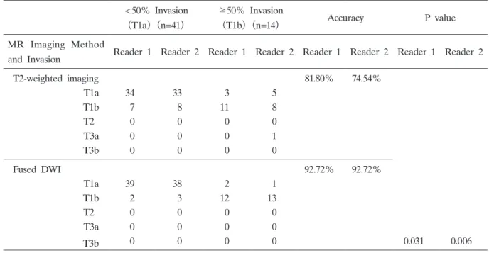
This study enrolled patients undergoing surgery at this hospital for cancer of the uterine body during the six years from January 2008 to February 2014. Cases in which images were unclear or the lesions were too small to be described by MRI examination were excluded, leaving 61 patients in the study. Using the results from pre-surgical MRI, a sequence comprising a T2-weighted axial view alone and a T2-weighted axial view to which a diffusion-weighted axial view had been added was created for each patient. Two radiologists then independently examined the image sequence to determine localized extension. Following surgery, the pre- surgical assessment was compared to the localized extension determined by histo- pathology of post-surgical samples to evaluate the effectiveness of adding diffusion- weighted imaging to the process. The first radiographic interpreterʼs rate of correct diagnosis using the T2-weighted axial view alone was 45 out of 55 cases(81.8%) , while using the T2-weighted axial view to which a diffusion-weighted axial view had been added gave a correct diagnosis rate of 51 out of 55 cases(92.7%) . The second radiographic interpreterʼs rate of correct diagnosis using the T2-weighted axial view alone was 41 out of 55 cases(74.5%) , while using the T2-weighted axial view with diffusion-weighted axial view added gave a correct diagnosis rate of 51 out of 55 cases(92.7%) . These differences were statistically significant based on the McNemar testing. This study confirmed that DWI is an effective means of diagnosing localized extension from images. It is anticipated that DWI will be used in the future clinical workplace to provide more accurate pre-surgical diagnoses.

Key words : diffusion-weighted imaging, localized extension, endometrial cancer Original

Department of Radiology, Showa University School of Medicine, 1—5-8 Hatanodai, Shinagawa-ku, Tokyo 142-8666, Japan.

To whom corresponding should be addressed.

(2)

Introduction

 Endometrial cancer is the seventh most common malignancy and the most common form of cancer treated in women by obstetrics and gynecology departments

1)

. Furthermore, adenocarcinoma originating in the endometrium is becoming more common, with the absolute number of cases noticeably increasing in Japan. Endometrial cancer occurs frequently in women in their 50s ; however, while there has been no significant change in the age profile of patients, the number of younger patients is increasing

2)

.

 Although a wide range of tumor tissue types can arise from endometrium, most are adenocarcinoma, with endometrioid adenocarcinoma being particularly common. During fiscal 2007, data from the Japan Society of Obstetrics and Gynecology revealed that 85% of malignant uterine body tumors were endometrioid adenocarcinoma.

 While prognostic factors in endometrial cancer include uterine factors such as tissue type, dif- ferentiation, muscle layer penetration, cervical penetration, vascular factors, abnormal endometrial hyperplasia, the presence of hormone receptors, DNA ploidy, and S-phase fraction, only the age at onset and depth of myometrial invasion are proven independent prognostic factors, indicating the importance of diagnosing myometrial invasion

3)

.

 The International Federation of Gynecology and Obstetrics(FIGO) staging and The  TNM Classification of Malignant Tumours(TNM) was revised in 2008, and in line with this, the classification of surgery and advancement has also been revised

4)

. At the same time, image- based diagnosis has become more important for diagnosing the clinical stage of uterine body cancers. Until now, magnetic resonance imaging(MRI) has been the most accurate method of obtaining such image diagnoses for pre-surgical evaluations of endometrial cancer and for assessing the depth of myometrial invasion

5-7)

, and a recent meta-analysis rated contrast-enhanced T1-weighted MRI as more effective than ultrasonography, CT, or non-contrast MRI

8)

. However, given the risk

9)

of nephrogenic systemic fibrosis(NSF) resulting when gadolinium contrast agent is administered to patients with renal failure, the demand for non-contrast MRI will rise in the future.

 Diffusion-weighted imaging(DWI) is a type of MRI sequence that captures the diffusion movement of water molecules, and is reportedly effective in distinguishing between benign and malignant tumors in the endometrium

10)

. Subsequent reports indicate that DWI is also effec- tive in distinguishing between benign and malignant tumors in various areas, and in identifying localized extension

11-20)

. We therefore sought to evaluate the effectiveness of DWI in identifying localized extension of uterine body cancer.

Materials and methods Patients

 Ethical approval for this retrospective study was granted by the institutional review board, and patient consent was not required.

 The scope of the study included patients who attended our hospital during the six years from

(3)

January 2008 to February 2014 and were subjected to T2-enhanced axial view and MRI, includ- ing DWI, prior to undergoing hysterectomy.

 In total, 55 patients(average age 59.20 years, range 33—92 years) were registered in the study, none of whom had undergone both chemotherapy and radiotherapy prior to surgery.

MRI examination

 All 55 patients were examined using a SIEMENS-manufactured body coil, of these, 33 had images taken using 3.0 TMRI(Magnetom Trio A Tim 3.0T, manufactured by SIEMENS) , 17 using 1.5 TMRI(Magnetom Avanto 1.5T, manufactured by SIEMENS) , and 5 using 1.5 TMRI

(Magnetom ESSENZA 1.5T, manufactured by SIEMENS) .

 All patients underwent MRI prior to surgery, with the MR sequences including both T2-weighted and diffusion-weighted images.

 T2-enhanced axial-view images were taken perpendicular to the body of the uterus, using the spin echo method, with the following scan parameters : TR / TE 6000-4500 / 103-94 msec ; slice thickness 5 mm ; field of view(FOV) 22 cm ; base resolution 384 ; voxel size 1.0 0.8 5.0 mm

(3.0T) , 1.1 0.8 5.0 mm(1.5T) .

 Diffusion-weighted images were taken perpendicular to the body axis, with the following scan parameters : TR / TE 4000-3400 / 97-79 msec ; slice thickness 5 mm ; field of view(FOV)

35 cm ; voxel size 1.0 0.8 5.0 mm(3.0T) , 1.1 0.8 5.0 mm(1.5T) ; b-values 50, 500, 1000, and 2000 s / mm

2

.

 The apparent diffusion coefficient map(ADC map) was automatically created using the manu- facturerʼs software program.

Image analysis

 The tumor areas showed a higher signal intensity on T2-enhanced images than normal myo- metrium and a lower signal intensity than normal endometrium in each case. With DWI and the ADC map, tumors showed a higher signal intensity than normal myometrium in each case.

 The diagnosis of local extension was also compared on T2-enhanced axial-view imaging, DWI, and ADC map. Two radiologists(with 4 and 33 years of experience in image-based diagnosis, respectively) independently determined localized extension of the tumors based on the UICC 7th edition TNM.

 The authors compared the results of T categorization using only the T2-enhanced axial view, according to the aforementioned categories, with those using the T2-enhanced axial view com- bined with DWI. When evaluating the DWI, the doctors also referred to the ADC map as a supplemental resource.

 The authors subsequently compared the results of histopathological testing following hysterec-

tomy, with the diagnosis regarding localized extension determined using each patientʼs pre-surgical

MRI examinations.

(4)

Statistical analysis

 Statistical analysis was implemented using the SPSS version17.0 software program and McNe- mar testing. P < 0.05 was considered to indicate a significant difference.

Results

 Of the 55 patients in this study with endometrioid adenocarcinoma, 41 were T1a and 4 were T1b. The length of the tumors ranged from 12 to 85 mm, with widths of 4-67 mm.

 The first radiographic interpreterʼs rate of correct diagnosis using the T2-weighted axial view alone was 45 out of 55 cases(81.8%) , while using the T2-weighted axial view to which a diffusion-weighted axial view had been added gave a correct diagnosis rate of 51 out of 55 cases

(92.7%) . In five cases of the T2-enhanced imaging and in two cases of DWI, the images were somewhat unclear due to artifact. In addition, in another five cases of the T2-enhanced imaging and in two cases of DWI, poor image detection due to myoma made them difficult to interpret.

 The second radiographic interpreterʼs rate of correct diagnosis using the T2-weighted axial view alone was 41 out of 55 cases(74.5%) , while using the T2-weighted axial view to which a diffusion-weighted axial view had been added, gave a correct diagnosis rate of 51 out of 55 cases(92.7%) . (Table 1) .

 With the T2-enhanced images, seven cases had somewhat unclear images due to artifact, while in another six cases, poor image detection due to myoma made the images difficult to interpret, and one case showed primary ovarian cancer. With the DWI, three cases showed somewhat unclear images due to artifact, and image interpretation was difficult in one case due to myoma.

 A difference in the radiologistʼs experience was thought responsible for the mismatch between the first and second radiographic interpreter.

Table 1.  Correlation of Histopathological Results in 55 Patients

<50% Invasion

(T1a)(n=41)

50% Invasion

(T1b)(n=14) Accuracy P value MR Imaging Method

and Invasion Reader 1 Reader 2 Reader 1 Reader 2 Reader 1 Reader 2 Reader 1 Reader 2

T2-weighted imaging 81.80% 74.54%

T1a 34 33 3 5

T1b 7 8 11 8

T2 0 0 0 0

T3a 0 0 0 1

T3b 0 0 0 0

Fused DWI 92.72% 92.72%

T1a 39 38 2 1

T1b 2 3 12 13

T2 0 0 0 0

T3a 0 0 0 0

T3b 0 0 0 0 0.031 0.006

(5)

 The McNemar analysis resulted in P values of 0.031 and 0.006 for the first and second radio- graphic interpreters, respectively. Both of these were below 0.05, within the range of significant difference.

Discussion

 Evaluating the stage of advancement of endometrial cancer is mainly carried out based on T2-enhanced imaging results ; however, using only these images makes accurate evaluation diffi- cult in many cases, necessitating the additional use of contrast MRI. In the case of local exten- sion, the reported rate of accurate diagnosis of myometrial invasion is 58〜77%

21-23)

.

 Many studies reported a significant improvement in the ability to diagnose myometrial invasion based on MRI results compared to T2-enhanced images alone. In particular, a contrast dynamic study is highly effective, improving accuracy significantly from 58〜77% with T2-enhanced images alone to 85〜93%

21-24)

. However, despite this improvement, it is believed that allergies to contrast agents and the fact that contrast agents cannot be used in patients with compromised renal function will mean that, in the future, it will become more important to implement pre- surgical diagnosis of endometrial cancer using non-contrast MRI, along with diagnosing localized extension prior to surgery using basic MRI technology. Accordingly, the use of DWI in pre- surgical evaluation provides an important supplementary sequence in the accurate diagnosis of endometrial cancer.

 MRI DWI provides roughly the same level of diagnostic ability as contrast MRI, and in many cases, it further enables the evaluation of tumor spread. However, according to the studies of Rechichi et al

25)

, DWI was more effective when a comparison was implemented using 1.5 TMRI between DWI and contrast MRI.

 In this study, endometrial cancer demonstrated higher signal intensity on DWI than normal endometrium, and the addition of diffusion-weighted imaging to the T2-enhanced axial view further improved the rate of accurate diagnosis. Thus, the present study clearly demonstrated the effectiveness of adding DWI to the T2-enhanced axial view.

 This study also revealed a case in which a mistaken evaluation of myometrial invasion in the periphery of the tumor led to a misevaluation of local extension using T2-enhanced images

(Fig. 1) . This was attributed to the fact that the signal-intensity difference between the tumor and normal muscle layer was unclear because of artifacts in the periphery of the tumor. Since the use of DWI together with the ADC map provides better, clearer contrast between the tumor and normal muscle layer than T2-enhanced images, it would contribute to a correct diagnosis.

 Accurate evaluation of myometrial invasion is known to be difficult for cases in which the tumors are large polyps and for cases complicated by uterine leiomyoma, uterine deformities, or a small uterus

26)

.

 The present study also revealed one case among those complicated by uterine leiomyoma that

was mistakenly diagnosed as local extension based on the T2-enhanced axial view alone (Fig. 2) .

  In this case, myoma was noted within the muscle layer on the cranial side of the tumor, and it is

believed that the boundary between the myoma and tumor was unclear in the T2-enhanced axial view.

(6)

 Furthermore, in cases complicated by adenomyosis, in which the tumor and the adenomyosis are connected, it is not possible to obtain visible contrast between the normal uterine muscle layer and the myoma, and this is considered to reduce diagnostic ability with regard to myome- trial invasion

27)

. In the past, the combined use of T2-enhanced images with DWI, using a 3T MRI, has allowed correct evaluation of myometrial invasion in cases complicated by adenomyosis and other factors that make the evaluation difficult

28)

.

 In this study, the T2-enhanced axial views were further taken perpendicular to the body of the uterus, while diffusion-weighted images were taken perpendicular to the trunk of the body. As a result, the cross-section of the two images was found to be different, making it difficult to arrive at a correct evaluation of local extension in the periphery of the tumor. Thus, we recommend ensuring that both T2-enhanced and diffusion-weighted images are taken using the same slice when assessing local extension.

Conflict of interest

 The authors have declared no conflict of interest.

Fig. 1.  40-year-old woman

The authors discovered a case in which a mistaken evaluation of myometrial invasion

in the periphery of the tumor (➡) led to a misevaluation of local extension using

T2 enhanced images. The T2 enhanced axial view (a)of the left side of the uterine

body does not clearly show the boundary between the myoma (▶) and the uterine

muscle layer, while diffusion-weighted imaging(DWI)(b)and ADCmap (c) show

a clear contrast. In this case, the patient was diagnosed as T1b when using the

T2 enhanced axial view alone, but determined to be T1a when diffusion-weighted

imaging was added, which was consistent with the pathological findings.

(7)

References

1) Amant F, Moerman P, Neven P, et al. Endometrial cancer. Lancet. 2005;366:491—505.

2) Japan Society of Obstetrics and Gynecology Gynecologic Oncology Committee; Gynecologic Oncology Committee Report. Nihon Sanka Fujimka Gakkai Zasshi. 2009;61:1540-1542.(in Japanese) .

3) Ronnnet BM.Endometrial carcinoma. In Kurman RJ ed. Blausteinʼs pathology of the female genital tract. 5th ed.

New York: Springer; 2002. pp 501-559.

4) Japan Society of Obstetrics and Gynecology, Japan Society of Pathology, Japan Radiological Society, et al. The General Rules for Clinical and Pathological Management of Uterine Corpus Cancer. 3rd ed. Tokyo: Kanehara publishing; 2012.

5) Manfredi R, Mirk P, Maresca G, et al. Local-regional staging of endometrial carcinoma: role of MR imaging in surgical planning. Radiology. 2004;231:372-378.

6) Hricak H, Rubinstein LV, Gherman GM, et al. MR imaging evaluation of endometrial carcinoma: results of an NCI cooperative study. Radiology. 1991;179:829-832.

7) Sironi S, Colombo E, Villa G, et al. Myometrial invasion by endometrial carcinoma: assessment with plain and gadolinium-enhanced MR imaging. Radiology. 1992;185:207-212.

Fig. 2.  62-year-old woman

One case was mistakenly diagnosed as local extension of tumor (➡) when using the T2 enhanced axial view(a) alone. In this case, the position of the myoma (▶) in relation to the muscle layer was difficult to identify, so a T2 enhanced saggital plane image has been provided for reference.

A uterine tumor at the base of the tumor can be seen in the T2 enhanced saggital plane view(b) , while the boundary between the myoma and uterine muscle layer at the base is unclear in the T2 enhanced axial view(a) . Diffusion-weighted imaging

(DWI)(c) and the ADCmap(d) show a clear contrast between the myoma and

the muscle layer. In this case, the patient was diagnosed as T1b when using the

T2 enhanced axial view alone, but determined to be T1a when diffusion-weighted

imaging was added, which was consistent with the pathological findings.

(8)

8) Kinkel K, Kaji Y, Yu KK, et al. Radiologic staging in patients with endometrial cancer: a meta-analysis. Radiology.

1999;212:711—718.

9) Sadowski EA, Bennett LK, Chan MR, et al. Nephrogenic systemic fibrosis: risk factors and incidence estimation.

Radiology. 2007;243:148-157.

10) Fujii S, Matsusue E, Kigawa J, et al. Diagnostic accuracy of the apparent diffusion coefficient in differentiating benign from malignant uterine endometrial cavity lesions: initial results. Eur Radiol. 2008;18:384-389.

11) Guo Y, Cai YQ, Cai ZL, et al. Differentiation of clinically benign and malignant breast lesions using diffusion- weighted imaging. J Magn Reson Imaging. 2002;16:172-178.

12) Woodhams R, Matsunaga K, Kan S, et al. ADC mapping of benign and malignant breast tumors. Magn Reson Med Sci. 2005;4:35-42.

13) Rubesova E, Grell AS, De Maertelaer V, et al. Quantitative diffusion imaging in breast cancer: a clinical prospec- tive study. J Magn Reson Imaging. 2006;24:319-324.

14) Nasu K, Kuroki Y, Nawano S, et al. Hepatic metastases: diffusion-weighted sensitivity-encoding versus SPIO- enhanced MR imaging. Radiology. 2006;239:122-130.

15) Cova M, Squillaci E, Stacul F, et al. Diffusion-weighted MRI in the evaluation of renal lesions: preliminary results.

Br J Radiol. 2004;77:851-857.

16) Thoeny HC, De Keyzer F, Oyen RH, et al. Diffusion-weighted MR imaging of kidneys in healthy volunteers and patients with parenchymal diseases: initial experience. Radiology. 2005;235:911-917.

17) Ichikawa T, Erturk SM, Motosugi U, et al. High-B-value diffusion-weighted MRI in colorectal cancer. AJR Am J Roentgenol. 2006;187:181-184.

18) Sato C, Naganawa S, Nakamura T, et al. Differentiation of noncancerous tissue and cancer lesions by apparent diffusion coefficient values in transition and peripheral zones of the prostate. J Magn Reson Imaging. 2005;21:258- 262.

19) Matsuki M, Inada Y, Tatsugami F, et al. Diffusion-weighted MR imaging for urinary bladder carcinoma: initial results. Eur Radiol. 2007;17:201-204.

20) Naganawa S, Sato C, Kumada H, et al. Apparent diffusion coefficient in cervical cancer of the uterus: comparison with the normal uterine cervix. Eur Radiol. 2005;15:71-78.

21) Yamashita Y, Harada M, Sawada T, et al. Normal uterus and FIGO stage I endometrial carcinoma: dynamic gadolinium-enhanced MR imaging. Radiology. 1993;186:495-501.

22) Ito K, Matsumoto T, Nakada T, et al. Assessing myometrial invasion by endometrial carcinoma with dynamic MRI.

J Comput Assist Tomogr. 1994;18:77-86.

23) Seki H, Kimura M, Sakai K. Myometrial invasion of endometrial carcinoma: assessment with dynamic MR and contrast-enhanced T1-weighted images. Clin Radiol. 1997;52:18-23.

24) Joja I, Asakawa M, Asakawa T, et al. Endometrial carcinoma: dynamic MRI with turbo-FLASH technique. J Com- put Assist Tomogr. 1996;20:878-887.

25) Rechichi G, Galimberti S, Signorelli M, et al. Myometrial invasion in endometrial cancer: diagnostic performance of diffusion-weighted MR imaging at 1.5-T. Eur Radiol. 2010;20:754-762.

26) Scoutt LM, McCarthy SM, Flynn SD, et al. Clinical stage I endometrial carcinoma: pitfalls in preoperative assess- ment with MR imaging. Work in progress. Radiology. 1995;194:567-572.

27) Tamai K, Togasi K, Ito T, et al. MR imaging findings of adenomyosis: correlation with histopathologic features and diagnostic pitfalls. Radiographics. 2005;25:21-40.

28) Lin G, Ng KK, Chang CJ, et al. Myometrial invasion in endometrial cancer: diagnostic accuracy of diffusion- weighted 3.0-T MR imaging--initial experience. Radiology. 2009;250:784-792.

[Received July 2, 2014 : Accepted July 16, 2014]

Table 1.  Correlation  of  Histopathological  Results  in  55  Patients
Fig. 1.  40-year-old  woman
Fig. 2.  62-year-old  woman

参照

関連したドキュメント

We note that this topos is Boolean, so it does not provide a counterexample to the assertion that every completely distributive Grothendieck topos has initial normal covers for all

Precisely, over a period of 120 months, the total number of new infections that will be generated from the two patches in the absence of optimal control is 1.2037× 10 4 , whereas,

At Geneva, he protested that those who had criticized the theory of collectives for excluding some sequences were now criticizing it because it did not exclude enough sequences

In this, the first ever in-depth study of the econometric practice of nonaca- demic economists, I analyse the way economists in business and government currently approach

An easy-to-use procedure is presented for improving the ε-constraint method for computing the efficient frontier of the portfolio selection problem endowed with additional cardinality

The simplest model developed here depends on only three independent parameters: the number of ordered mutations necessary for a cell to become cancerous, the fraction of the

Keywords: continuous time random walk, Brownian motion, collision time, skew Young tableaux, tandem queue.. AMS 2000 Subject Classification: Primary:

As an application, in Section 5 we will use the former mirror coupling to give a unifying proof of Chavel’s conjecture on the domain monotonicity of the Neumann heat kernel for