Prognostic Factors for Recurrence after Tegafur-uracil Plus Leucovorin Adjuvant Chemotherapy in Patients with Colorectal Cancer
Makoto W ATANABE
*, Masahiko M URAKAMI , Yoshiaki O ZAWA , Satoru G OTO , Akira F UJIMORI , Koji O TSUKA
and Takeshi A OKI
Abstract : To evaluate prognostic factors for recurrence after tegafur-uracil plus leucovorin (UFT / LV) adjuvant chemotherapy in patients with colorectal cancer
(CRC) . Consecutive patients with CRC who received UFT / LV as adjuvant che- motherapy at Showa University Hospital between June 2005 and December 2008 were included in the study, 5-year disease-free survival (DFS) and overall survival
(OS) rates were estimated, and prognostic factors for recurrence were analyzed using the Cox proportional hazards model for multivariate analysis. Of 92 patients included in the study, 17 (18.5%) had disease recurrence. The 5-year DFS and OS rates were 82.2% and 91.9%, respectively. In the multivariate analysis, preoperative CA19-9 level > 37 U / ml, emergency operation, and T4 lesions were independent significant prognostic factors after treatment with UFT / LV adjuvant chemotherapy.
The three independent prognostic factors ― T4 lesions, emergency operation, and high preoperative CA19-9 levels ― may be useful for decision-making regarding whether patients should receive 5-fluouracil-based or L-oxaliplatin-based adjuvant chemotherapy. As this was a single-institution study with a small number of patients, our findings need to be confirmed in larger multicenter studies..
Key words : adjuvant chemotherapy, colorectal cancer, UFT / LV, long-term outcomes, prognostic factors
Introduction
In patients with cancer, postoperative adjuvant chemotherapy reduces disease recurrence and improves overall survival. Such benefits for patients with Stage Ⅲ colorectal cancer (CRC) are great enough to recommend adjuvant chemotherapy
1). Based on the results from the Japan Clinical Oncology Group (JCOG0205)
2)and the National Surgical Adjuvant Breast and Bowel Protocol (NSABP C-06)
3), a regimen of uracil and tegafur plus leucovorin (UFT / LV) has been widely used as standard postoperative adjuvant chemotherapy for Stage Ⅲ CRC in Japan.
L-oxaliplatin (L-OHP) -based adjuvant chemotherapy such as FOLFOX (oxaliplatin plus infusional leucovorin and fluorouracil)
4)and 5-fluouracil (5-FU) -based adjuvant chemotherapy such as capcitabine
5)or 5-FU / LV are also used as standard adjuvant chemotherapy for Stage Ⅲ Original
Department of Surgery, Division of General and Gastroenterological Surgery, Showa University School of Medicine, 1—5-8 Hatanodai, Shinagawa-ku, Tokyo 142-8666 Japan.
*
To whom corresponding should be addressed.
CRC in Japan
6). Furthermore, based on the results of the Adjuvant Chemotherapy Trial of TS-1 for Colon Cancer (ACTS-CC) trial
7), which demonstrated non-inferiority of S-1 compared with UFT / LV for Stage Ⅲ colon cancer, S-1 will also be a new adjuvant chemotherapy option for colon cancer. However, clinically useful predictors that can help select adjuvant chemotherapy regimens have not been identified. Although various findings in patients with Stage Ⅱ CRC, including poorly differentiated histology, T4 lesions, perforation, and inadequately sampled lymph nodes (n <13) , have been considered for use in selection of adjuvant chemotherapy
8-10), there is no international consensus. Therefore, we evaluated long-term outcomes and prognostic factors for recurrence after UFT / LV adjuvant chemotherapy in patients with Stage Ⅱ or Ⅲ CRC and identified potential selection criteria for UFT / LV as adjuvant chemotherapy.
Materials and methods Patients
From June 2005 to December 2008, consecutive patients who received oral UFT / LV as adjuvant chemotherapy only for CRC at Showa University Hospital were prospectively enrolled. The main inclusion criteria for treatment with UFT / LV adjuvant chemotherapy were : histologically proven Stage Ⅱ or Ⅲ colorectal adenocarcinoma, an Eastern Cooperative Oncology Group performance status (PS) of 1, no prior chemotherapy or radiotherapy for CRC, and adequate bone marrow, renal and hepatic function.
This study was conducted in accordance with the Declaration of Helsinki. Written informed consent was obtained from all patients, with the approval of the ethics committee of Showa University Hospital.
Treatment protocol
We administered chemotherapy to all eligible patients between 3 and 6 weeks after surgery.
UFT (300 mg / m
2/ day) and LV (75 mg / body / day) were administered orally on days 1—28, followed by a 7-day rest period ; this 35-day cycle was repeated up to five times. The daily UFT and LV doses were divided into three doses that were given 8 h apart with water.
Patients were instructed to avoid consuming food during the hour before and the hour after each dose. Additional details and toxicity assessments have been described elsewhere
11).
Patient follow-up and recurrence of disease
After completing chemotherapy, patients were scheduled for follow-up as outpatients every 3
months during the first 3 years, every 6 months during the next 2 years, and annually thereafter,
as per the 2010 Japanese Society for Cancer of the Colon and Rectum (JSCCR) guidelines
6).
Levels of serum tumor markers (carcinoembryonic antigen [CEA] and carbohydrate antigen
19-9 [CA19-9]) were measured every 3 months for the first 3 years and every 6 months for
the next 2 years. Computed tomography (CT) scans of the chest, abdomen, and pelvis were
performed every 6 months for 5 years. Colonoscopies were performed every 12 months for 3
years.
Local recurrence was defined as clinical or radiologic tumor regrowth within the previous pelvic treatment field, peritoneum, or anastomosis. Distant recurrence was defined as tumor growth in any other area. Overall survival (OS) was calculated from the date of surgery to the date of death or last follow-up. Disease-free survival (DFS) was defined as the period from the date of surgery to the date of recurrence or last follow-up.
Clinical and pathological variables
The patient characteristics that we recorded included age, sex, PS, tumor location, preoperative CEA and CA19-9 levels, histologic type, tumor size, depth of tumor, lymphatic and venous invasion, number of lymph node (LN) metastases, degree of LN metastases, pathological stage, surgical approach and procedure, level of LN dissection, number of LNs examined, and postoperative complications. DFS, OS, and the relationship between clinicopathological variables and development of recurrence were analyzed. The level of LN dissection and histologic types were evaluated according to the Japanese Classification of Colorectal Carcinoma, Second English Edition
12). Depth of tumor, degree of LN metastases, and pathological stage were categorized according to the Seventh Edition of the TNM Classification (TNM7th)
13).
Statistical analysis
Continuous variables are expressed as medians and ranges. Categorical variables are expressed as numbers and percentages. The relationship between clinicopathological variables and development of recurrence were assessed by univariate analyses using Cox proportional hazards regression models and are expressed as hazard ratios and 95% confidence intervals (CIs) . All factors with a p value of <0.05 were introduced into multivariate Cox regression analyses with a semi-manual backward (likelihood ratio) variable selection. The 5-year DFS and OS rates were estimated using the Kaplan—Meier method. Finally, based on the number of prognostic factors for recurrence, DFS and OS were compared using a log-rank test and Wilcoxon test. All statistical analyses were performed using JMP
ⓇPro version 11.0.0 software (SAS Institute, Cary, NC, USA) and p values of < 0.05 were considered statistically significant.
Results
Patient characteristics and surgical outcomes
A total of 92 patients were enrolled in the study. Patient characteristics and surgical outcomes are summarized in Table 1. Emergency operations were performed for two patients (2.2%) due to tumor perforation.
Evolution of disease
The median follow-up period was 70 months (range, 9—120 months) . Seventeen patients
(18.5%) developed disease recurrence ; their characteristics are listed in Table 2. Eight of
them (47.1%) had local recurrence (median time to relapse, 23.3 months) ― three developed
peritoneal recurrences, and five developed anastomotic recurrences. The other nine (52.9%)
developed distant recurrences (median time to relapse, 15.1 months)― three, three, two and one developed metastases of the liver, lungs, distant lymph nodes, and omentum, respectively. When recurrences were categorized according to stage (TNM7th) , four patients (23.5%) were in stage
ⅡA, two (11.8%) were in Ⅱ B, three (17.6%) were in Ⅱ C, two (11.8%) were in ⅢA, four
(23.5%) were in ⅢB, and two (11.8%) were in ⅢC.
As the first treatment for recurrence, eight patients underwent an operation, five patients received L-OHP-based adjuvant chemotherapy such as FOLFOX or CPT-11 plus infusional leucovorin and fluorouracil (FOLFIRI) , three patients received S-1 plus CPT-11, and one patient received best supportive care. At last follow-up, five patients were still receiving chemotherapy.
Age (years) Median (range) 67 (30-84)
Sex M / F 60 / 32
PS (ECOG), N (%) 0 70 (76.1)
1 22(23.9)
Tumor location, N (%) Right-side colon (C, A, T)29 (31.5)
Left-side colon (D, S) 27 (29.3)
Rectum 36 (39.1)
Preoperative CEA level (ng / ml), N (%) 5.1 45 (48.9)
>5.1 32 (34.8)
Unknown 15 (16.3)
Preoperative CA19-9 level (U / ml), N (%) 37 66 (71.7)
>37 9 (9.8)
Unknown 17 (18.5)
Histologic typesa, N (%) Papillaly 1 (1.1)
Tublar 82 (89.1)
poorly 6 (6.5)
Mucinous 3 (3.3)
Tumor size (mm) Median (range) 45 (17-90)
Depth of tumor (TNM 7th), N (%) T1 1 (1.1)
T2 5 (5.4)
T3 63 (68.5)
T4a 14 (15.2)
T4b 9 (9.8)
Lymphatic invasion, N (%) Negative 21 (22.8)
Positive 71 (77.2)
Venous invasion, N (%) Negative 15 (16.3)
Positiv 77 (83.7)
No. of LN metastases, (N) Median (range) 0 (0-7)
LN metastasis (TNM 7th), N (%) N0 52 (56.5)
N1a 23 (25.0)
N1b 13 (14.1)
N2a 3 (3.3)
N2b 1 (1.1)
Stage (TNM7th), N (%) ⅡA 43 (46.7)
ⅡB 4 (4.4)
ⅡC 5 (5.4)
ⅢA 12 (13.0)
ⅢB 22 (23.9)
ⅢC 6 (6.5)
Operation, N (%) Elective 90 (97.8)
Emergency 2 (2.2)
Surgical approach, N (%) Laparoscopic 40 (43.5)
Laparotomy 52 (56.5)
Surgical procedure, N (%) Colectomy 57 (62.0)
HAR 11 (11.9)
LAR 11 (11.9)
uLAR 7 (7.6)
Hartmann 1 (1.1)
APR 5 (5.4)
Level of LN dissectiona, N (%)D1 2 (2.2)
D2 36 (39.1)
D3 54 (58.7)
No. of LNs examined, (N) Median (range) 20 (4-52)
Operating time (min) Median (range) 200 (95-735)
Operative blood loss (ml) Median (range) 120 (3-1070)
Complications, N (%) None 70(76.1)
Superficial SSI 7 (7.6)
Organ / space SSI 2 (2.2)
Paralytic ileus 11(11.9)
Remote infections 2 (2.2)
Length of hospital stay (day) Median (range) 11 (4-51)
Remote infections were pneumonia and a urinary tract infection PS Peformance status, ECOG Eastern Cooperative Oncology Group,
C Cecum, A Ascending colon, T Transverse colon, D Descending colon, S Sigmoid colon
CEA Carcinoembryonic antigen, CA19-9 Carbohydrate antigen 19-9 HAR High anterior resection, LAR Low anterior resection, uLAR Ultra-low anterior resection, APR Abdominoperineal resection,
LN Lymph node, SSI Surgical site infection
a Japanese Classification of Colorectal Carcinoma, Second English Edition (12)
Table 1. Patient characteristics and surgical outcomes (N = 92)
At the time of the final analysis, eight patients (8.7%) had died due to disease progression.
The 5-year DFS and OS rates for the entire study population were 82.2% and 91.9%, respectively (Fig. 1a, b) .
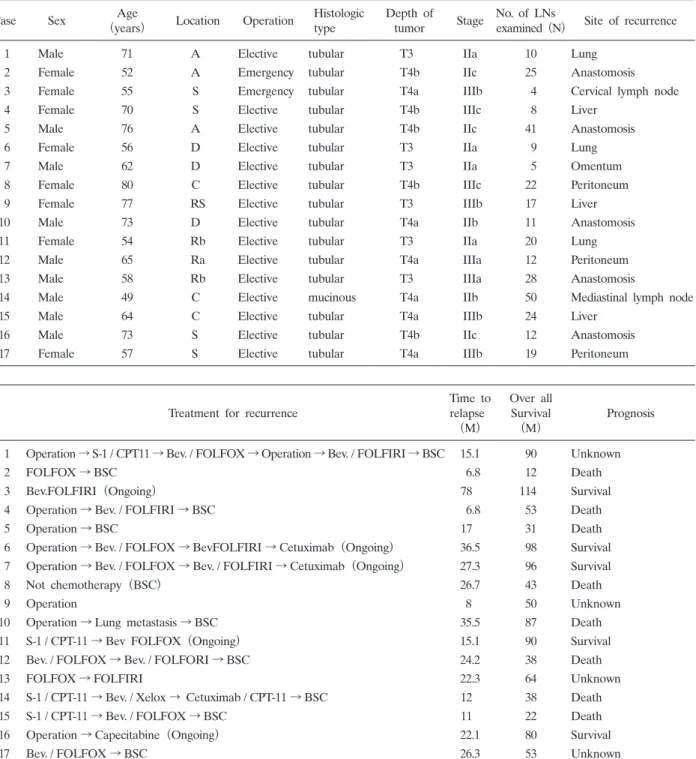
Table 2. Characteristics of patients with disease recurrence
(median follow-up period, 70 months [range, 9-120 months])
Case Sex Age
(years) Location Operation Histologic type
Depth of
tumor Stage No. of LNs
examined (N) Site of recurrence
1 Male 71 A Elective tubular T3 IIa 10 Lung
2 Female 52 A Emergency tubular T4b IIc 25 Anastomosis
3 Female 55 S Emergency tubular T4a IIIb 4 Cervical lymph node
4 Female 70 S Elective tubular T4b IIIc 8 Liver
5 Male 76 A Elective tubular T4b IIc 41 Anastomosis
6 Female 56 D Elective tubular T3 IIa 9 Lung
7 Male 62 D Elective tubular T3 IIa 5 Omentum
8 Female 80 C Elective tubular T4b IIIc 22 Peritoneum
9 Female 77 RS Elective tubular T3 IIIb 17 Liver
10 Male 73 D Elective tubular T4a IIb 11 Anastomosis
11 Female 54 Rb Elective tubular T3 IIa 20 Lung
12 Male 65 Ra Elective tubular T4a IIIa 12 Peritoneum
13 Male 58 Rb Elective tubular T3 IIIa 28 Anastomosis
14 Male 49 C Elective mucinous T4a IIb 50 Mediastinal lymph node
15 Male 64 C Elective tubular T4a IIIb 24 Liver
16 Male 73 S Elective tubular T4b IIc 12 Anastomosis
17 Female 57 S Elective tubular T4a IIIb 19 Peritoneum
Treatment for recurrence
Time to relapse
(M)
Over all Survival
(M) Prognosis
1 Operation → S-1 / CPT11 → Bev. / FOLFOX → Operation → Bev. / FOLFIRI → BSC 15.1 90 Unknown
2 FOLFOX → BSC 6.8 12 Death
3 Bev.FOLFIRI(Ongoing) 78 114 Survival
4 Operation → Bev. / FOLFIRI → BSC 6.8 53 Death
5 Operation → BSC 17 31 Death
6 Operation → Bev. / FOLFOX → BevFOLFIRI → Cetuximab(Ongoing) 36.5 98 Survival 7 Operation → Bev. / FOLFOX → Bev. / FOLFIRI → Cetuximab(Ongoing) 27.3 96 Survival
8 Not chemotherapy(BSC) 26.7 43 Death
9 Operation 8 50 Unknown
10 Operation → Lung metastasis → BSC 35.5 87 Death
11 S-1 / CPT-11 → Bev FOLFOX (Ongoing) 15.1 90 Survival
12 Bev. / FOLFOX → Bev. / FOLFORI → BSC 24.2 38 Death
13 FOLFOX → FOLFIRI 22.3 64 Unknown
14 S-1 / CPT-11 → Bev. / Xelox → Cetuximab / CPT-11 → BSC 12 38 Death
15 S-1 / CPT-11 → Bev. / FOLFOX → BSC 11 22 Death
16 Operation → Capecitabine (Ongoing) 22.1 80 Survival
17 Bev. / FOLFOX → BSC 26.3 53 Unknown
A Ascending colon, S Sigmoid colon, D Descending colon, RS Rectosigmoid, Ra Rectum (above the peritoneal reflection) , Rb
Rectum (below the peritoneal reflection) , M Months, FOLFOX Oxaliplatin plus infusional leucovorin and fluorouracil, FOLFIRI
CPT-11 plus infusional leucovorin and fluorouracil, Bev. Bevacizumab, BSC Best supportive care
Prognostic factors for recurrence
Results of the univariate and multivariate analyses using the Cox proportional hazard model to identify significant prognostic factors for recurrence are presented in Table 3. In the univariate analysis, five variables were identified as significant prognostic factors for recurrence affecting DFS : preoperative CA19-9 level > 37 U / ml (hazard ratio [HR] , 6.520 ; 95% CI, 1.961—19.650 ; p = 0.0036) , emergency operation (HR, 8.978 ; 95% CI, 1.404—32.284 ; p =0.025) , D1 LN dissection
(HR, 6.977 ; 95% CI, 1.093—25.034 ; p = 0.042) , T4 lesions (HR, 7.553 ; 95% CI, 2.860—22.015 ; p <0.0001) , and >3 LN metastases (HR, 5.661 ; 95% CI, 1.296—17.570 ; p = 0.025) . In the multivariate analysis, preoperative CA19-9 level > 37 U / ml (HR, 7.826 ; 95% CI, 1.562—33.271 ; p = 0.016) , emergency operation (HR, 3.560e +9 ; 95% CI, 1.323—1.164e + 20 ; p = 0.038) , and T4 lesions (HR, 5.571 ; 95% CI, 1.472—22.184 ; p = 0.012) were independent significant prognostic factors after treatment. Both DFS and OS were significantly different according to the number of independent prognostic factors for recurrence (number of prognostic factors 0 vs 1 : HR,
Fig. 1. Disease-free survival (DFS) and overall survival (OS) curves for all patients and for patients in Stage Ⅱ and Ⅲ .
(a) DFS curve for all patients ; the 5-year DFS rate was 82.2%.
(b) OS curve for all patients ; the 5-year OS rate was 91.9%.
(c) DFS curves for Stage Ⅱ and Stage Ⅲ patients ; the 5-year DFS rates for Stage Ⅱ and Stage Ⅲ patients were 82.5% and 81.9%, respectively. There was no significant difference in 5-year DFS between Stage Ⅱ and Stage Ⅲ patients (log-rank test ; p = 0.696) .
(d) OS curves for Stage Ⅱ and Stage Ⅲ patients ; the 5-year OS rates for Stage Ⅱ and
Stage Ⅲ patients were 94.7% and 89.1%, respectively. There was no significant
difference in 5-year OS between Stage Ⅱ and Stage Ⅲ patients (log-rank test ; p =
0.674) .
Table 3. Univariate and multivariate analyses using the Cox proportional hazard model to identify significant prognostic factors for recurrence
Univariate analysis Multivariate analysis Factor Hazard ratio (95% CI) P value Hazard ratio (95% CI) P value Age (years)
> 65 0.639 (0.232-1.667) 0.36
65 1
Sex
Male 0.584 (0.223-1.556) 0.274
Female 1
Preoperative CEA level (ng / ml)
> 5.1 1.312 (0.422-3.952) 0.627
5.1 1
Preoperative CA19-9 level (U / ml)
> 37 6.520 (1.961-19.650) 0.0036 11.038 (2.517-47.262) 0.0025
37 1 1
Operation
Emergency 8.978 (1.404-32.284) 0.025 15.002 (1.732-101.219) 0.017
Elective 1 1
Tumor location
Colon 2.561 (0.905-9.102) 0.078
Rectum 1
Surgical approach
Laparotomy 1.792 (0.663-6.640) 0.258
Laparoscopic 1
Tumor size
> 50 0.783 (0.284-2.040) 0.618
50 1
LN dissection
D1 6.977 (1.093-25.034) 0.042 2.058 (0.088-23.763) 0.592
D2, D3 1 1
Histologic types
Mucinous, poorly 1.695 (0.266-6.070) 0.513
Tubular, Papirally 1
Depth of tumor
T4 7.553 (2.860-22.015) < 0.0001 4.956 (1.172-21.230) 0.03
T3 1 1
Lymphatic invasion
Positive 1.053 (0.297-2.977) 0.928
Negative 1
Venous invasion
Positive 1.539 (0.434-9.767) 0.545
Negative 1
No. of LNs examined
> 13 1.952 (0.733-5.107) 0.175
13 1
No. of LN metastases
> 3 5.661 (1.296-17.570) 0.025 4.101 (0.572-19.610) 0.14
3 1 1
Stage
Ⅱ 1.208 (0.453-3.162) 0.698
Ⅲ 1
Postoperative complications
+ 1.948 (0.676-5.131) 0.207
1
CEA Carcinoembryonic antigen, CA19-9 Carbohydrate antigen 19-9
6.976 ; 95% CI, 2.580—21.973 ; log-rank test, p < 0.0001 ; Wilcoxon test, p < 0.0001) (Fig. 2) . Discussion
To our knowledge, this is the first report on the analysis of prognostic factors for recurrence after UFT / LV adjuvant chemotherapy for patients with CRC. In this study, common sites of recurrence after UFT / LV adjuvant chemotherapy included the anastomosis (29.4% [5 / 17]) , and other local sites, the liver and the lungs (17.6% [3 / 17] each) . According to the JSCCR, the recurrence rates after curative resection are 2.4% for the anastomosis, 23.1% for other local
Fig. 2. Disease-free survival (DFS) and overall survival (OS)
curves based on the number of prognostic factors for recurrence.
*(a) DFS curves based on the number of prognostic factors for recurrence
(b) OS curves based on the number of prognostic factors for recurrence
*