INTRODUCTION
Vasculitis is defined as inflammation and necrosis with leukocytic infiltration of the vessel wall. Reac-tive pathological damage to the wall and surrounding tissues is also seen (1). Some patients with vasculitis present with non-specific symptoms including fever, weight loss and elevated erythrocyte sedimentation rate (ESR) or C-reactive protein (CRP). Tradition-ally, nuclear medicine investigation of a patient with fever or inflammatory reaction of unknown focus has been done by67Ga-citrate scintigraphy. It is known
that18F-fluorodeoxy glucose (FDG) accumulates in
both malignant cells and inflammatory tissue. It is also reported that FDG-PET is superior to67Ga-citrate
scintigraphy in detecting an inflammatory focus (2). As FDG accumulates in normal organs such as the brain, myocardium, liver and bowel, the lack of a spe-cific anatomical location sometimes leads to equivo-cal PET findings. Within the last 5 years, PET/CT has almost replaced PET-alone system. PET/CT
sys-tem can produce metabolic images and anatomic im-ages in a single session with a good coregistration. PET/CT improves the anatomic localization of ab-normalities identified by PET and reduces the num-ber of false-positive studies by accurate identification of the physiologic accumulation of FDG in normal organs.
We report three patients with vasculitis demon-strating abnormal FDG uptake in the arterial wall with FDG-PET/CT.
FDG-PET/CT protocol and image interpretation The patients were required to fast for 6 hours and avoid strenuous work or exercise for 24 hours before the PET/CT scan. After intravenous admini-stration of 3.7 MBq/kg FDG, the patients were asked to stay on bed rest in a separated booth. All patients were examined with a PET/CT scanner (Aquiduo, Toshiba, Japan) 1 hour after FDG injection. They were imaged from the top of the head to the mid-dle of thigh. Imaging time was approximately 20 min. The attenuation-corrected PET image, non-attenuation-corrected PET image and CT image were reviewed, and the attenuation-corrected PET image and CT image were coregistered using Aqueri-asNET viewer (TeraRecon, Inc).
CASE REPORT
FDG-PET/CT for diagnosis and follow-up of vasculitis
Hideki Otsuka, Naomi Morita, Kyo Yamashita, and Hiromu Nishitani
Department of Radiology, Institute of Health Biosciences, The University of Tokushima Graduate School, Tokushima, Japan
Abstract : We report three cases of vasculitis evaluated by FDG-PET/CT. Vasculitis is de-fined as inflammatory changes and necrosis in the aterial wall. The patients presented with non-specific symptoms such as fever up or elevated inflammatory markers. FDG-PET/CT clearly demonstrated intense FDG uptake in vessel walls. A 72-year-old female patient with a one month history of pyrexia had abnormal laboratory data suggesting an inflammatory process. FDG-PET/CT was very useful for the diagnosis of vasculitis. Steroid therapy was introduced. Normalization of laboratory data and symptomatic improvement correlated with normalization of FDG uptake in the vessels. J. Med. Invest. 54 : 345-349, August, 2007 Keywords : FDG-PET/CT, vasculitis, arteritis, inflammation, therapy monitoring
Received for publication April 16, 2007 ; accepted May 18, 2007. Address correspondence and reprint requests to Hideki Otsuka, Department of Radiology, Institute of Health Biosciences, The Uni-versity of Tokushima Graduate School, Kuramoto-cho, Tokushima 770-8503, Japan and Fax : +81-88-633-7174.
CASE REPORTS
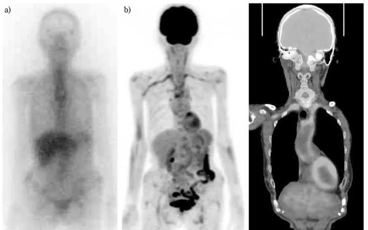
Case 1 (Fig. 1)A 77-year-old female with heart failure was ad-mitted to our hospital. She had low blood pressure in her upper extremities bilaterally and elevated CRP (4.1 mg/dl, normal!0.3 mg/dl).67Ga-citrate
scin-tigraphy did not show any abnormal finding. FDG-PET/CT scan demonstrated intense FDG uptake in the walls of the thoracic aorta, abdominal aorta, carotid arteries and iliac arteries. Contrast-enhanced CT images showed thickened vascular walls. Stenosis of the subclavicular arteries due to thickened wall caused the low blood pressure in her arms. Large-vessel vasculitis was confirmed by laboratory data and FDG-PET/CT and CT findings. Steroid therapy was started. On follow up CT, thickened wall remained and inflammatory markers were unstable.
Case 2 (Fig. 2)
A 72-year-old female presented with a one month history of fever. Laboratory tests showed elevated C-reactive protein (CRP) of 12.63 mg/dl and
eryth-rocyte sedimentation rate (ESR) of 40 mm/h (nor-mal range 3-15 mm/h). The diagnosis of vasculitis was confirmed by PET/CT and 30 mg/day of pred-nisolone (PSL) was started. One month later, symp-toms had improved and abnormal uptake in the ar-terial wall disappeared on repeated FDG-PET/CT. Case 3 (Fig. 3)
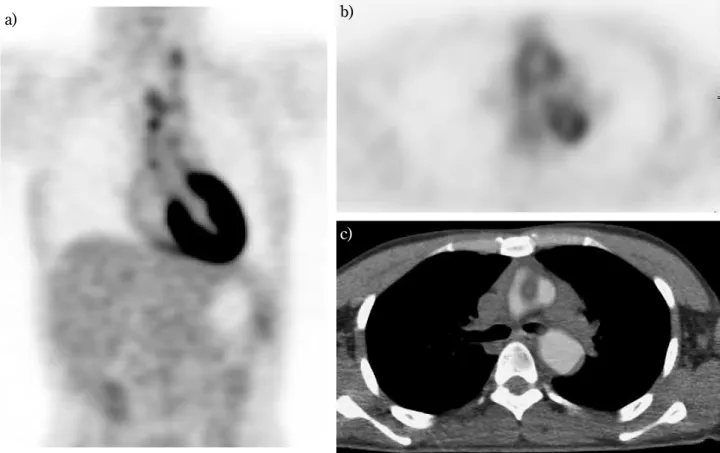
A 32-year-old female was referred with an 8-month of fever of unknown origin (FUO). She presented with general fatigue and elevated inflammatory mark-ers (CRP 5.1 mg/dl, ESR 70mm/h). FDG-PET/CT showed increased FDG uptake in the thoracic aorta and its branches (brachiocephalic artery and carotid arteries), which suggested vasculitis and contrast-enhanced CT was recommended. Contrast-contrast-enhanced CT demonstrated thickened arterial wall, correspond-ing to intense FDG uptake. These clinical findcorrespond-ings confirmed the diagnosis of vasculitis. Her symptoms improved and laboratory data normalized with the administration of steroids. On follow up CT, no re-markable change was seen in thickened arterial wall.
a) b) c)
Fig. 1. A 77-year-old female. a)67Ga-citrate scintigraphy shows no abnormal uptake. b) MIP image of FDG-PET. c) MPR image of
PET/CT. FDG-PET/CT scan demonstrated intense FDG uptake in the walls of the thoracic aorta, abdominal aorta, carotid arteries and iliac arteries.
a) b)
Fig. 2. A 72-year-old female. a) FDG-PET image. The diagnosis of vasculitis is confirmed. b) FDG-PET image after steroid ther-apy. Abnormal uptake in the arterial wall disappeared on repeated FDG-PET.
b) a)
c)
Fig. 3. A 32-year-old female. a) Coronal view of FDG-PET. b) Axial view of FDG-PET. c) Axial view of PET/CT image. FDG-PET/ CT shows increased FDG uptake in the thoracic aorta and its branches (brachiocephalic artery and carotid arteries)
DISCUSSION
Vasculitis is classified into three types according to vessel size. The common large-vessel vasculitides are giant cell arteritis (GCA) and Takayasu disease. Large-vessel vasculitis is considered to be the cause in about 17% of all FUO patients (3, 4).67Ga-citrate had
been considered to be a good tracer to investigate these patients, but the spatial resolution of67Ga
scin-tigraphy is not sufficient. One of our patient was ex-amined by67Ga scintigraphy, which showed no
ab-normal uptake in the arterial walls. Blockmans first reported the use of FDG-PET to investigate GCA patients (5). FDG accumulates in inflammatory tis-sue due to the over-expression of glucose transporter (GLUT, mainly GLUT-1, 3) and over-production of glycolytic enzymes in inflammatory tissue. FDG-PET is highly effective in detecting vasculitis any-where in the body and has high sensitivity (77-100%) and specificity (89-100%) (6-10). If using PET-alone system, FDG uptake are often considered to be equivocal because of the lack of anatomical local-ization. There have been some literatures that re-ported coregistration of PET and contrast-enhanced CT images with image fusion software (8, 11). In-tense FDG uptake clearly correlated with thickened and well-enhanced arterial wall on CT. Meller, et al. reported that FDG-PET images identified more vas-cular regions than MRI did (12). The misregistration of metabolic and anatomic images is sometimes prob-lematic. In our cases, we could evaluate both the ac-tivity of arterial inflammation and anatomical local-ization with minimal misregistration using PET/CT system.
Assessment of the response to therapy has a cen-tral role in patient management. For the patients with vasculitis, ESR or CRP is commonly used to assess response to steroid therapy. CT and MRI fre-quently show residual abnormal findings even after symptoms have completely resolved and sometimes show discrepancies with laboratory data. FDG cumulation in the arterial wall reflects vasculitis ac-tivity, and correlates well with blood inflammatory markers. In our follow up PET/CT examination, normalization of FDG uptake clearly correlated with clinical improvement and normalization of labora-tory findings. Some patients with vasculitis have non-specific symptoms and proper diagnosis at an early stage with PET/CT allows early treatment, which will lead to a better patient outcome.
PET/CT has the potential to diagnose and monitor the response to therapy both in malignant tumors
and inflammatory diseases by integrating anatomic and metabolic images.
REFERENCES
1. Johnston SL, Lock RJ, Gomples MM : Takayasu arteritis : a review. J Clin Pathol 55 : 481-486, 2002
2. Meller J, Altenvoerde G, Munzel U : Fever of un-known origin : prospective comparison of [18F]
FDG imaging with a double-head coincidence camera [DHCC] and Ga-67 citrate SPECT. Eur J Nucl Med 27 : 1617-1625, 2002
3. Vanderschueren S, Knockaert DC, Adriaens-sens T : From prolonged febrile illness to fever unknown origin : the challenge continues. Arch Intern Med 12 : 163 : 1033-1041, 2003
4. Knockaert DC, Vanderschueren S, Blockmans D : Fever of unknown origin in adults : 40 years on. J Intern Med 252 : 263-275, 2003
5. Blockmans D, Maes A, Stroobants S : New ar-guments for a vasculitic nature of polymyalgia rheumatica using positron emission tomogra-phy. Rheumatology 38 : 444-447, 1999
6. Bleeker-Rovers CP, Bredie SJ, van der Meer JW, Corstens FH, Oyen WJ : F-18-fluorodeoxyglucose positron emission tomography in diagnosis and follow-up of patients with different types of vas-culitis. Neth J Med 61 : 323-329, 2003
7. Webb M, Chambers A, Al-Nahhas A : The role of F-18-FDG-PET in characterizing disease ac-tivity in Takayasu arteritis. Eur J Nucl Med Mol Imaging 31 : 627-634, 2004
8. Kobayashi Y, Ishii K, Oda K, Narian T, Tanaka Y, Ishikawa K : Aortic wall inflammation due to Takayasu arteritis imaged with 18F-FDG PET coregistered with enhanced CT. J Nucl Med 46 : 917-922, 2005
9. Walter MA, Melzer RA, Schindler C, Muller-Brand J, Tyndall A, Nitzsche EU : The value of [18F]FDG-PET in the diagnosis of large vessel vasculitis and the assessment of activity and extent of disease. Eur J Nucl Med Mol Imag-ing 32 : 674-681, 2005
10. Blockmans D, de Ceuninck L, Vanderschueren S, Knockaert D, Mortelmans L, Bobbaers H : Repetive18F-fluorodeoxyglucose positron
emis-sion tomography in giant cell arteritis : a pro-spective study of 35 patients, Arthritis Rheum 55 : 131-137, 2006
Ohtomo K : Abnormal accumulation of [18F] fluorodeoxyglucose in the aortic wall related to inflammatory changes : three case reports. Ann Nucl Med 20 : 361-364, 2006
12. Meller J, Strutz F, Siefker U, Scheel A, Sahlmann CO, Lehmann K : Early diagnosis and follow-up of aortitis with [18F]FDG PET and MRI. Eur J Nucl Med 30 : 730-736, 2003