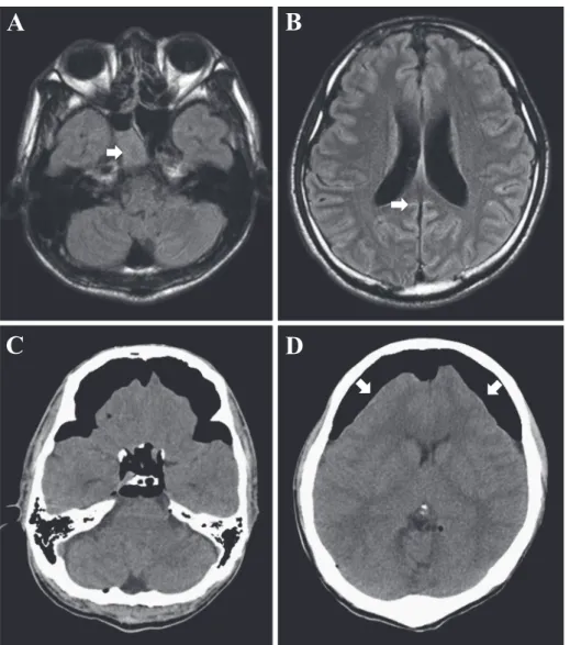
53:478 はじめに 細菌性髄膜炎は中枢性感染症の中でも死亡率も高く,また 脳浮腫や脳ヘルニア,脳血管障害,水頭症,聴力障害などの 脳神経障害,脳膿瘍など合併症や後遺症の割合も高く,重篤 な疾患である1).合併症として,気脳症をともなうこともあり, さらに緊張性気脳症が出現することもある2)3).緊張性気脳 症は,一般には緊急な外科的処置を要する事も多いため,早 期の診断と対応が必要な疾患である4).緊張性気脳症の診断 は,合併する神経症状と画像診断が必要であり,“Mount Fuji sign” と名称された非常に特徴ある画像所見が報告されてい る5)∼7).しかしながら,この特徴ある所見に関しては神経内 科医の間で周知されているとはいえない.われわれは,蝶形 骨洞炎から細菌性髄膜炎を併発し,経過中に緊張性気脳症に いたったまれな 1 例を経験したので報告する. 症 例 症例:39 歳,男性 主訴:頭痛・嘔吐 既往歴:特記事項なし. 家族歴:特記事項なし. 現病歴:頭痛が出現し軽快せず,第 3 病日近医受診し, MRI 検査で蝶形骨洞の副鼻腔炎を指摘された.第 5 病日より, 39C の発熱,頭痛,嘔吐が加わり,他院受診し,抗生剤内服, 解熱鎮痛剤内服処方された.第 6 病日に症状軽快しないため, 当院紹介入院となった.受診時,36.9C で意識は清明,頭痛, 嘔気,嘔吐あり,神経学的には,項部硬直をみとめ,Kernig 徴候陽性,Laseague 徴候陽性であったが,その他,神経学 的には異常をみとめなかった.血液検査では WBC 28,890/mm3, CRP 32.6mg/dl,プロカルシトニン 10 mg/ml(基準値 0.5 未満) と炎症所見をみとめた.脳脊髄液検査では細胞数 10,186 mm3 (多核球 91%),蛋白 240 mg/dl,糖 21.0 mg/dl(同時血糖 147 mg/dl)で細菌性髄膜炎の所見であった.しかし,後日報告 された,血液培養,髄液培養では起炎菌は検出されなかった. 頭部 MRI では蝶形骨洞内に膿の貯留および副鼻腔炎の所見 を呈し(Fig. 1A),FLAIR 画像では両側頭頂葉内側の脳溝に 髄液高蛋白を反映した高信号をみとめた(Fig. 1B).以上より 蝶形骨洞炎に続発した細菌性髄膜炎と判断し,メロペネム 6 g/ 日とデカドロン 9.9 mg/ 日で加療開始した.開始後臨床 所見はすみやかに治療に反応し,頭痛,発熱は消失,検査所 見も順調に改善した.しかし入院 4 日目(第 10 病日),意識 レベルの低下,不全対麻痺が出現し,起立,歩行不能となっ た.頭部 CT では非常に特徴ある “Mount Fuji sign” をみとめ, 緊張性気脳症が推測された(Fig. 1C, D).瘻孔の存在が推測 され,検討したが CT 画像上は瘻孔の確認はできなかったし, また髄液漏もみとめられなかった.外科的処置による治療を 考慮したが,髄膜炎そのものは改善しつつあり,注意深く観 察しながら,安静臥床と充分な補液をおこない,抗菌薬投与 を継続したところ,しだいに症状と頭部画像所見は改善した. 一時,少量の硬膜下血腫が合併したが,気脳症の改善ととも
短 報
細菌性髄膜炎の経過中に“Mount Fuji sign”画像所見を呈した
緊張性気脳症の 1 例
姫野 隆洋
1)竹島 慎一
1)久保 智司
1)原 直之
1)高松 和弘
1)栗山 勝
1)*
要旨: 症例は 39 歳男性である.蝶形骨洞炎から波及し細菌性髄膜炎を発症した.抗菌薬で順調に経過してい たが,意識障害,対麻痺が出現し,頭部 CT で Mount Fuji sign をみとめ,緊張性気脳症の合併と診断した.髄膜炎 は順調な経過なので,外科的処置はせず,保存的に治療し治癒した.緊張性気脳症の発生機序は,蝶形骨洞から 脳内への瘻孔,髄液採取時の一時的な髄液圧低下,外気流入とチェックバルブ機構などを推定したが詳細な機序 は不明.気脳症は細菌性髄膜炎の合併症の一つであるが,蝶形骨洞炎からの細菌性髄膜炎では,気脳症の合併に 注意が必要である.緊張性気脳症の診断での特徴的な Mount Fuji sign の重要性を強調した.
(臨床神経 2013;53:478-481)
Key words: 細菌性髄膜炎,緊張性気脳症,蝶形骨洞炎,Mount Fuji sign
*Corresponding author: 脳神経センター大田記念病院脳神経内科〔〒 720-0825 広島県福山市沖野上町 3 丁目 6-28〕
1)
脳神経センター大田記念病院脳神経内科 (受付日:2012 年 11 月 8 日)
細菌性髄膜炎の経過中に“Mount Fuji sign”画像所見を呈した緊張性気脳症の 1 例 53:479 に消失した.その後起立歩行開始し,第 66 病日には後遺症 なく独歩退院となった. 考 察 細菌性髄膜炎をふくむ鼻性頭蓋内合併症の発症原因として は,①急性副鼻腔炎や慢性副鼻腔炎の急性増悪に引き続いて おこる自然発症,②副鼻腔や頭蓋底の手術後に発症する術後 発症,③交通事故後などの外傷後発症の三つに分類されてい る.近年,術後発症や,外傷後発症は減少してきており,自 然発症が多いと報告されている8).また責任病巣に関しては, 副鼻腔炎そのものが前頭洞および櫛骨洞が多く,蝶形骨洞炎 はまれであるため9),蝶形骨洞炎からの髄膜炎にいたる例の 頻度は少ない8).本例は手術歴や外傷歴はなく,蝶形骨洞炎 が脳内へ穿破したことによる自然発症型の続発性細菌性髄膜 炎と考えられた.起炎菌としては連鎖球菌,ブドウ球菌が検 出される症例が多いが,本例のように,培養陰性の例も 25%にみとめられている8).本例では,前医で蝶形骨洞炎を 指摘され,抗菌薬内服処方されており,そのため無菌化して いた可能性もある.
Fig. 1 Brain MRI and CT findings.
A, B (Brain MRI on admission): A; T1 weighted axial image (Axial, TR 8,500 ms, TE 91 ms) demonstrated
sinusitis with accumulation of fluid (arrow) in the right sphenoid sinus. B; Axial fluid attenuated inversion recovery (FLAIR) image demonstrated high intensity areas in cerebral sulci of bilateral parietal lobes. These findings were resulted from bacterial meningitis. C, D (Brain CT on the tenth hospital day) Axial CT showed revealed pneumocephalus with compression of frontal lobes and the widening of the interhemispheric space between the tips of the frontal lobes. These findings were known as “Mount Fuji sign” (arrows), which was a important sign of “tension” pneumocephalus.
臨床神経学 53 巻 6 号(2013:6) 53:480
ところで本例では治療経過中,第 10 病日に高度気脳症を 合併したが,この際,軽度の意識障害,不全対麻痺症状をと もない持続するため,緊張性気脳症の状態が考えられた.画 像所見でも緊張性気脳症に特徴的とされる Mount Fuji sign5)∼7) と呼ばれる所見をみとめたため,緊張性気脳症が確認された. この画像所見は 1988 年,Ishiwata らより最初に指摘され,硬 膜下腔の空気による両側前頭葉の高度圧迫所見と左右前頭極 間の拡大が生じ,その画像が富士山のシルエットに類似する ことから名称されたユニークな画像所見である10).外傷後 や脳外科術後に出現したばあい,緊急で除圧させる必要があ り,その画像所見の意義が論じられている5)∼7)10).本症では, 基礎疾患が細菌性髄膜炎であり,抗菌薬により良好に反応し ていたため,外科的処置はおこなわず,内科的治療のみで加 療したところ徐々に改善し治癒した. 細菌性髄膜炎の合併症としては脳膿瘍や,脳血管障害,水 頭症などがよく知られており,気脳症も生ずるが,本例のよ うに鼻性頭蓋内合併症としての緊張性気脳症を呈した例は, 文献上では糖尿病合併の 11 歳女児例が報告されているのみ である2).髄膜炎による緊張性気脳症の併発に関しては,ガ ス産生菌の感染のみならず,肺炎球菌や連鎖球菌髄膜炎によ り発症した例が報告3)されているが,本例では起炎菌の確 定はできなかった.蝶形骨洞炎による瘻孔形成の可能性,髄 液採取での一時的髄液圧の低下,空気圧の不均衡で外気の流 入,チェックバルブ機構による流入した外気の貯留などが推 定されたが,詳細な機序は不明である.また蝶形骨洞の副鼻 腔炎の頻度は少ないが,構造上骨破壊がおこりやすいとされ, 頭蓋内への瘻孔形成が生じやすいことも関連するものと考え られた. 本報告の要旨は,第 93 回日本神経学会中国・四国地方会で発表し, 会長推薦演題に選ばれた. ※本論文に関連し,開示すべき COI 状態にある企業,組織,団体 はいずれも有りません. 文 献
1) Pfister H, Roos KL. Bacterial meningitis. In: Roos KL, editor. Principles of neurologic infectious diseases. New York: McGraw-Hill; 2005. p. 13-28.
2) Kaur S, Seth A, Narula MK. Pneumocephalus: A rare complication of meningitis. Indian J Pediatr 2012; 11 May. EPub 3) Ohe Y, Maruyama H, Deguchi I, et al. An adult case of
peumocephalus and peumococcal meningitis associated with the sphenoid sinusitis. Intern Med 2012;51:1129-1131.
4) Prüss H, Klingebiel R, Endres M. Tension pneumocephalus with diplegia and deterioration of consciousness. Case Rep Neurol 2011;3:48-49.
5) Michel SJ. The Mount Fuji sign. Radiology 2004;232:449-450. 6) Beiko J, McDonald P. Tension pneumocephalus—the Mount
Fuji sign. Can J Neurol Sci 2005;32:538-539.
7) Sadeghian H. Mount Fuji sign in tension pneumocephalus. Arch Neurol 2000;57:1366.
8) 北村剛一,大塚康司,高田大輔ら.鼻性頭蓋内合併症例. 日鼻誌 2006;45:172-176.
9) Tan HK, Ong YK. Acute isolated sphenoid sinusitis. Ann Acad Med Singapore 2004;33:656-659.
10) Ishiwata Y, Fujitsu K, Sekino T, et al. Subdural tension pneumocephalus following surgery for chronic subdural hematoma. J Neurosurg 1988;68:58-61.
細菌性髄膜炎の経過中に“Mount Fuji sign”画像所見を呈した緊張性気脳症の 1 例 53:481
Abstract
Tension pneumocephalus complicated from bacterial meningitis
—A report of case presenting “Mount Fuji sign” in brain CT—
Takahiro Himeno, M.D.
1), Shinnichi Takeshima, M.D.
1), Satoshi Kubo, M.D.
1),
Naoyuki Hara, M.D.
1), Kazuhiro Takamatsu, M.D.
1)and Masaru Kuriyama, M.D. Ph.D.
1)1)Department of Neurology, Brain Attack Center Ota Memorial Hospital