Screening of Edible Plants for Urease
Inhibitors and Chemical Structures of the
Inhibitors
著者
Shabana Samah Ahmed Sabry Abdel Hamid
内容記述
学位授与大学: Osaka Prefecture University(大阪
府立大学), 学位の種類: 博士(応用生命科学), 学
位記番号: 論生命第28号, 学位授与年月日:
2010-03-31, 指導教員: 林英雄.
大阪府立大学博士 (応用生命科学) 学位論文
Screening of Edible Plants for Urease Inhibitors
and Chemical Structures of the Inhibitors
食用植物を対象としたウレアーゼ阻害物質の検索と
その化学構造
Shabana Samah Ahmed Sabry Abdel Hamid
2010
Screening of Edible Plants for Urease Inhibitors
and Chemical Structures of the Inhibitors
by
Shabana Samah Ahmed Sabry Abdel Hamid
Thesis submitted for the partial fulfillment
of the requirment for the degree of
Doctor of Philosophy
in Applied Life Sciences
Division of Applied Life Sciences
Graduate School of Life and Environmental Sciences
Osaka Prefecture Universiy
1-1 Gakuen-cho, Sakai, Osaka 599-8531, Japan
2010
i
Contents
Contents i
List of Figures iii
List of Tables v
List of
Abbreviation
vi
Chapter 1 Introduction 1
Chapter 2 Screening of the edible plants, seaweeds, and foodstuffs for inhibitory activity against Jack bean urease 5
2.1. Introduction 5
2.2. Samples of edible plants used in screening 7
2.3. Result of screening 9
2.3.1. Preliminary screening results 9
2.4. Experimental section 11
2.4.1. Sample preparation 11
2.4.2. Preparation of Jack bean urease enzyme 11
2.4.3. Urease inhibitory assay 12
Chapter 3 Isolation of urease inhibitor from Allium cepa (onion) 13
3.1. Introduction 13
3.2. Isolation of compound 1 (quercetin
4`-O-β-D-glucopyranoside) 15
3.3. Identification of active compound 1 17
3.4. Urease inhibitory activity of compound 1 18
ii
Chapter 4 Isolation of phenolic urease inhibitors from Psidium guajava
(guava leaves) 25
4.1. Introduction 25
4.2. Isolation of active compounds 2, 3 and 4 from Psidium
guajava 27
4.3. Identification of active compound 2 29
4.4. Identification of active compound 3 34
4.5. Identification of active compound 4 38
4.6. Urease inhibitory activity of compounds 2-4 41
4.7. Experimental section 42
Chapter 5 Urease inhibitory activity of quercetin glycosides 44 5.1. Biological activity of quercetin glycosides 44 5.2. Urease inhibitory activity of quercetin glycosides 44
5.3. Experimental section 47
Chapter 6 Terpenoid urease inhibitors isolated from Psidium gujava 49
6.1. Introduction 49
6.2. Isolation of compound 5 49
6.3. Identification of active compound 5 51
6.4. Isolation of compound 6 58
6.5. Identification of compound 6 58
6.6. Urease inhibitory activity of compounds 5 and 6 63
6.7. Experimental section 64 Summary 66 Reference Acknowledgment Publication 68 75 76
iii
List of Figures
Fig. 1.1. Some synthetic urease inhibitors 3
Fig. 1.2. Some natural urease inhibitors 4
Fig. 3.2.1. Isolation procedure of active compound 1 from bulbs of
Allium cepa 16
Fig. 3.3.1. 1H-NMR spectrum of compound 1 (400 MHz, CD3OD) 19
Fig. 3.3.2. 13C-NMR spectrum of compound 1 (100 MHz, CD3OD) 19
Fig. 3.3.3. Correlation between anomeric proton H-1``and C-4` in
HMBC spectrum of compound 1 21
Fig. 3.3.4. Comparison of sugar moiety of compound 1 with authentic
sugars on TLC 21
Fig. 3.3.5. Structure of compound 1 22
Fig. 3.4.1. Inhibitory activity of compound 1 22 Fig. 4.2.1. Isolation procedure of phenolic urease inhibitors from leaves
of Psidium guajava 28
Fig. 4.3.1. Structures of compounds 2-4 31
Fig. 4.3.2. 1H-NMR spectrum of compound 2 (400 MHz, CD3OD) 33
Fig. 4.3.3. 13C-NMR spectrum of compound 2 (100 MHz, CD3OD) 33
Fig. 4.4.1. 1H-NMR spectrum of compound 3 (400 MHz, CD3OD) 37
Fig. 4.4.2. 13C-NMR spectrum of compound 3 (100 MHz, CD3OD) 37
Fig. 4.5.1. 1H-NMR spectrum of compound 4 (400 MHz, acetone-d6) 40
Fig. 4.5.2. 13C-NMR spectrum of compound 4 (100 MHz, acetone-d6) 40
Fig. 5.2.1. Structures of quercetin glycosides 48 Fig. 6.2.1. Isolation procedure of compound 5 from leaves of Psidium
guajava 50
Fig. 6.3.1. 1H-NMR spectrum of compound 5 (400 MHz,
acetone-d6) 55
iv
Fig. 6.3.3. Partial structures of compound 5 56
Fig. 6.3.4. 1H-1H COSY and HMBC correlation of compound 5 56 Fig. 6.3.5. Correlation between H-1` to C-5, C-6, C-2`, C-3`, C-8`, C-9`
in HMBC spectrum of compound 5 57
Fig. 6.3.6. Correlation between H-15` to C-2`, C-7` and H-14` to C-3` in
HMBC spectrum of compound 5 57
Fig. 6.3.7. Structure of compound 5 (guajadial) 58 Fig. 6.4.1. Isolation procedure of compound 6 from leaves of Psidium
guajava 60
Fig. 6.5.1. 1H-NMR spectrum of compound 6 (400 MHz, acetone-d6) 62
Fig. 6.5.2. 13C-NMR spectrum of compound 6 (100 MHz, acetone-d6) 62
v
List of Tables
Table 2.2.1. Foodstuff samples examined for urease inhibition 8 Table 2.3.1. Inhibitory activity of methanol extracts against Jack bean
urease 9
Table 2.3.2. Inhibitory activity of ethyl acetate extract, butanol extract
and aqueous residue against Jack bean urease 10
Table 3.1.1. Taxonomic data of Allium cepa 14
Table 3.3.1. 1H-NMR and 13C-NMR data of compound 1 20 Table 4.1.1. Taxonomic data of Psidium guajava 25 Table 4.3.1. 1H-NMRand 13C-NMR data of compound 2 32 Table 4.4.1. 1H-NMRand 13C-NMR data of compound 3 36 Table 4.5.1. 1HNMR and 13C NMR data of compound 4 39 Table 4.6.1. Urease inhibitory activity of compounds 2-4 41 Table 5.2.1. Inhibitory activity of quercetin and its derivatives against
Jack bean and Lactobacillus fermentum ureases 47 Table 6.3.1. 1H-NMRand 13C-NMR data of compound 5 54 Table 6.5.1. 1H-NMRand 13C-NMR data of compound 6 61 Table 6.6.1. Urease inhibitory activity of compounds 5 and 6 63
vi
List of Abbreviations
CC Column chromatography
COSY Correlated Spectroscopy
EI-MS Electron impact mass spectroscopy
FAB-MS Fast atom bombardment mass spectroscopy HMBC Heteronuclear multiple bond connectivity HMQC Heteronuclear multiple quantum coherence ODS Octadesylsilane
1
Chapter 1
Introduction
James B. Sumner crystallized the enzyme urease (urea amidohydrolase EC 3.5.1.5) from Jack bean, Canavalia ensiformis, in 1926; showing for the first time that enzymes
can be crystallized,1) and in 1978 Jack bean urease was shown to contain nickel ions.2) A variety of ureases are found in bacteria, fungi, higher plants, and in soil as a soil
enzyme.3) In the mammalian body, no urease activity has been detected, but urea-splitting bacteria colonizing the human body have urease activity.4)
Medically, bacterial ureases are important virulence factors implicated in the pathogenesis of many clinical conditions, such as pyelonephritis, hepatic coma, peptic
ulceration, and the formation of infection-induced urinary stones.5) In agriculture, high urease activity causes significant environmental and economic problems by releasing
abnormally large amounts of ammonia into the atmosphere during urea fertilization. This further induces plant damage primarily by depriving plants of essential nutrients
and secondly through ammonia toxicity.6) Many urease inhibitors have been described in the past, such as hydroxamic acids, which were found to be potent and specific
inhibitors of urease activities for enzymes of plant and bacterial origin.4) Fluorofamide and hydroxyureas are also urease inhibitors, but some examples could not be used in
vivo because of their toxicity or instability; for example, acetohydroxamic acid was demonstrated to be teratogenic in rats.7) Thus, current efforts are focused on seeking
novel urease inhibitors with good bioavilability and low toxicity among synthetic isoflavones, which showed inhibitory activity against Helicobacter pylori urease.7)
2
Biscoumarins are naturally occurring compounds that can be also synthesized, and
synthetic biscoumarins were reported to show urease inhibitory activity.8) Synthetic cyclic β-triketone, hydroxyl urea, cystamine, and triketone oximes were also reported to
show urease inhibitory activity.9) Many classes of synthetic hetero-compounds were reported as urease inhibitors, such as oxadiazoles, thiadiazoles, and triazoles.10)
Although certain synthetic compounds have shown potential urease inhibition,6-10) no natural product with this activity had been discovered as that point. A diterpene ester
with a myrsinol type skeleton was isolated from Euphorbia decipiens as the first naturally occurring urease inhibitor,11) while two diterpene polyesters were isolated
from this plant later and were reported to show urease inhibitory activity.12) In addition, datisdirin is a flavone isolated from Datisca cannabina that showed Jack bean urease
inhibitory activity.13)
Recently, attention has been focused on exploring the novel biological properties of
phytochemicals isolated from edible plants, because several hundred native plant species are used globally in daily diets as vegetables, fruits, spices, and condiments.
Many of them are also employed for traditional medicines. Edible plants are rich in phytochemicals, such as vitamins, terpenoids, flavonoids, alkaloids, organosulfur
compounds, pigments, and other phenolics, which exhibit various biological activities. Catechins in green tea extract were reported to inhibit H. pylori urease strongly.14) In
addition, a H. pylori urease inhibitor has been isolated from Rubus coreanus Miquel, and characterized as a proteinaceous substance.15) Some of the synthetic and natural
compounds that showed urease inhibitory activity are shown in Fig. 1.1 and Fig. 1.2, respectively.
3
Thus, edible plants may be a safe resource for urease control. From this standpoint,
this study was initiated to isolate urease inhibitory compounds from edible plants and foodstuffs. One hundred and twenty five samples of edible plants, seaweeds, and
foodstuffs were bought in both fresh and dry states through a vegetable store in Sakai city, Osaka, Japan, and a vegetable store in Cairo, Egypt, during April 2007-January
2008. These samples were screened for their inhibitory activity against Jack bean urease using a urease inhibitory assay.
During this work quercetin 4`-O-β-D-glucopyranoside (1) was isolated from Allium
cepa as a urease inhibitor, and three quercetin derivatives (2, 3, and 4) were isolated
from Psidium guajava in addition to one meroterpenoid (5) and ursolic acid (6) as urease inhibitory compounds.
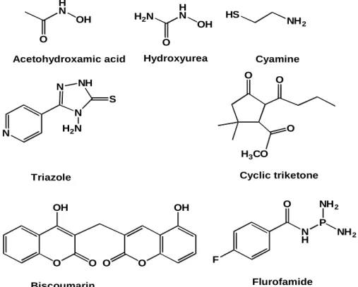
H N O OH H2N HS NH2 H N O OH O O N N NH N S H2N O O OH O O OH N H O F P NH2 NH2 Cyclic triketone Acetohydroxamic acid Hydroxyurea Cyamine
Triazole
Biscoumarin Flurofamide H3CO
O
4 AcO AcO HO HO O H AcO N O O O O HO OH OH O HO OH OH OH OH OH Diterpene ester with myrsinol type skeleton
Gallocatechin Datisdirin Diterpene polyester O O H OAc HO O AcO H OAc OAc
Fig. 1.2. Some natural urease inhibitors
5
Chapter 2
Screening of the edible plants, seaweeds, and foodstuffs for
inhibitory activity against Jack bean urease
2.1. Introduction
Plants have always played a major role in the treatment of human and animal
diseases as a therapeutic resource for traditional medicine. Ancient peoples have used plants to cure a variety of human ailments, and the use of medicinal plants for curing
disease was documented as early as 3000 B.C., with the ancient Egyptians placing great confidence in plants for curing many diseases. The use of herbs to treat disease is
almost universal among non-industrialized societies. A number of traditional areas have come to dominate the practice of herbal medicine at the end of twentieth century,
mostly based on Chinese, Indian, and Greek herbal medicine.
A wealth of plants are used by local populations for the treatment of several
ailments, for example; rosemary (Rosmarinus officinalis) tea to treat asthma, tonsillitis, lack of appetite, gout, and nasal obstruction; blaeberry (Vaccinium myrtillus L.) to treat
elevated triglycerides levels in blood; and maize (Zea mays) to treat bladder inflammations.16) Roselle (Hibiscus sabdariffa) is used in many folk medicines as a
mild laxative, to increase urination, and as a tonic tea to aid digestive and kidney function.17) Some epidemiological studies have suggested that a high consumption of
fruits and vegetables among edible plants of Thailand could help prevent several chronic diseases including cancer.18) Furthermore, a natural product isolated from grape
6
used as medicinal plants, of which most utilize roots, stems, leaves, flowers, buds, or
fruits containing seeds. We most commonly eat the seeds (e.g. maize, wheat, coffee, and various nuts), fruit (e.g. tomato and apple), leaves (e.g. lettuce, spinach, and cabbage),
or roots (e.g. carrot and beets) of edible plants, and we also eat the stem of many plants (e.g. asparagus). There are also a few edible petioles (leaf stalks), such as celery, as well
as some edible flowers.
Edible plants are important sources of minerals, fiber, and vitamins, which provide
essential nutrients for human health. It is well known that edible plants contain secondary metabolites, such as flavonoids, terpenes, alkaloids, glycosides, and others,
which are responsible for the curative action of the plant. For example, the oils of aromatic plants, many of which are used for flavors in the food and beverage industry,
are recognized as having several therapeutic applications, such as anti-inflammatory, antioxidant, cytotoxic, and biocide activities against broad range of organisms.20)
Bioactive compounds in raspberry, strawberry, blueberry, bilberry, and cranberry demonstrated high antimicrobial activity against human pathogens, including
Salmonella, Bacillus cereus, and Staphylococcus.21)
Studies on flavonoids from fruits show their importance as antioxidants, which can
help to prevent problems including cardiovascular disease and diabetes.21) Diets rich in fruits and vegetables are associated with a decreased risk of certain cancers and
cardiovascular diseases, and this has been attributed to the high concentrations of antioxidants in such foods, including ascorbic acid, α-tocopherol, and polyphenolics.22)
When edible plants were screened for antibacterial activity against Streptococcus
mutans, several spices from the Labiatae, Liliaceae, and Zingiberaceae families showed
7
Recently, natural products isolated from edible plants have become of greater
interest due to their availability and limited side-effects or toxicity. From this point of view, we tried to isolate natural urease inhibitors from edible plants and foodstuffs. In
the course of our search, the methanol extracts of edible parts of plants and foodstuffs were screened for their urease inhibitory activity using Jack bean urease.
2.2. Samples of edible plants used in screening
Edible parts of vegetables, fruits, spices, and powders were screened for urease inhibitory activity. The samples used in the present study are shown in Table 2.2.1.
8
Table 2.2.1. Foodstuff samples examined for urease inhibition
Foodstuff samples Vegetables and
fruits
Brassica rapa, Brassica oleracea capitata group, Solanum melongena, Cucumis sativus, Brassica campestris, 3 types of Solanum lycopersicum, Spinacia oleracea, Raphanus sativus var. longipinnatus (root and leaves), Cryptotaenia japonica, Petroselinum crispum, Capsicum annuum (green, yellow and red) Nageia nagi, Abelmoschus esculentus, Phaseolus vulgaris, Chrysanthemum coronarium, Pisum sativum, Brassica rapa var. japonica, Aralia elata, Brassica rapa var. perviridis, Allium tuberosum, Momordica charanita, Amorphophallus konjac, Miscanthus sinensis, Vicia faba, Zingiber officinalis, Zingiber mioga, Allium wakegi, Asparagus officinalis, Brassica oleracea italica group, Sprout of Raphanus sativus, Dioscorea opposite, Dioscorea japonica (Thunb.), Pholiota nameko, Citrus reticulate, Nelumbo nucifera, Pleurotus eryngii, Pteridium aquilinum var. latiusculum, Perilla frutescens, Petasites japonicus, Diospyros kaki, Pyvus communis, Lilium auratum, Arctium lappa, Citrus limon, Citrus sudachi, Citrus unshiu, Citrus aurantium, seeds of Vigna radiate, Vigna radiate
(sprout), Ananas comosus, Malus domestica, Allium cepa, sprout of Vigana
mungo L. (Hepper), Prunus cerasus, Citrullus lanatus, Actinidia deliciosa, Dioscorea batatas, Lens culinaris, Lupinus albus, Carum carvi, Eruca sativa, Foeniculum vulgar, Cymbopogon nardus, Persea Americana, Citrus paradise, Citrus sinensis, Tangor of Citrus reticulate x Citrus sinensis, Solanum tuberosum, Musa acuminate, Benincasa hispda, Cucurbita maxima, Actinidia deliciosa, Triticum aestivum, Brassica Oleracea var. capitata (f.Rubra), Perilla frutescens var. crispa, Daucus carota, Arachis hypogaea, Coriandrum sativum, Pimpinella anisum, Oryza sativa, Zea mays, Allium sativum, Corchorus olitorius, Apium graveolens, Cucurbita pepo, Allium chinense, Allium fistulosum, Diospyros kaki, Pyrus communis, Lilium auratum, Actium lappa, Brassica napus, Auricularia auricula- judae, Raphanus sativus var. longipinnatus, Brassica rapa
var. komatsuna
Spices, tea and powders
Cinnamomum zeylanicum, Psidium guajava,Coffee arabica, Glycyrrhiza glabra, Cuminum cyminum, Rosa odorata, Hibiscus sabdariffa, Matricaria chamomilla,
cymbopogon (lemongrass),chili powder
9 2.3. Result of screening
2.3.1. Preliminary screening results
The edible parts of plant-derived foodstuff samples were extracted with methanol. Preliminary screening on the methanol extracts at a concentration of 500 ppm was performed by measuring the inhibitory activity against Jack bean urease. Jack bean urease has been widely used as an enzyme for inhibitor screening. The screening results shown in Table 2.3.1 indicated that five samples showed high activity against Jack bean urease in the range of 71-100%. These five samples were from mung bean, pineapple, cinnamon, avocado peel, and guava. Ten samples with moderate activity in the range of 31-70% were from apple, red onion, pepper, black gram, licorice, cummin, peel of sudachi, lemon peel, watermelon, and coffee beans, and three samples, cherry, satsuma, and bitter orange showed low activity in the range of 20-30%. Seaweeds such as wakame, hitoegusa, and konbu showed only slight inhibitory activity.
Table 2.3.1. Inhibitory activity of methanol extracts against Jack bean urease
The inhibitory activity of methanol extracts was measured at a concentration of 500 ppm.
No Species names Inhibitory activity (%)
1 Sprout of Vigna radiata 96
2 Ananas comosus 91
3 Cinnamomum zeylanicum 83
4 Psidium guajava 81
5 peel of Persea americana 71
6 Malus domestica 69
7 Allium cepa 51
8 Capsicum annuum 41
9 Vigna mungo (L) Hepper 58 10 Glycyrrhiza glabra 50
11 Cuminum cyminum 40
12 peel of Citrus sudachi 43
13 peel of Citrus limon 38
14 Citrullus lanatus 36
15 Coffea arabica 51
16 Prunus cerasus 27
17 Citrus unshiu 24
10
The methanol extracts of active samples were concentrated and then extracted with
ethyl acetate and butanol, successively. Inhibitory activity of ethyl acetate extract, butanol extract and aqueous residue at a concentration of 1250 ppm was listed in Table
2.3.2.
Table 2.3.2. Inhibitory activity of ethyl acetate extract, butanol extract and aqueous residue against Jack bean urease
The assay results in Table 2.3.2 indicated that the aqueous extract of all active
samples showed high inhibitory activity in the range of 71-98%. BuOH extracts of seven samples (sprout of mung bean, pineapple, cinnamon, guava, apple, onion, and
watermelon) showed high inhibitory activity in the range of 85-99%, and ethyl acetate extracts of three samples (guava, cinnamon, and watermelon) showed high inhibitory
activity in the range of 80-92%.
Although the aqueous residue and butanol extract of the active samples showed
high inhibitory activity they were discarded due to their contents of amino acids, which
Species names Inhibitory activity (%)
EtOAc extract BuOH extract Aqueous extract
Sprout of Vigna radiata 6 99 98
Ananas comosus 69 89 94 Cinnamomum zeylanicum 92 96 93 Psidium guajava 80 90 83 Malus domestica 26 85 98 Allium cepa 24 91 97 Capsicum annuum 64 63 98
peel of Citrus sudachi 37 58 95
peel of Citrus limon 67 54 84
Coffea arabica 0 64 71
Citrullus lanatus 89 99 75
11
may interfere with the assay and give false results as previously demonestrated by
Kawai.24)
From the urease inhibitory assay data and the availability of the raw materials, the
leaves of Psidium guajava and the bulbs of Allium cepa were selected as research materials for further purification and isolation of active urease inhibitors.
2.4. Experimental section 2.4.1. Sample preparation
The edible plants and plant-derived foodstuff samples were bought in a fresh state at a vegetable store in Sakai city, Osaka, Japan during April-August 2007 and from a
store in Qalyubia, northern Cairo, Egypt, in January 2008. Samples were cut into small pieces and immediately soaked in methanol at room temperature for 7 days then filtered.
The filtered samples were evaporated to dryness, dissolved in methanol at 10 mg dry weight/ml, and subjected to the urease inhibitory assay.
2.4.2. Preparation of Jack bean urease enzyme
1- Solution A: Freshly prepared phosphate buffer pH 7.0 (20 mM of KH2PO4 was
adjusted by drop-wise addition of 0.02 M NaOH to pH 7.0). One ml of EDTA (0.5 M) was added to the phosphate buffer solution and then the volume was completed to 100
ml by using milli-Q water.
2- Solution B: 40 µl BSA (bovine serum albumin 5%) was diluted with 960 µl of
solution A to give a BSA concentration of 0.2%.
3- From solution B, 500 µl was added to 49.5 ml of solution A and the new solution was
used to dissolve 7.5 mg of Jack bean urease.
4- The solution of enzyme was transferred into microfuge tubes, (700 µl per tube) and
12 2.4.3. Urease inhibitory assay
Materials
Urease inhibitory assay was carried out on Jack bean urease, which was purchased
from Toyobo, Japan. Amount of ammonia released was measured by UV mini-1240 uv-vis Spectrophotometer (Shimazu Seisakusyo, Japan). Sodium nitroprusside dihydrate,
MES (2-morpholinoethanesulfonic acid) and sodium hypochlorite solution were obtained from Wako pure chemical industries, Japan.
Method
Reaction mixtures composed of 0.2 ml (0.47 U) of Jack bean urease solution and
1.2 ml of buffer (100 mM MES buffer, pH 6.0) were incubated with 0.1 ml of a sample solution at 37○C for 5 min in test tubes. After incubation, 0.5 ml (0.2 mM) of urea was
added to the reaction mixture and incubated for 20 min. Urease activity was determined by measuring ammonia production using the indophenol method described by
Weatherburn.25) One ml each of phenol reagent (1% w/v phenol and 0.005% w/v sodium nitroprusside) and alkali reagent (1% w/v NaOH and 0.075% active chloride
NaOCl) were added to each test tube. The increase in absorbance at 640 nm was measured after 30 min. All reactions were performed in triplicate in a final volume of 4
ml. Percentage inhibitions were calculated using the formula [100 – (OD sample / OD
13
Chapter 3
Isolation of urease inhibitor from Allium cepa (onion)
3.1. Introduction
Allium is the onion genus with 600-750 species, making it one of the largest plant
genera in the world. Allium was classified in family Alliaceae; previously some
botanical authorities have included it in the lily family (Liliaceae).26) Members of the genus include many economically important crops and garden vegetables, including
onions (A. cepa), shallots (A. oschaninii), leeks (A. ampeloprasum), scallions (A.
ascalonicum), and herbs such as garlic (A. sativum). These species produce chemical
compounds (mostly cysteine sulfoxide) that give them a characteristic onion or garlic taste and odor.
A. cepa L. also known as the garden onion or bulb onion is one of the oldest
cultivated plants, and it is now used for both food and medical purposes. Taxonomic
data of A. cepa is shown in Table 3.1.1. Although A. cepa is known only in cultivation, some wild species occur in central Asia, and the most closely related species include
Allium vavailovii Popov & Vved and Allium asarense R.M. Fritsch & Matin from Iran.
As one of the oldest vegetables under cultivation, it is found in large number of recipes
and preparations spanning almost all world cultures. A historical view showed the importance of onions to the ancient world, where its production started in 3000 B.C.,
and it was introduced to Europe by the Phoenicians around 2000 years ago.26, 27) In ancient Egypt, onion was believed to be a sacred food, and it was worshipped by the
14
eternal life.26, 27) Onions were even used in Egyptian burials.26) It was also widely
consumed by the Romans and Greeks, who liked its taste and knew about its curative properties.26, 27) Onions are a rich source of a number of phyto-elements, which make it
an important component of the Mediterranean diet. It is also useful for the treatment or prevention of number of diseases including coronary heart disease, obesity,
hypercholesterolemia, type 2 diabetes, hypertension, cataract, and disturbances of gastrointestinal tract.28) Notably a population-based case control study showed that the
consumption of Allium vegetables was associated with a reduced risk of cancer.29)
Table 3.1.1. Taxonomic data of Allium cepa
As mentioned in chapter 2, it had been shown that a methanol extract of onion
exhibited a moderate inhibitory activity against Jack bean urease. In this research, one
quercetin derivative (compound 1) was isolated from A. cepa as a urease inhibitor. This chapter deals with the isolation, identification and urease inhibitory activity of
compound 1. Kingdom Plantae (unranked) Angiosperms (unranked) Monocots order Asparagales family Alliaceae Genus Allium Species A. cepa
15
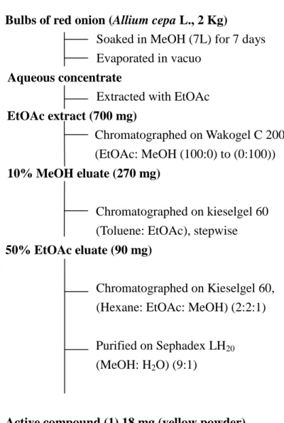
3.2. Isolation of compound 1 (quercetin 4`-O-β-D-glucopyranoside)
The methanol extract of A. cepa (2 kg) was evaporated in vacuo, and the aqueous concentrate obtained was divided into ethyl acetate and butanol extracts and aqueous
residue. The three extracts were subjected to the urease inhibitory assay. The active ethyl acetate extract was purified by a combination of column chromatography on
Wakogel C-200, silica gel G-60, and Sephadex LH-20 successively to yield an active compound 1 (18 mg) as shown in Fig. 3.2.1.
16
Bulbs of red onion (Allium cepa L., 2 Kg) Soaked in MeOH (7L) for 7 days Evaporated in vacuo
Aqueous concentrate
Extracted with EtOAc EtOAc extract (700 mg)
Chromatographed on Wakogel C 200 (EtOAc: MeOH (100:0) to (0:100)) 10% MeOH eluate (270 mg)
Chromatographed on kieselgel 60 (Toluene: EtOAc), stepwise 50% EtOAc eluate (90 mg)
Chromatographed on Kieselgel 60, (Hexane: EtOAc: MeOH) (2:2:1)
Purified on Sephadex LH20
(MeOH: H2O) (9:1)
Active compound (1) 18 mg (yellow powder)
17 3.3. Identification of active compound 1
Compound 1 was isolated as a yellow powder and showed bluish green color with FeCl3 on TLC, indicating that compound 1 was a phenolic compound. The molecular
formula of compound 1 was established to be C21H20O12 from FAB-MS data (m/z 465
[M+1]+) together with 1H- and 13C-NMR spectral data (Table 3.3.1).
The 1H-and 13C-NMR spectra of compound 1 (Figs. 3.3.1 and 3.3.2) showed signals of two aromatic protons with meta coupling at δH 6.11 (H-6, d. J = 1.6 Hz; δC 100.32)
and δH 6.31 (H-8, d. J = 1.6 Hz; δC 95.46) corresponded to a tetrasubstituted benzene
ring. Another 1,3,4-trisubstituted benzene ring was shown by the signals at δH 7.68
(H-2`, d. J = 2.4 Hz; δC 117.48), δH 7.63 (H-6`, dd. J = 8.4, 2.4 Hz; δC 122.24), and δH
7.23 (H-5`, d. J = 8.4 Hz; δC 118.6). These data together with 13C-NMR signals
attributed to a ketone carbonyl (C-4, δC 178.39) and two oxygenated olefinic carbons
[(C-2, δC 149.05) and (C-3, δC 138.9)] in addition to five oxygenated aromatic carbons
[(C-5, δC 163.46), (C-7, δC 166.73), (C-9, δC 159.20), (C-3`, δC 147.79), and (C-4`, δC
148.83)] and two quaternary aromatic carbons [(C-10, δC 105.53) and (C-1`, δC 128.59)]
strongly indicated that compound 1 was a flavonol. This flavonol was confirmed to be quercetin by comparing its signals with those of standard reference and values reported
previously.30)
Other signals in the 1H-NMR and 13C-NMR spectra of 1 could be assigned to a sugar unit, as evident from an anomeric proton signal at δH 4.87 (H-1``, d. J = 7.2 Hz; δC
104.41) and signals of four oxygenated methines at δH 3.50 (H-2``, m; δC 75.82), δH 3.47
(H-3``, m; δC 78.54), δH 3.41 (H-4``, m; δC 72.31) and δH 3,42 (H-5``, m; δC 79.36) in
addition to one oxygenated methylene at δH 3.9 (H-6``a, dd. J = 2.4, 11.6 Hz) and δH 3.7
18
anomeric proton H-1`` (J = 7.2 Hz) a β-configuration of hexopyranose was deduced. On
the basis of all these data, compound 1 was deduced to be quercetin β-glucoside by comparing with those previously reported.30)
The connectivity of the sugar unit was established from the HMBC spectrum showing a correlation between anomeric proton H-1`` (δH 4.87) and C-4` of quercetin
(δC 148.83) (Fig. 3.3.3). Acid hydrolysis of 1, followed by comparing the sugar unit
with the authentic samples of D-glucose, D-galactose, and D-mannose using cellulose
TLC and a solution of butanol: isopropyl alcohol: water (4:1:5) as elution system determined the sugar moiety to be glucose31) as shown in Fig. 3.3.4.
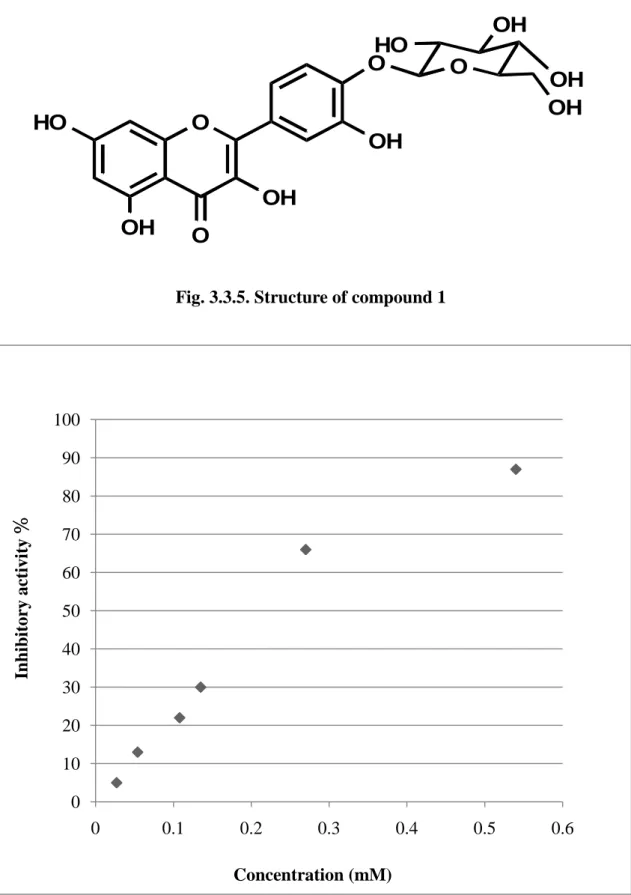
On the basis of all spectroscopic data and comparison of MS, 1H-NMR, and
13
C-NMR data with those reported previously,32, 33) compound 1 was identified as
quercetin 4`-O-β-D-glucopyranoside as shown in Fig. 3.3.5. Although compound 1 was previously isolated from A. cepa32) and Picea abies,33) the urease inhibitory activity of
this compound is still unreported.
3.4. Urease inhibitory activity of compound 1
Compound 1 showed 87% inhibitory activity against Jack bean urease at a concentration of 540 µM and 50% inhibitory activity at 190 µM (Fig. 3.4.1).
19
Fig. 3.3.1.1H-NMR spectrum of compound 1 (400 MHz, CD3OD)
Fig. 3.3.2. 13C-NMR spectrum of compound 1 (100 MHz, CD3OD)
PPM
8 7 6 5 4 3
DFILE C:\Documents and Settings\NPC students\デスクトップ\Compound-01-1H-002-FID.als COMNT auto
DATIM Fri Dec 07 11:56:24 2007 OBNUC 1H EXMOD NON OBFRQ 399.65 MHz OBSET 124.00 KHz OBFIN 10500.00 Hz POINT 32768 FREQU 7992.01 Hz SCANS 32 ACQTM 4.1001 sec PD 2.9000 sec PW1 6.70 usec IRNUC 1H CTEMP 18.7 c SLVNT CD3OD EXREF 0.00 ppm BF 0.12 Hz RGAIN 13 auto 7 . 6 3 4 7 . 6 2 9 7 . 5 8 8 7 . 5 8 3 7 . 5 6 7 7 . 5 6 1 7 . 1 7 9 7 . 1 5 7 6 . 2 5 8 6 . 2 5 3 6 . 0 6 4 6 . 0 5 9 4 . 8 2 4 4 . 8 2 1 4 . 8 0 6 3 . 8 5 5 3 . 8 5 0 3 . 8 2 5 3 . 8 2 0 3 . 6 6 7 3 . 6 5 3 3 . 6 3 7 3 . 6 2 3 3 . 4 7 7 3 . 4 5 5 3 . 4 4 1 3 . 4 3 9 3 . 4 3 7 3 . 4 2 1 3 . 4 1 3 3 . 4 0 5 3 . 3 9 8 3 . 3 9 4 3 . 3 8 9 3 . 3 8 1 3 . 3 7 6 3 . 3 5 8 3 . 3 3 6 3 . 3 3 5 3 . 3 1 3 3 . 2 4 9 3 . 2 1 8 3 . 2 1 4 3 . 2 1 0 3 . 2 0 6 3 . 2 0 2 PPM 175 150 125 100 75 50 25 0 auto 178.390 166.734 163.463 159.209 149.048 148.833 147.793 138.904 128.595 122.242 118.607 117.484 105.531 104.416 100.326 95.469 79.369 78.543 75.817 72.314 63.434 50.836 50.630 50.415 50.200 49.994 49.779 49.564 49.350 O O HO OH OH OH O OH OH OH HO O 2 3 4 5 6 7 8 9 10 1` 2` 3` 4` 5` 6` 1`` 2``3`` 4`` 5``6``
20
Table 3.3.1.1H-NMR and 13C-NMR data of compound 1
Taken in CD3OD at 400 MHz for 1H-NMR and at 100 MHz for 13C-NMR
position δc δH (integral, mult., J HZ) 1H-1H COSY
HMBC (H to C) 2 149.05 3 138.90 4 178.39 5 163.46 6 100.32 6.11(1H, d. J = 1.6 Hz) 6, 8,10,5,7,4 7 166.73 8 95.46 6.31(1H, d. J = 1.6 Hz) 6, 8,10,9,7,4 9 159.20 10 105.53 1` 128.59 2` 117.48 7.68 (1H, d. J = 2.4 Hz) 6,6`,1`,4`,2 3` 147.79 4` 148.83 5` 118.60 7.23 (1H, d. J = 8.4 Hz) H6`` 6,10,2`,1`,3`,4` 6` 122.24 7.63 (1H, dd. J = 8.4, 2.4 Hz) H5`` 6,5`,4` 1`` 104.41 4.87 (1H, d. J = 7.2 Hz) H2`` 4`,6,2``,5`` 2`` 75.82 3.50 (1H, m) H1``, H3`` 3``,1`` 3`` 78.54 3.47 (1H, m) H2``, H4`` 1``,4``,2`` 4`` 72.31 3.40 (1H, m) H3``, H5`` 3``,6`` 5`` 79.36 3.42 (1H, m) H4``, 2H6`` 6`` 6`` 63.43 3.90 (1H, dd. J = 2.4, 11.6 Hz), 3.70 (1H, dd. J = 5.6, 11.6 Hz) H5`` 4``,5``
21
Fig. 3.3.3. Correlation between anomeric proton H-1``and C-4` in HMBC spectrum of compound 1
Fig. 3.3.4. Comparison of sugar moiety of compound 1 with authentic sugars on TLC 5.00 4.75 4.50 PPM 160 140 120 PPM H-1`` C-4` H-1`` to C-4`
22
Fig. 3.3.5. Structure of compound 1
Fig. 3.4.1. Inhibitory activity of compound 1 0 10 20 30 40 50 60 70 80 90 100 0 0.1 0.2 0.3 0.4 0.5 0.6 In h ib itory acti vi ty % Concentration (mM)
O
O
HO
OH
OH
OH
O
OH
OH
OH
HO
O
23 3.5. Experimental section
General experimental procedure
Mass spectra were recorded on a JMS-700 mass spectrometer with glycerol as a matrix along with a Kratos PCKompact MALDI instrument. 1H-NMR and 13C-NMR
spectra were obtained with a JEOL JNM A-400 spectrometer. Chemical shifts were given in a δ (ppm) scale. Column chromatography (CC) separation were carried out on
Wakogel C-200 (Wako Pure Chemical Industries), silica gel G-60 (Kieselgel 70-200 and 230-400 mesh, Merck), Chromatorex ODS (Fuji Silysia, Japan) and Sephadex LH-20
(Amersham Bioscience, Sweden). TLC analysis was performed on cellulose (Merck), precoated silica gel 60 F254 plates and RP-18 F254s (0.2 mm, Merck) and spots were
visualized by UV absorption at 254 nm and fluorescence at 365 nm, or by spraying with FeCl3 or 10% H2SO4 in ethanol followed by heating.
Urease inhibitory assay
The assay was carried out according to a procedure described in sections 2.4.2 and
2.4.3.
Plant material
Fresh A. cepa bulbs were purchased at a vegetable store in Sakai city, Osaka, Japan during August 2007.
Isolation of compound 1
Fresh bulbs of red onion A. cepa (2 Kg) were extracted with MeOH (7 L) for 7 days.
The methanol extract was evaporated in vacuo, and aqueous concentrate was partitioned between ethyl acetate and butanol to afford three extracts, which were subjected to
urease inhibitory assay against Jack bean urease at a final concentration of 1250 ppm. The ethyl acetate extract (700 mg) was subjected to Wakogel C-200 (ethyl
24
acetate-methanol by 10% stepwise) chromatography to obtain an active fraction (10%
methanol eluate), which showed 70% inhibitory activity at a concentration of 250 ppm. This eluate (270 mg) was subjected to silica gel G-60 column chromatography (CC)
eluted with toluene-ethyl acetate by a 10% stepwise gradient to yield an active fraction (50% ethyl acetate eluate, 90 mg), which showed 79% inhibitory activity at a
concentration of 250 ppm.
These two active fractions were re-subjected to silica gel G-60 CC, eluted with
n-hexane-ethyl acetate-methanol (2:2:1, v/v) to yield two active fractions, which were
combined and re-applied to Sephadex LH-20 eluted with MeOH:H2O (9:1 v/v) to yield
an active compound 1 (18 mg).
Compound 1 (quercetin 4`-O-β-D-glucopyranoside), yellow powder; 1H- and
13
C-NMR data (Table 3.3.1); FAB-MS m/z 465 [M+H] +; FAB-MS m/z (rel. int.) 465
(20), 277(15), 185 (100), 93 (95), 75 (25), 57 (15), 45 (12).
Acid hydrolysis of compound 1
Acid hydrolysis of compound 1 was performed as described previously.31)
Compound 1 (5 mg) was treated with 2N HCl (1h of reaction time, in a sealed flask, at
100○C), and after cooling the reaction mixture was extracted with ethyl acetate. Sugar in the neutralized acidic phase was analyzed on cellulose TLC by comparison with the authentic samples using butanol: isopropyl alcohol: water (4:1:5) as a mobile phase, and
25
Chapter 4
Isolation of phenolic urease inhibitors from Psidium guajava
(guava leaves)
4.1. Introduction
Psidium guajava is an important food crop and medicinal plant in the tropical and
subtropical countries. It belongs to genus Psidium which contains about 100 species of
tropical shrub and small trees. P. guajava, which is considered a native to Mexico,43) extends throughout the South America, Europe, Africa and Asia, where it has been used
widely. It grows in all the tropical and subtropical areas of the world, adapts to different climatic conditions but prefers dry climates.43) Taxonomic data of P. guajava is shown
in Table 4.1.1. P. guajava is a small tree of 10 m high and with thin, smooth, patchy, peeling bark. Leaves are opposite, short-petiolate and the blade oval with prominent
pinnate veins is 5-15 cm long.
Table 4.1.1. Taxonomic data of Psidium guajava Kingdom Plantae Division Magnoliophyta Class Magnoliopsida Subclass Rosidae Order Myrtales Family Myrtaceae Subfamily Myrtoideae Tribe Myrteae Genus Psidium Species P. guajava L.
26
The folk use of guava has been documented in the indigenous groups of Mexican
Indians, Maya, Nahuatl, Zapotec and Popoluca. Although, a decoction of the leaves could be used for treatment of cough, communities of Nahuatl and Maya origin, used
guava leaves decoction to treat digestive suffering associated with severe diarrhea (a frequent disease in rainy weather).34)
On the other hand, guava leaves are commonly used in Mexico to treat gastrointestinal and respiratory disturbances, anti-inflammmatory medicine,35) and
applied on wounds, ulcers and for rheumatic pain, in addition the leaves could be chewed to relieve toothache.34) A combined decoction of the leaves and bark is given to
expel the placenta after childbirth.36) A water leaf extract is used to reduce blood glucose level in diabetics and the hot tea was very common among the local people of
Veracruz.35)
Moreover, the leaves were traditionally used in South African folk medicine to
manage, control, and/or treatment of a plethora of human ailments, including diabetes mellitus and hypertension.37) Also, guava leaves has been used widely in the traditional
medicine of Latin America and Caribbean in the treatment of diarrhea and stomach-aches due to indigestion.38)
Recent studies revealed that P. guajava leaves contain a lot of phytochemical compounds which were responsible for biological activities. Leaves contain essential oil
with the main components being α-pinene, β-pinene, limonene, menthol, terpenyl acetate, isopropyl alcohol, longicyclene, caryophyllene, humuline, selinene, cardinene,
and curcumene.39) Flavonoids and saponins combined with oleanolic acid have been isolated from the leaves.40) In addition, the leaves contain triterpenic acids, tannin, and a
27
some acids like guavanoic acid, guavacoumaric acid, jacoumaric acid, and 2α-hydroxyursolic acid were isolated.42)
Recent ethno-pharmacological studies showed that P. guajava is used in many parts
of the world for the treatment of number of diseases, such as diabetes, hypertension, caries, wounds, pain relief, reducing fever and as anti-inflammatory agent.43) Also recent
studies proved that guava leaves could be used for treatment of heart problems, constipation, cough, digestive problems, gastritis, lung problems, vaginal discharge,
vertigo, vomiting and antiamoebic.44)
As mentioned in chapter 2, it had been shown that methanol extract of guava leaves
exhibited high inhibitory activity against Jack bean urease.
During the present study, three quercetin derivatives compound 2, compound 3, and
compound 4 were isolated from ethyl acetate extract of P. guajava as urease inhibitors. This chapter deals with the isolation, identification and urease inhibitory activity of
these compounds.
4.2. Isolation of active compounds 2, 3 and 4 from Psidium guajava
The methanol extract of P. guajava (2 kg) was evaporated in vacuo, and the
aqueous concentrate obtained was partitioned with ethyl acetate and butanol to afford three extracts. The active ethyl acetate extract was purified by combination of column
chromatography on Wakogel C-200, silica gel G-60, Chromatorex ODS, and Sephadex
LH-20 successively to yield active compounds 2, 3, and 4, which were identified as guaijaverin, avicularin and quercetin, respectively,as shown in Fig. 4.2.1.
28 Guava leaves (2 Kg)
Soaked in MeOH (10 L) for 7 days Evaporated in vacuo
Aqueous concentrate
Extracted with EtOAc and BuOH EtOAc extract 250 ml (7.2 g) (250 mg) 20-40 % EtOAc eluates (430 mg) 10 % MeOH eluate Chromatographed on
silica gel 60 n-hexane: EtOAc: MeOH (2.5: 2: 1)
Eluate with Fr. No. 9 (100 mg)
Silica gel 60
n-hexane:EtOAc: MeOH
(2: 4: 1)
Silica gel 60
EtOAc: MeOH stepwise
ODS (MeOH: H2O) (1:1)
Compound 2 (3 mg)
Eluate with Fr. No. 4 (75mg) ODS (MeOH: H2O) (9:1) Silica gel 60 EtOAc: MeOH (7: 1) Sephadex LH20 MeOH: H2O (1: 1) Compound 3 (8 mg)
Silica gel 60 (n-hexane: acetone) (8: 2), (1: 1), (0: 100) 50% Acetone eluate (175 mg) Silica gel 60 (n-hexane: acetone) (6.5: 3.5)
Flashed on silica gel 60 (n-hexane: EtOAc)
ODS (MeOH: H2O)
Compound 4 (10 mg)
Fig. 4.2.1. Isolation procedure of phenolic urease inhibitors from leaves of Psidium guajava Chromatographed on Wakogel-200
29 4.3. Identification of active compound 2
Compound 2 was isolated as a yellow powder; and it was expected to be a phenolic compound as it showed a blue color with FeCl3 on TLC. The molecular formula of
compound 2 was established to be C20H18O11 from FAB-MS data (m/z 435 [M+1]+)
together with 1H-and 13C-NMR spectral data (Table 4.3.1).
Compound 2 was confirmed to be a quercetin derivative by comparing its 1H- and
13
C-NMR data with those previously mentioned for compound 1. The 1H- and 13C-NMR
spectra (Figs. 4.3.2 and 4.3.3) showed signals similar to those of compound 1 in the downfield part of the spectrum.
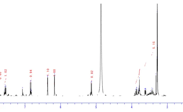
The upfield part of the spectrum exhibited a signal for an anomeric proton of sugar at δH 5.14 (H-1``, d. J = 6.8 Hz; δC 104.64), and this signal together with three signals of
oxygenated methine at δH 3.87 (H-2``, dd. J = 8.4, 6.8 Hz; δC 72.87), δH 3.62 (H-3``, dd.
J = 8.4, 3.2 Hz; δC 74.13) and δH 3.80 (H-4``, m, overlapped; δC 69.11), and one
oxygenated methylene at δH 3.42 (H-5``a, dd. J = 13.2, 3.2 Hz; δC 66.94) and δH 3.82
(H-5``b, m, overlapped) could be assigned to an aldopentose sugar moitey.
The connectivity of the sugar unit was established from slight up-field shift of (C-3, δC 135.63) in addition to downfield shift of (C-2, δC 158.67) and (C-4, δC 179.45),
indicating that the connectivity of sugar unit was at position (C-3, δC 135.63) of
quercetin.46)
On the other hand, the sugar moiety was deduced to be arabinose by comparing its signals with those previously reported.30, 45-46) An α-configuration of arabinose was
confirmed from comparing its 1H- and 13C-NMR data with those reported previously.47) A 1C4-conformation of α-L-arabinopyranoside was deduced from the signal of anomeric
30
hypothesis of α-L-arabinopyranoside was also supported by the axial orientation of the
H-2`` (δH 3.87, dd. J = 8.4, 6.8 Hz) and H-3`` (δH 3.62, dd. J = 8.4, 3.2 Hz) as (JH2``-H3``=
8.4 Hz) in addition to equatorial orientation of H-4`` (δH 3.80, m, overlapped) which
showed small vicinal coupling constant with the two protons of 5`` [H-5``a (δH 3.42, dd.
J = 13.2, 3.2 Hz) and H-5``b (δH 3.82, m, overlapped)].47)
Based on all spectroscopic data and comparison of MS, 1H-NMR, and 13C-NMR data with those previously reported,45, 48-49) compound 2 was identified as quercetin
3-O-α-L-arabinopyranoside (guaijaverin) as shown in Fig. 4.3.1. All 1H- and 13C-NMR data showed a competability with those previosly reported.45, 48)
Compound 2 was previously isolated from guava leaves Psidium guajava49) and apple pomace.45) Guaijaverin as bioactive compound from P. guajava was reported to be
a potential antiplaque agent against Streptococcus mutans,48) but its urease inhibitory activity is still unreported.
31 HO O OH OH O OH O OH OH HO O HO O O OH OH O OH O HO OH OH O OH OH OH O OH Compound 2 Compound 4 Compound 3 HO 3 4 5 6 7 8 9 10 1` 2` 3` 4` 5` 6` 1`` 2`` 3`` 4`` 5`` 2 3 4 5 6 7 8 9 10 1` 2` 3` 4` 5` 6` 2 3 5 6 7 8 9 10 1` 2` 3` 4` 5` 6` 1`` 2`` 3`` 4`` 5`` 2 4
32
Table 4.3.1.1H-NMRand13C-NMRdata of compound 2
Taken in CD3OD at 400 MHz for 1H-NMR and at 100 MHz for 13C-NMR
position δc δH (integral, mult. J HZ)
2 158.67 3 135.63 4 179.45 5 163.03 6 99.92 6.18 (1H, d. J = 2.0 Hz) 7 166.19 8 94.73 6.37 (1H, d. J = 2.0 Hz) 9 158.43 10 105.58 1` 123.02 2` 117.44 7.73 (1H, d. J = 2.4 Hz) 3` 145.97 4` 149.94 5` 116.17 6.85 (1H, d. J = 8.4 Hz) 6` 122.88 7.55 (1H, dd. J = 8.4, 2.4 Hz) 1`` 104.64 5.14 (1H, d. J = 6.8 Hz) 2`` 72.87 3.87 (1H, dd. J = 8.4, 6.8 Hz) 3`` 74.13 3.62 (1H, dd. J = 8.4, 3.2 Hz) 4`` 69.11 3.80 (1H, m, overlapped) 5`` 66.94 3.42 (1H, dd. J = 13.2, 3.2 Hz) 3.82 (1H, m, overlapped)
33
Fig. 4.3.2.1H-NMR spectrum of compound 2 (400 MHz, CD3OD)
Fig. 4.3.3. 13C-NMR spectrum of compound 2 (100 MHz, CD3OD)
PPM
7 6 5 4 3
auto
C:\Documents and Settings\NPC students\デスクトップ\guava 4 HNMR als.als
0 . 6 0 1 . 0 2 0 . 9 4 1 . 1 0 1 . 0 0 0 . 8 2 5 . 1 5 7 . 7 2 3 7 . 7 1 7 7 . 5 8 2 7 . 5 6 2 7 . 5 5 6 7 . 5 4 0 7 . 5 3 5 7 . 0 7 1 6 . 8 5 3 6 . 8 4 1 6 . 8 3 2 6 . 8 2 0 6 . 3 6 7 6 . 3 6 1 6 . 1 7 4 6 . 1 6 9 5 . 1 4 2 5 . 1 2 5 4 . 8 6 6 3 . 8 9 2 3 . 8 7 5 3 . 8 7 1 3 . 8 5 4 3 . 8 1 7 3 . 8 0 9 3 . 7 9 3 3 . 7 8 5 3 . 6 3 3 3 . 6 2 5 3 . 6 1 2 3 . 6 0 4 3 . 4 7 1 3 . 4 3 4 3 . 4 0 9 3 . 3 2 2 3 . 2 9 2 3 . 2 8 7 3 . 2 8 3 3 . 2 7 9 3 . 2 7 5 PPM 200 175 150 125 100 75 50 25 auto
C:\Documents and Settings\NPC students\デスクトップ\final 4g 13 C of guajaverin.als
1 7 9 . 4 5 3 1 6 6 . 1 8 6 1 6 3 . 0 3 1 1 5 8 . 6 6 9 1 5 8 . 4 3 0 1 4 9 . 9 4 6 1 4 5 . 9 7 3 1 3 5 . 6 3 8 1 2 3 . 0 2 4 1 2 2 . 8 8 4 1 1 7 . 4 4 0 1 1 6 . 1 6 8 1 0 5 . 5 8 6 1 0 4 . 6 4 4 9 9 . 9 2 7 9 4 . 7 3 1 7 4 . 1 2 9 7 2 . 8 7 3 6 9 . 1 0 7 6 6 . 9 4 2 4 9 . 8 4 3 4 9 . 6 3 6 4 9 . 4 2 1 4 9 . 2 8 1 4 9 . 2 0 7 4 9 . 0 0 0 4 8 . 7 8 5 4 8 . 5 7 0 4 8 . 3 5 6
34 4.4. Identification of active compound 3
Compound 3 was isolated as a yellow powder that showed blue color with FeCl3 on
TLC; therefore it was suggested to be a similar compound to those previously
mentioned. The molecular formula of compound 3 was established to be C20H18O11
from FAB-MS data (m/z 435 [M+1]+) together with 1H- and 13C-NMR spectral data
(Table 4.4.1). The 1H- and 13C-NMR spectra of compound 3 (Figs. 4.4.1 and 4.4.2) showed signals similar to those of compounds 1 and 2, thus compound 3 was confirmed
to be a quercetin derivative.
The 1H- and 13C-NMR spectra exhibited other signals for anomeric proton at δH
5.41(H-1``, br s; δC 109.52), in addition to three oxygenated methines at δH 4.28 (H-2``,
dd. J = 1.2, 2.8 Hz; δC 83.29), δH 3.85 (H-3``, dd. J = 2.8, 5.2 Hz; δC 78.70) and δH 3.80
(H-4``, m; δC 88.04), and oxygenated methylene at δH 3.46 (H-5``a, dd. J = 12.0, 3.6 Hz)
and δH 3.42 (H-5``b, dd. J = 4.8, 12.0 Hz; δC 62.55), these signals could be assigned to
be a sugar unit. The up-field shift of (C-3, δC 134.88) and downfield shift of (C-2, δC
159.28) and (C-4, δC 179.95) indicated that the 3-OH of quercetin was glycosilated.46)
The sugar moiety was confirmed to be arabinose with an α-configuration by comparing its signals with those reported previously.30, 45-46) An α-L-arabino-furanoside was
deduced from the signal of anomeric proton H-1`` (5.41, br s) as zero value of a coupling constant,the values of the vicinal coupling constant JH1``- H2`` = 0 Hz, JH 2``- 3`` =
2.8 Hz, and JH 3``- 4`` = 5.2 Hz together with a signal of anomeric carbon at 109.52.45-47)
From all spectroscopic data and comparison of MS, 1H-NMR, and 13C-NMR data
with those reported previously,45,47,49) compound 3 was identified as quercetin 3-O-α-L-arabinofuranoside (avicularin) as shown in Fig. 4.3.1. All 1H- and 13C-NMR
35
Although compound 3 was previously isolated from guava leaves49) and apple
36
Table 4.4.1.1H-NMRand13C-NMRdata of compound 3
Taken in CD3OD at 400 MHz for 1H-NMR and at 100 MHz for 13C-NMR
position δc δH (integral, mult. J HZ)
2 159.28 3 134.88 4 179.95 5 163.06 6 100.01 6.15 (1H, d. J = 2.0 Hz) 7 166.45 8 94.86 6.33 (1H, d. J = 2.0 Hz) 9 158.60 10 105.50 1` 123.10 2` 116.81 7.47 (1H, d. J = 2.0 Hz) 3` 146.37 4` 149.87 5` 116.43 6.84 (1H, d. J = 8.4 Hz) 6` 122.95 7.43 (1H, dd. J = 8.4, 2.0 Hz) 1`` 109.52 5.41 (1H, br s) 2`` 83.29 4.28 (1H, dd. J = 1.2, 2.8 Hz) 3`` 78.70 3.85 (1H, dd. J = 2.8, 5.2 Hz) 4`` 88.04 3.80 (1H, m) 5`` 62.55 3.46 (1H, dd. J = 12.0, 3.6 Hz) 3.42 (1H, dd. J = 4.8, 12.0 Hz)
37
Fig. 4.4.1.1H-NMR spectrum of compound 3 (400 MHz, CD3OD)
Fig. 4.4.2. 13C-NMR spectrum of compound 3 (100 MHz, CD3OD)
PPM
7 6 5 4 3 2
auto
C:\Documents and Settings\NPC students\デスクトップ\compound 5 g2 HNMr.als
1 . 8 8 0 . 9 2 0 . 8 5 1 . 0 0 0 . 9 7 0 . 9 7 1 . 9 3 1 . 9 4 7 . 4 7 9 7 . 4 7 4 7 . 4 5 8 7 . 4 5 2 7 . 4 3 7 7 . 4 3 1 6 . 8 7 6 6 . 8 6 2 6 . 8 4 1 6 . 8 1 8 6 . 3 4 9 6 . 3 3 8 6 . 3 3 3 6 . 1 5 3 6 . 1 4 8 5 . 4 3 1 5 . 4 1 6 4 . 9 5 0 4 . 9 4 7 4 . 9 4 5 4 . 9 4 3 4 . 9 4 1 4 . 8 6 2 4 . 2 8 8 4 . 2 8 5 4 . 2 8 0 4 . 2 7 8 3 . 9 0 7 3 . 8 7 3 3 . 8 6 6 3 . 8 6 0 3 . 8 5 3 3 . 8 3 4 3 . 8 2 3 3 . 8 1 3 3 . 8 1 1 3 . 8 0 1 3 . 6 9 0 3 . 4 9 1 3 . 4 8 2 3 . 4 7 3 3 . 4 6 1 3 . 4 5 7 3 . 4 5 2 3 . 4 4 5 3 . 4 2 7 3 . 4 1 5 3 . 3 4 1 3 . 3 2 5 3 . 3 1 6 3 . 3 0 0 3 . 2 8 6 3 . 2 8 1 3 . 2 7 4 3 . 2 6 9 3 . 2 6 5 3 . 2 6 1 3 . 2 5 7 3 . 2 5 2 3 . 2 3 6 3 . 2 3 2 3 . 1 2 2 1 . 8 6 6 PPM 200 175 150 125 100 75 50 25 0
C:\Documents and Settings\NPC students\デスクトップ\compound guava 5g 13C als.als
1 7 9 . 9 5 0 1 6 6 . 4 4 8 1 6 3 . 0 6 4 1 5 9 . 2 7 7 1 5 8 . 6 0 2 1 4 9 . 8 6 7 1 4 6 . 3 6 8 1 3 4 . 8 8 4 1 2 3 . 1 0 3 1 2 2 . 9 4 6 1 1 6 . 8 1 3 1 1 6 . 4 3 4 1 0 9 . 5 1 9 1 0 5 . 5 0 1 1 0 0 . 0 1 0 9 4 . 8 5 6 8 8 . 0 4 0 8 3 . 2 8 9 7 8 . 7 0 4 6 2 . 5 5 1 4 9 . 8 4 8 4 9 . 6 3 4 4 9 . 4 2 0 4 9 . 2 1 4 4 9 . 0 0 0 4 8 . 7 8 6 4 8 . 5 7 2 4 8 . 3 5 8
38 4.5. Identification of active compound 4
Compound 4 was isolated as yellow needles and it showed blue color with FeCl3 on
TLC. The structure of compound 4 was confirmed by 1H- and 13C-NMR data (Table
4.5.1) to be quercetin as shown in Fig. 4.3.1. The 1H- and 13C-NMR spectra of compound 4 (Figs. 4.5.1 and 4.5.2) showed signals with good agreement with those
39
Table 4.5.1.1HNMR and13C NMRdata of compound 4
Taken in acetone-d6 at 400 MHz for 1H-NMR and at 100 MHz for 13C-NMR
position δc δH (integral, mult. J HZ)
2 147.94 3 136.34 4 176.15 5 161.91 6 98.74 6.24 (1H, d. J = 1.6 Hz) 7 164.57 8 94.05 6.52 (1H, d. J = 1.6 Hz) 9 157.35 10 103.72 1` 123.40 2` 115.82 7.80 (1H, d. J = 2.0 Hz) 3` 145.41 4` 146.52 5` 115.36 6.96 (1H, d. J = 8.8 Hz) 6` 121.04 7.67 (1H, dd. J = 8.8, 2.0 Hz)
40
Fig. 4.5.1. 1H-NMR spectrum of compound 4 (400 MHz, acetone-d6)
Fig. 4.5.2. 13C-NMR spectrum of compound 4 (100 MHz, acetone-d6)
PPM
10 8 6 4 2
DFILE C:\Documents and Settings\NPC students\デスクトップ\quercetine final 2.als COMNT auto
DATIM Mon Jan 04 17:24:27 2010 OBNUC 1H EXMOD NON OBFRQ 399.65 MHz OBSET 124.00 KHz OBFIN 10500.00 Hz POINT 32768 FREQU 7992.01 Hz SCANS 321 ACQTM 4.1001 sec PD 2.9000 sec PW1 6.70 usec IRNUC 1H CTEMP 13.5 c SLVNT ACETN EXREF 0.00 ppm BF 1.00 Hz RGAIN 20 auto PPM 200 150 100 50 0
DFILE C:\Documents and Settings\NPC students\デスクトップ\compound 7 quercetin 13 C fid.als
COMNT auto
DATIM Sat Sep 26 16:45:59 2009
OBNUC 13C EXMOD BCM OBFRQ 100.40 MHz OBSET 125.00 KHz OBFIN 10500.00 Hz POINT 32768 FREQU 27210.88 Hz SCANS 711 ACQTM 1.2042 sec PD 3.8000 sec PW1 6.70 usec IRNUC 1H CTEMP 21.0 c SLVNT ACETN EXREF 29.80 ppm BF 1.00 Hz RGAIN 20 auto 2 0 6 . 3 7 3 2 0 6 . 1 7 4 2 0 5 . 9 6 8 1 7 6 . 5 3 5 1 6 4 . 9 5 3 1 6 2 . 2 9 4 1 5 7 . 7 4 2 1 4 8 . 3 2 5 1 4 6 . 9 1 2 1 4 5 . 7 9 7 1 3 6 . 7 1 8 1 2 3 . 7 3 3 1 2 1 . 4 2 8 1 1 6 . 2 0 7 1 1 5 . 7 4 4 1 0 4 . 1 0 5 9 9 . 1 2 4 9 4 . 4 3 2 3 0 . 3 7 8 3 0 . 1 8 8 2 9 . 9 9 8 2 9 . 8 0 0 2 9 . 6 1 0 2 9 . 4 2 0 2 9 . 2 2 2
41 4.6. Urease inhibitory activity of compounds 2-4
Urease inhibitory activity of compounds 2-4 was evaluated against Jack bean urease. The IC50 values of compounds 2-4 were determined and shown in Table 4.6.1.
Compound 4 (quercetin) showed the strongest inhibitory activity. The activity was decrease by glycosilation as in case of compound 2 (guajaverin) and compound 3
(avicularin). Quercetin was reported to prove gastric cell death by inhibiting apoptotic signaling by H. pylori vacuolating cytotoxin.50) The inhibitory activity of quercetin has
been previously reported,51) but the inhibitory activity of compounds 2 (guajaverin) and 3 (avicularin) against urease is reported for the first time in this study.
Table 4.6.1. Urease inhibitory activity of compounds 2-4 Compounds Urease inhibitory activity (IC50 µM)
Guajaverin (2) 120
Avicularin (3) 140 Quercetin (4) 80
42 4.7. Experimental section
General experimental procedure
The general experimental procedure was carried out in the way almost the same as described in section 3.5.
Urease inhibitory assay
The assay was carried out according to a procedure described in sections 2.4.2 and
2.4.3.
Plant material
Psidium guajava leaves were collected from a farm in Qalyubia, northern Cairo,
Egypt, in April 2008. The fresh leaves were washed and carefully dried in the sun.
Extraction and isolation of active compounds 2-4
The dried guava leaves P. guajava (2 kg) were soaked in methanol (10 L) for 7 days.
The methanol extract was evaporated in vacuo, and the aqueous concentrate obtained was partitioned with ethyl acetate and butanol. An active ethyl acetate extract (7.2 g)
was subjected to chromatography on Wakogel C-200 (n-hexane-ethyl acetate-methanol by 10% stepwise) to obtain 20-40% ethyl acetate eluates and 80% methanol eluate.The
80% methanol eluate (250 mg) was subjected to column chromatography on silica gel G-60 using n-hexane-ethyl acetate-methanol (2.5:2:1) to obtain two active fractions.
An active fraction with 81% inhibitory activity was subjected to silica gel G-60 using n-hexane-ethyl acetate-methanol (2:4:1), from which the active fraction was
subjected to another column of silica gel G-60 using ethyl acetate-methanol stepwise
and finally purified using ODS (water-methanol, stepwise) to obtain compound 2 (3 mg). The other active fraction showing 48% inhibitory activity was subjected to ODS column chromatography using water-methanol (9:1) to obtain an active fraction, which
43
was subjected to silica gel G-60 using ethyl acetate-methanol (7:1, stepwise) and finally
purified on Sephadex LH20 using MeOH-H2O (1:1) to obtain compound 3 (8 mg). The
20-40% ethyl acetate eluates (430 mg) were purified using silica gel G-60
(n-hexane-acetone) (8:2, 1:1, and 0:100) to obtain an active fraction of 50% acetone eluate, which was subjected to silica gel G-60 using (n-hexane-acetone) (6.5:3.5) to
obtained an active fraction. This fraction was flash-chromatographed on silica gel G-60 using n-hexane-ethyl acetate (1:1) and finally purified on ODS column chromatography
using (water-methanol, stepwise) to obtain an active compound 4 (10 mg).
Compound 2 (quercetin 3-O-α-L-arabinopyranoside), yellow powder; 1H- and
13
C-NMR (Table 4.4.1); FAB-MS m/z 465 [M+H] +; FAB-MS m/z (rel. int.) 435 (1), 369
(10), 277 (25), 185(100), 93 (100), 75 (35).
Compound 3 (quercetin 3-O-α-L-arabinofuranoside), yellow powder; 1H- and
13
C-NMR (Table 4.5.1); FAB-MS m/z 465 [M+H] +; FAB-MS m/z (rel. int.) 435 (2.5), 461 (5), 369 (12), 277 (50), 275 (6), 185 (100), 183 (5), 93 (100), 75(85).
Compound 4 (quercetin), yellow needles; 1H- and 13C-NMR (Table 4.6.1).
44
Chapter 5
Urease inhibitory activity of quercetin glycosides
5.1. Biological activity of quercetin glycosides
Quercetin, a flavonol found in fruits and vegetables and other plants, has been proven to possess a beneficial impact on health. It is one of the most potent antioxidants
among polyphenols.52) Quercetin derivatives have also been demonstrated to display antiviral, antibacterial, anticarcinogenic, and anti-inflammatory effects.53) The
anticarcinogenic properties of quercetin result from its significant impact on an increase in the apoptosis of mutated cells, inhibition of DNA synthesis, inhibition of cancerous
cell growth, decrease and modification of cellular signal transduction pathways,54) and inhibition of both tyrosinase activity and protein expression.55)
Reports are available on antibacterial activity of guaijaverin against Staphylococcus
aureus,49) Bacillus cereus, and Salmonella enteritidis.40) Guaijaverin is a potential antiplaque agent against Streptococcus mutans.48) Furthermore, flavonols have been shown to possess effective glucosyltransferase inhibitory activity and displaying distinct
biological activities.56)
5.2. Urease inhibitory activity of quercetin glycosides
Urease inhibitory activity of compounds 1, 2, 3, and 4 together with two standard quercetin glycosides, quercitrin (quercetin 3-O-α-L-rhamnoside) and isoquercitrin (quercetin 3-O-β-D-glucoside), shown in Fig. 5.2.1 was evaluated using both
45
L. fermentum is an acid urease that may be similar to many bacterial acidic
ureases.57) It was used for comparison because it exhibits activity in the same pH range as H. pylori urease. This fact may guide us in this research for natural urease inhibitors,
especially for dangerous bacterial ureases such as an enzyme of H. pylori, which cause gastric ulcers and have been linked to stomach cancer.
The inhibitory activities of quercetin glycosides against two ureases were compared with acetohydroxamic acid as a standard urease inhibitor, and are summarized in Table
5.2.1.
In the case of Jack bean urease, quercetin (4) showed the strongest inhibitory
activity with an IC50 value of 80 µM. The glycosilation of the 3-hydroxy and 4`-hydroxy
groups was suggested to decrease the inhibitory activity. This suggestion was supported
by the inhibitory activity of guaijaverin (2) and avicularin (3), which were slightly more active than other glycosides. Quercetin 4`-O-β-D-glucopyranoside (1) showed less
inhibitory activity than compounds (2) and (3). These results suggested that the glycosilation of the 3-hydroxy group decreased the activity, and glycosilation of
4`-hydroxy decreased the activity to a greater extent. This illustrated the reson that quercetin 3-O-β-D-glucopyranoside (isoquercitrin) showed greater inhibitory activity
than compound (1).
The activity of quercetin (4) has been already reported,51) and kampferol, apigenin, luteolin, and baicalein have been also reported to exhibit inhibitory activity against urease,51) indicating that 5- and 7-hydroxy groups on the A-ring of aglycones play an
important role in exhibiting the inhibitory activity.
Sugar ring could affect the inhibitory activity, as illustrated from the greater