A Pathological Study on Cerebral Lesions in
Diver's Decompression Sickness (DCS)
著者
KITANO Motoo, URAGO Atsushi, HAYASHI Ko,
KAWASHIMA Mahito, FUNAKOSHI Keisuke, YAMADA
Kunio, TOKUFUJI Shin-ichiro
journal or
publication title
南太平洋研究=South Pacific Study
volume
10
number
2
page range
275-285
South Pacific Study Vol.10, No. 2, 1990 275
A Pathological Study on Cerebral Lesions in Diver' s
Decompression Sickness (DCS)
Motoo Kitano1^ Atsushi Urago2), Ko Hayashi3), Mahito Kawashima4), Keisuke Funakoshi5), Kunio Yamada6), and Shin-ichiro Tokufuji7)
Abstract
Histopathological analysis was done on brains of four autopsy cases of acute decompression sicknes (DCS). The findings of the brains in four cases of acuteDCS was summarized as follows: 1) marked engorgement of the meningeal veins with marked stasis of blood with or without
presence of intravascular air bubbles, 2) marked edema of the cerebral parenchyma with perivascular hemorrhage, 3) pallor spots around the venules and blood capillaries in the brain,
especially in the deeper cortex and subcortical white matter, and 4) relatively-sharply defined foci of edematous necrosis in the deep layer of the white matter of the cerebrum, especially in the periventricular layer.
As for the causes of the cerebral lesions in DCS, bubble embolization seemed to be the most
important factor. Alteration of permeability of the blood vessels caused by trapping of very
small bubble emboli should contribute significantly to the histogenesis of pallor spots.
Pathological-anatomical analysis ascribed that the pathogenesis of focal edematous necrosis to
marked disturbance of venous circulation due to bubble embloization with a superimposing severe shock. For the latter lesion of the brain, the etiology seemed identical with that of the spinal cord
lision in DCS with apparent distruptions in the circulation of veins in and around thespinal cord. Key words: Decompression sickness (DCS), Cerebral lesions, Pallor spots, Edematous necrosis,
Bubble embolism.
1) Department of Oral Pathology, Kagoshima University Dental School* and Depertment of Critical Care Medicine, Kagoshima University Medical Hospital.
2) Department of Oral Pathology, Kagoshima University Dental School. 3) Hayashi MedicalHospital.
4) Kawashima Orthopedic Hospital and Department of Oral Pathology, Kagoshima University Dental School. 5) Department of Oral Pathology, Kyushu Dental College.
6) Undersea Medical Center,Japan Marine SelfDefence Force. 7) Department of Clinical Pathology, Kyushu Rosai Hospital.
276 Kitano et al: A PathologicalStudy on Cerebral Lesions in Diver* s DCS
Introduction
Decompression sickness (DCS) is an illness most frequently seen in divers and caisson workers. Highly dissolved air gasses in blood and tissues under hyperbaric environmentbecome bubbles if the speed of reduction of the enviromental pressure is too fast to be eliminated from the lungs.
Histopathologic studies on changes of the brain in autopsy cases of DCS have been studied
only by few investigators 1)5)6). Accordingly, theexact mechanisms by which the brain is injured
are still obscure. The conventional veiw would ascribe the tissue damage in DCS to arterial bubble embolization with a consequent obstruction of arterioles and bloodcapillaries 6\
Recenrly, another hypothesis has been proposed by Chryssanthou and associates !) from the results of their experimental work. They suggest that the vascular permeability of the brain can be altered by the action of chemical agents released in the course of reactions initiated by interface
activity of the blood and air bubbles. This alteration of the vascular permeability will then cause
some injurious changes of the brain.
We have already described in a previous paper 9)n)12) the pathogenesis of the spinal cord damage in the autopsy cases of DCS and concluded that the circulatory disturbance of the venous system played animportant role in thedevelopment and progression of theparenchymal changes. This paper is based upon four autopsy cases of DCS ofJapanese divers (Table). The purposes
of this study are to describe the histopathologic changes of the brains and to discuss the pathogenesis of the cerebral lesions.
Cases and Autopsy Findings
Case 1: A 38-year-old male helmet diver dived to a depth of 40 meters for four hours, and
surfaced in20 minutes. While surfacing, helost his consciousness and expired soonafter.
Autopsy findings : At the time of autopsy, foamy blood with numerous air bubbles was
flowing out from incised sites of the heart, vena cavae, portal veins of the omentum and
mesenterium, and superficial veins of the brain. In addition, the blood seemd to be somewhat
concentrated with an increased viscosity. Severe cyanosis was noted in the body surface.
Extensive and marked congestion, edema, and intraalveolar hemorrhage were found in the lungs.
The other visceral organs were also congested. There were many dilated sinusoids in the bone
marrow tissue of the femur containing a characteristic vacuoles which were unstainable by all dyes
used in our laboratory in the lumens of the sinusoids (Fig. 1).
The brain weighed 1,580 grams and showed a marked edematous swelling and congestion.
Histologic examination disclosed marked erythrocytic stasis within the small blood vessels
including the blood capillaries accompanied by a perivascular hemorrhage of the gray and white
matters of the brain. Perivascular hemorrhage was widely seen in the brain stem including the midbrain, pons, and medulla oblongata. Coexisistence of numerous pallor spots was another
South Pacific Study Vo白0, No, 2, 1990 277
Fig.上CharacteriStically dilated sinusoids SuggcStlng lodgmcnt of air bubbles of the bone
marrowofthefemoralhcad. Cascl. H-E X 130.
spots. Axons in the affected areas were spared. NeⅣe cells and glial cells underwent regressive
changes. The spots trended to be scattered in the deeper cortex and also in the subcortical white
matter.
me spinal cord also showed an edematous swelling. Perivascular hemorrhage was
occasionally seen in the spinal cord parenchyma.
CaSe 2・・ A 28-year-Old male scuba diver collectlng Shellfish at sea bottom of a depth oE 40
meters・ He rcpcatcd diving and surfacing seven times, and began complainlng Of dyspnea and
numbness in his lower limbs immediately after the last surfacing. Approximately ten minutes
later, he fell into a shock state. He expired in an ambulance eight hours after the onset of the SymptOmS・
Autopsy findings : Cyanosis was extensive and marked in the body surface・ Although there
wcrc no visible air bubbles in the blood within the heaれ and large blood vessels at the time of autopsy, the synovial fluid within the joint cavities of the hip and knee was foamy containing numerous air bubbles. Marked congestion, edema and intraalveolar hemorrhage were obseⅣed in the lungs. In addition, the lungs showed a number of fat emboli within the intrapulmonary
blood vessels. The bone marrow of the femoral shaft showed an extensive and marked foamy appearance with progressive necrotic changes. From these findings, it may be assumed that the
bone marrow was an important tissue that seⅣed as a soure of embolic fat in the lungs 。)・
The brain weighed 1,500 grams and showed marked congestion and edema・ Perivascular
hemorrhage was also obseⅣed. Rare faction of the parenchymal tissue and swelling of the
astroglial cells occurred in some perivacular layers. me pallor spots seen in Case 1 were also
pr'esent in a great MAY number in the deeper cortex and subcortical white matter (Fig・ 2)・ Serial
sections of the brain disclosed that the pallor spots were located around the blood capillaries or venules (Fig. 3). The axons were spared, and the ncⅣc cells and glial cells were regressive in these
areas.
The spinal cord was swollen with a marked edeme and perivcntricular hemorrhage・ These
changes seemed to be closely related to a huge engorgement of the epidural veins around the Splnal cord. Blood within the lumens almost entirely coagulated and contained a number of fat droplets 脚H
278 Kitano et al: A Pathological Study on Cerebral Lesions in Diver' s DCS
Fig. 2 : Innumerable pallor spots of the cortical gray matter and white matter of the frontal lobe of the cerebrum. The spots are difficult to be stained with various dyes nsed routinely used in our laboratory. Case 2, PTAH X 130.
:•'•%
•" -X
Fig. 3 : Largermagnification of pallor spots. A capillary runs in the affected area. Case 2,
H-E X -165.
Case 3: A 36-year-old male scuba diver four times to a depth of 60 meters for about 40
minutes each time accompanied by an interruption of 15 minutes. He complained of pains in both legs after surfacing from the last dive. He twice tried a conventional treatment (water again: fukashi) for his DCS. However, his symptoms never improved and repetitious vomitting occurred. About 20 hours later, he was transferred to a hospital. Physical examinations on the admission disclosed a complete sensory loss and flaccied paralysis of the bilateral lower extremities, as well as hypesthesia and weakness of muscular power of the bilateral upper extremities. Vesicorectal disturbance and slight dyspnea were also observed. He was then received into a recompression chamber and treated by recompression. When the atmospheric pressure inside the chamber raised to reach 5 ATA, he had a marked hematemesis and his general condition became worse. So, the recompression therapy had to be discontinued. The next morning he fell into a severe shock state and had a sustained high fever up to 42 degrees centigrade. He expired five days after the onset of the disease without any signs of recovery from the shock. The clinical diagnosis was DCS manifesting spinal cord damage with severe shock.
Autopsy findings : In gross inspection, there were no visible air bubbles in the blood within the blood vessels. Hemorrhagic and erosive esophagitis was found. Marked congestion, edema and aspiration of blood associated with marked traheobronchitis and slight bronchopneumonia were noted. A small number of thrombi and fat emboli were observed in the pulmonary vessels.
South Pacific Study Vol.10, No. 2, 1990 279
The other organs were markedly congested. The bone marrow of the right femoral head showed
extensive necrosis and contained numerous air bubbles ' ',
The spinal cord showed extensive areas of edema and necrosis, especially in the thoracal
segments (Fig. 4)9). The necrotic lesions trended to be limited to the lateral and posterior funiculi of the cord. There were several thrombi within the small veins in and around the affected areas.
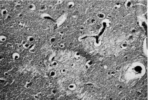
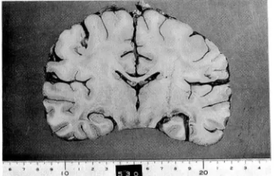
The brain weighed 1,320 grams and showed marked congestion and moderate edema (Fig. 5). There were innumerable pallor spots up to 0.5 mm in the diameter around the blood capillaries and venules of the cerebral parenchyma, especially in the deeper cortex and subcortical white
matter. Regressive changes in the nerve cells snd glial cells in the affected areas were observed.
Decrease in the number of Purkinje cells and the nerve cells in the granular layer of the cerebellar cortex was observed and was fully corresponded to the presence ofa longstading severe anoxia due
to shock. There were few number of thrombi in the small blood vessels of the brain. Perivascular hemorrhage was widely seen.
^--*«^- •
1
Jag?-t^
Fig. 4 : Ninth thoracal segment of the spinal cord showing extensive and marked edemaouts
necrosis. The lateral and posterior funiculi are particularly involved in the lesion characteristic to DCS. The gray matter is well preserved.
Case 3, H-E X 8.
Fig. 5 : Cut surface of the brain of Case 3. Marked congestion of the super surface veins and
280 Kitano et al: A Pathological Study on Cerebral Lesions in Diver' s DCS
Several, small, up to 3 mm in diameter, relatively-sharply demarcated foci of necrosis were
present inthe deep layer ofthe white matter ofthe cerebrum, especially inthe periventricular layer (Figs. 6,7,8,9). They showed tissue rarefaction with amarked edema. Destruction ofthe myelin sheaths and anomalous swelling of the axons were characteristic. Mesenchymal cell reaction was
scarce in and around the necrotic focy, but slight hemorrhage was seen in the necrotic tissues.
The necrotic foci seemed to be closely related to markedly dilated veins around the foci (Figs.
6,7,8). The lumens of these veins were empty suggesting lodgment of air bubbles which were occasionally entrapped and encircled by thrombotic material (Fig. 7). In some of the necrotic
foci couples ofcapillaries containing thrombi were also present (Fig. 8).
Fat embolism was not observed by Oil-red-O staining applied on frozen sections of the
cerebral tissue.
Fig. 6 : Aperiventricular focus of edematous necrosis. Dilated veins suggesting lodgment of air
bubbes surround the focus. Case3, H-E X 35.
Fig. 7.: A vein containing vacuoles ( most possibly air bubbles ) enveloped by thrombi in the
periphery of a necrotic focus. Axonal swelling is prominent ir, the perivascular layer. Case 3, H-E X 165.
Fig. 8
South Pacific Study Vol.10,No. 2, 1990
1 -'
V,VK ,
r ^ c
f&
-: Capillaries withthrombi in and around a necrotic focus. Case 3, H-E X 100.
281
Fig. 9 : Schematic drawing of localization of the foci of edematous necrosis. Dark spots show
Case 4: A20-year-old male scuba diver had ahistory of diving to adepth of50 meters for 20
minutes. About 20 minutes after the surfacing, he complained of dyspnea and numbness of both
legs and was transferred to a hospital. Physical examinations disclosed a complete sensory loss
up to the 3-4th cervical dermatome. Pyramical signs were seen in all the extremities showing
flaccied paralysis and in the respiratory muscles resulting in inability of automatic respiratory
movement. Vesicorectal dysturbance was also noted. His consciousness was maintained ingood condition and there was no cranial nerve dysfunction. He showed some improvement with
recompression therapy. However, 14 days after the onset of the disease, he died of acute
respiratory failure.Autopsy finding : Intravascular air bubbles were not visible to the naked eye. Slight to
moderate congestion was noted in the general visceral organs. Cystitis was marked. A small
number of thrombi were found in the intrapulmonary blood vessels and the lungs showed slight
bronchopneumonia. There was no detectable fat emboli in the lungs and other organs.
The brain showed marked congestion and edema, and weighed 1,575 grams. There were a
relatively few pallor spots in the cerebral parenchyma, especially in the deeper cortex. Axonal
and cellular damage in the affected areas was also noted. Very tiny foci of hemorrhage werescatteredin the perivascular layers.
The spinal cord had an extensive and marked necrotic change with edema in the cervical and
thoracal segments. The necrotic change was limited to the white matter of the cord, expecially in
the lateral and posterior funiculi. There were many thrombosed veins in and around the necrotic
foci of the spinal cord and also in the epidural space of the vertebral canal 9).
282 Kitano et al: A PathologicalStudy on Cerebral Lesions in Diver* s DCS
Discussion
The pathogenesis of cerebral damage in decompression sickness has been a subject of controversy. The conventional view would ascribe tissue damage to arterial bubble embolization
with consequent obstruction of arterioles and blood capillaries 6). Recently another hypothesis
was proposed that the venous obstruction leading to venous infarction would be the most causal factor for the development and progression of damage of various organs and tissues in DCS
4)7)9)10
The common findings of the brains in four cases of decompression sickness in the present study can be summarized as follows ; 1) marked engorgment of the meningeal veins with a
marked stasis of blood with or withoutpresence of intravascular air bubbles, 2) marked edema of the cerebral parenchyma with perivascular hemorrhage, and 3) pallor spots around the venules and blood capillaries in the brain, especially in the deeper cortex and subcortical white matter.
The pallor spots seen around the blood capillaries and venules seemed to have resulted from
an alteration of permeability of the blood vessels l\ The spots were small in size, and distributed
in the deeper cortex and subcortical white matter. The size and location ofthe spots suggest that very small emboli which are almost-undoutedly air bubbles should be trapped in the deeper cerebral cortex 6^.
It is generally accepted that air bubbles are initially liberated from tissues and blood during decompression. The bubbles arising in the interstitium create tissue injury and also enter the circulation. Vascular permeability increases due to intimal damage by direct mechanical actions
of intravascular bubbles resulting in plasma loss and interstitial edema. In the blood vessels, the
air bubbles begin to exert direct mechanical effects such as bubble emblization, intimal damage,
etc., and indirect effects due to surface activity at interfaces. The
bubble-blood-interaction as discussed by many researchers 1)4)15)18) tends to alter the secondary and tertially
configuration of blood proteins, leading to an aggregation of blood platelets and an activation ofthe clotting system of blood and to release of vasoconstrictive substances, and finally to cause a
disturbance of blood circulation due to increase ofvascular permeability hemoconcentration, and intravascular coagulations of blood. When the air bubbles arising in the interstitium enter thecirculation, other products of tissue and celluar disintegration, such substances as lipid, free fatty
acids, peptides, histaminelike substances, potasium ion, etc. also enter. They all may play a role
as agents of increase of vascular permeability together with intravascular air bubbles 6).
Another problem having to be considered and discussed is the development of multiple foci
of edematous necrosis which are presset in the deep layer of the white matter, especially in the
periventricular areas. The foci were only seen in Case 3, and there has been no report of such acase of DCS revealing focal edematous necrosis of the brain in world literature. However, it
seemed to be a very important event in DCS. The associated findings were a considerable
number of veins and venules containing thrombi ordilating with an empty lumens suggesting the
lodgment of air bubbles in and around the necrotic foci.We previously reported that the spinal cord damage in DCS is caused by severe circulatory
disturbance, especially by reduction ofvenous return from the spinal cord parenchyma 9)11). The
histopathologic features in the spinal cord in DCS are very much akin to the change in the brain of
South Pacific StudyVol.10,No. 2, 1990 283
Case 3, that is the necrosis associated with marked edema. Edematous necrosis seems to be created from venous obstruction, rather than arterial obstruction 9>13)15). There appears to be an
intimate topographic correlation of vascular changes of the veins and venules with perenchymal lesions of the spinal cord in DCS. Therefore, pathogenesis of thecerebral lesions in DCS may be also the same as that of spinal cord lesions, in which dyshoric changes of veins and blood capillaries of venous side contribute to the parenchymal changes.
Thenext problem is how theairbubbles reach themedullary blood vessels, and stagnate for a long time within them. It is generally accepted that air bubbles are initially liberated from tissues and blood during decompression. Bubbles arising in the interstitium also enter the circulation.
The lipoid-rich tissues of the central nervous system including the cerebrum is a predictable site of
creation of intraparenchymal air bubbles, because the solubility of nitrogen gas in the lipid-, and
lipoid-rich tissues is much higher than in the non-fatty tissues 2^lsK The fibrin thrombi seen in
and around the necrotic foci of this case seemed to be closely associated with intravascular air bubbles. The fibrin thrombi should contribute to the stagnation of the air bubbles within the lumen of the blood vessels 10). However, it must be remembered that the present case affected bya longstanding severe shock before the death. The retardation ofthe venous return from the deep layer ofthe brain must be aggravated througt right cardiac failure due to severe shock.
Although many hypothesis are proposed in the literature on the pathogenesis of the tissue
damage in decompression sickness, the following two mechanisms seem most important ; 1)
direct tissue damage by the autochthonous bubble formation in the parenchyma 2)6)7), and 2)
indirect tissue damage due to the circulatory disturbance l^^l6\ Distribution of the parenchymal change in the Case 3 was restricted to the area around the veins and venules. Suchadistribution ofthe parenchymal lesions seems to be incompatible with only the direct action of autochthonous air bubbles to the parenchyma. Thus, the present study supports the later
possibility.
In conclusion, as for the causes of the cerebral lesions in DCS bubble embolization is the
most important factor. Alteration of permeability of the blood vessels and the activation of blood clotting system contribute signigicantly to the histogenesis of cerebral lesions. When
marked disturbance of the venous return occurs, the deep layer of the white matter especially the
periventricular areas tend to be affected by focal edematous necrosis. For such lesions of the
brain, the pathogenesis seems identical with that of the spinal cord lesions in DCS with apparentcirculatory distrubance of the venous system.
Acknowledgements
We are grateful to Prof. Riki Okeda (Dpt. Neuropathol., Med. Res. Inst., Tokyo Med.
Dent. Univ.) for invaluable advice and discussions concerning this work. We also thak Dr.
Charles E. Lehner (Dpt. Preventive Med., Wisconsin-Madison Univ. Med. School) for a critical
284 Kitano et al: A Pathological Study on Cerebral Lesions in Diver' s DCS
References
1) Chryssanthou, C, et al., 1977. Blood-brain and blood-lung barrier alteration by dysbaric
exposure. Undersea Biomed. Res., 4: 117-129.
2) Francis, T.Jr., et al., 1988. Is there a role for the autochthonous bubble in the pathogenesis of spinal cord decompression sickness? J. Neuropathol. Exp. Neurol., 47: 457-487.
3) Gersh, I., et al., 1944. Tissue and vascular bubbles after decompression from high pressure atmospheres — Correlation of specific gravity with morphological changes. J. Cell Comp.
Physiol., 24: 35-70
4) Hallenbeck, J. M., et al., 1975., Mechanisms underlying spinal cord damage in decompression
sickness. Neurol., 25: 308-316.
5) Hayashi, K., 1974. Clinical and experimental studies on decompression sickness (in Japanese,
with English abstract). Fukouka Acta Med., 65: 889-908.
6) Haymaker, W., 1957. Decompression sickness. In Handbuch der speziellen pathologischen
Anatomie und Histologic 13 Band. Nerven-system. ed. Olubarsch, F. Henke, R. Rossle. pp 1600-1672, Springer-Verlag, Berlin.
7) Hills, B. A. and James, P. B., 1982. Spinal decompression sickness : Mechanical studies and a
model. Undersea Biomed. Res. 9: 185-201.
8) Kawashima, M., et al., 1977. Histopathology of the early stage of osteonecrosis in divers. Undersea Biomed. Res., 4: 409-417.
9) Kitano, M., et al., 1977. Three autopsy cases of acute decompression sickness — Consideration
of pathogenesis about spinal cord damage in decompression sickness. Orthop. Traumatol.,
26: 269-276.
10) Kitano, M., et al., 1978. Experimental studies on decompression sickness — Consideration about hypercoagulability of blood decompression sickness. Orthop. Traumatol., 27:653-658. 11) Kitano, M. and Hayashi, K., 1981. Acute decompression sickness — Report of an autopsy
case with widespread fat embolism., Acta Pathol. Jpn., 31: 269-276.
12) Kitano, M., et al., 1987. Late menifestation of spinal cord lesions in decompression sickness :
Histopathologic analysis ofan autopsy case. Underwater Hyperbaric Physiol., IX: 219-228.13) Okeda, R., and Shibata, T., 1973. Radiation encephalopathy. Acta Pathol. Jpn., 23:
867-883.
14) Philp R. B., 1972. Interactions between gas bubbles and components of blood : Implications in
decompression sickness. Aerospace Med., 43: 946-953..15) Suzuki, K., et al., 1984. Vascular changes of Methotrexate-related disseminated necrotizing
leukoencephlopathy. Acta Neuropathol. (Bed), 65: 145-149.
16) Takagi, M., and Mano, Y., 1982. Acute decompression sickness —Report of an autopsy case.
Bull. Tokyo Med. Dent. Univ., 29: 71-76.
17) Vernon, H. M., 1907. The solubility of air in fats, and relation to caisson disease. Proc. Roy.
Soc. London, 79:366-371.
18) Warren, B. A., 1973. The ultrastructural morphology ofair embolism : Platelet adhesion to the interface and endothelial damage. Brit. J. Exp. Pathol., 54: 163-172.
South Pacific Study Vol.10, No. 2, 1990 285
Table : Four Autopsy Casesof Acute Decompression Sickness in male divers Duration of
Age Profession illness Clinical Diagnosis
Case 1 38 yrs helmet diver 0 hrs Chokes
Case 2 28 yrs scuba diver 8 hrs Chokes (+ spinal cord injury?)
Case 3 36 yrs scuba diver 5 days Spinal cord injury with severe shock Case 4 20 yrs scuba diver 14 days Spinal cord injury (died of
respiratory failure)
acute