INTRODUCTION
Insulin elicits many biological responses such as cellular metabolism and gene expression. One of the most important metabolic responses induced by insulin is the stimulation of glucose uptake in muscle and adipose tissues. This effect occurs as a consequence of the translocation of GLUT4 from the intracellular pool to the cell surface (1-4). Defect of this function is thought to be one of the main causes of non-insulin-dependent diabetes mellitus (NIDDM). Therefore, elucidating the molecular mechanism of GLUT4 translocation is important
for understanding the etiology of NIDDM.
To examine the mechanisms of GLUT4 trans-location, we developed a sensitive and quantitative method to measure directly c-myc epitope-tagged GLUT4 (GLUT4myc) on the cell surface (5). Using this system, we found that phosphatidylinositol (PI) 3-kinase (p85/p110 heterodimer type) plays a key role in GLUT4 translocation triggered by insulin, by platelet-derived growth factor, and by epidermal growth factor in cultured cells (6 -8).
Activation of PI3-kinase is essential for insulin-stimulated GLUT4 translocation and glucose uptake (7). However, the downstream mediators of PI3-kinase for GLUT4 translocation are still unknown. One candidate molecule is Akt (Protein kinase B or RAC/PK) (9-11). Akt is the cellular homologue of a viral oncogene, v-Akt, and, has therefore also been termed c-Akt (12). It has a PH (pleckstrin homology) domain in the N-terminus which binds
Overexpression of wild-type Akt1 promoted insulin-stimulated
p70S6 kinase (p70S6K) activity and affected GSK3β
regulation, but did not promote insulin-stimulated GLUT4
translocation or glucose transport in L6 myotubes
Satoshi Noda
*, Kazuhiro Kishi
†, Tomoyuki Yuasa
†, Hideki Hayashi
†, Tetuo Ohnishi
†,
Ikuko Miyata
†, Hiromu Nishitani
*, and Yousuke Ebina
† *Department of Radiology, The University of Tokushima School of Medicine ; and†
Division of Molecular Genetics, Institute for Enzyme Research, The University of Tokushima, Japan
Abstract : We have developed a simple, direct and sensitive method to detect GLUT4 on the cell surface. Using this system, we found that PI3-kinase plays a key role in the signaling pathway of insulin-stimulated GLUT4 translocation. One of the down stream effectors of PI3-kinase is serine-threonine kinase Akt (protein kinase B, RAK-PK), but the involvement of Akt in insulin-stimulated GLUT4 translocation is controversial. To investigate whether Akt1 regulates insulin-stimulated GLUT4 translocation and glucose uptake in L6 myotubes, we established L6 myotubes stably expressing c-myc epitope-tagged GLUT4 (GLUT4myc) and mouse wild type (WT) Akt1. We found that overexpression of WT Akt1 promoted insulin-stimulated p70S6 kinase (p70S6K) activity and increased the basal activity of GSK3β, but did not promote insulin-stimulated GLUT4 translocation or glucose uptake. These data supported the result that Akt is not a main signaling molecule to transmit the signal of insulin-stimulated GLUT4 translocation or glucose uptake from insulin-activated PI3-kinase. J. Med. Invest. 47 : 47-55, 2000
Key words : AKT, p70S6 kinase, GSK3β, GLUT4
Received for publication November 30, 1999 ; accepted December 17, 1999.
Address correspondence and reprint requests to Yousuke Ebina, M.D., Ph.D., Division of Molecular Genetics, Institute for Enzyme Research, The University of Tokushima, Kuramoto-cho, Tokushima 770-8503, Japan and Fax : +81-88-633-7437.
The Journal of Medical Investigation Vol. 47 2000 47 47
to other kinases (13). This PH domain shares structural similarity with PKC (protein kinase C) isozymes and PKA (cyclic AMP-dependent protein kinase) (12). There are three isoforms of Akt ; Akt1 (PKBα), Akt 2 (PKBβ), and Akt 3 (PKBγ) (14-16).
Akt is rapidly activated by insulin and by certain growth factors (17, 18). It is fully activated by phosphorylation of two key regulatory amino acid residues, Thr308
and Ser473
(19). Phosphatidylinositol 3,4,5-triphosphate-dependent protein kinase (PDK1) phosphorylates Akt catalytic domain Thr308
and activates Akt (20). There are two types of constitu-tively active mutants of Akt ; viral Gag protein fused type (21, 22) and myristoylation signal sequence tagged type (23-25). Overexpression of these active Akt promoted p70S6 kinase activity (10, 26), GLUT4 translocation and glucose uptake (23, 24). Overexpression of wild type Akt inhibited GSK3β activity (17). However, Kitamura et al . reported that Akt is not required for insulin-stimulated GLUT4 translocaiton or glucose uptake (27).
Therefore, we examined the effects of overex-pressed WT Akt1 on insulin-stimulated GLUT4 translocation, glucose uptake, p70 S6kinase activity, and GSK3β activity in L6-GLUT4myc myotubes.
MATERIALS AND METHODS
Cells and materialsThe parent cell line used in this study was L6-GLUT4myc (28, 29). The HA-tagged mouse wild-type Akt1 (30) was subcloned into a mammalian expres-sion vector, pCXN (31). This plasmid was cotransfected into L6-GLUT4myc cells with pSV2-hph (32), a hygromycin B phosphotransferase expression plasmid using lipofectamine reagent, and selected with hygromycin B (Sigma). Two independent clones were established, #203 and #2, to avoid clonal deviations.
Cell surface anti-c-myc antibody binding assay (GLUT4myc translocation assay)
Cells in 24-well plates were incubated in 500µl of Krebs-Ringer-HEPES buffer (KRHB) (5) for 20 min at 37℃ and then with given concentrations of ligands for 10 min at 37℃. GLUT4myc translocation was measured as described (29).
2-Deoxyglucose Uptake Measurement
Cells in 24-well plates were treated with given
concentrations of ligands for 10 min at 37℃. 2-Deoxyglucose uptake was measured as described previously (5).
Cell lysate and Immunoprecipitation
For Akt phosphorylation and kinase assays, cells in 6 -well plates were incubated in KRH buffer for 30 min at 37℃, incubated in the absence or presence of 10-7
M insulin for 5 min at 37℃, lysed with buffer containing 1% Nonidet P-40, sonicated and centrifuged at 15,000 rpm for 15 min as described previously (33), and the supernatants were incubated with appropriate polyclonal antibodies for 2h at 4℃. The immunocomplexes were precipitated with protein A-Sepharose CL-4B (Amersham Pharmacia Biotech). For p70S6K, GSK3β phosphorylation and kinase assays, cells in 6 -well plates were deprived of serum for 4 h, incubated in KRH buffer for 30 min at 37℃, incubated in the absence or presence of 10-7
M insulin for 10 min at 37℃, lysed in a solution containing 50mM Hepes (pH7.5), 150mM NaCl, 1% TritonX-100, 20µM P-APMSF, 1mg/ml Bacitracin, 5mM EDTA, 5 mM EGTA, 1 mM Na pyrophosphate, 1 mM Na3VO4, and 20 mM NaF, and immunoprecipitated
as described above.
Immunoblotting
Cell lysates were boiled for 5 min in Laemmli sample buffer and subjected to SDS-polyacrylamide gel electrophoresis ; the separated proteins were transferred to nitrocellulose filter, probed with the antibodies indicated below and detected as described previously (34).
Antibodies
A monoclonal antibody (9E10) against human c-myc was obtained from the American Type Culture Collection. Phosphospecific Akt (Ser473), p70S6K (Thr389), and GSK 3α/β (Ser21/9) antibody were purchased from New England Biolabs, Inc.. Anti-Akt1, anti-p70S6K, and anti-GSK3β were prepared by im-munizing rabbits with keyhole limpet hemocyanin-coupled peptides, the COOH-terminal 20 amino acids of Akt1 (CVDSERRPHFPQFSYSASGTA), COOH-terminal 31 amino acids of p70S6K (MAG VFDIDLDQPEDAGSEDELEEQQNLNESC), and NH3-terminal 23 amino acids of GSK3β (CDTNA GDRGQTNNAASASASNST).
S. Noda et al. Effect of Akt1 on GLUT4 translocation
48 S. Noda et al. Effect of Akt1 on GLUT4 translocation
Analysis of Akt, p70S6K, and GSK3
α/β
Phos-phorylationsCells lysates were separated by 7% SDS-polyacrylamide gel electrophoresis and immunoblotted with each Phosphospecific antibody as described above.
Akt, p70S6K, and GSK3
β
kinase assaysFor Akt kinase assays, the immunoprecipitates were washed three times with washing buffer (i) (140mM NaCl, 20mM Tris-HCl (pH 8.0), 1% Nonidet P-40, and 1 mM DTT), and once with kinase buffer (50mM Tris-HCl (pH7.5), 10mM MgCl2, 1mM DTT),
and incubated for 30 min at 30℃ in a reaction mixture containing 1µM Protein kinase inhibitor, 160µM“Crosstide”(GRPRTSSFAEG) as substrate, 50mM Tris-HCl (pH7.5), 10mM MgCl2, 1mM DTT,
5µM unlabeled ATP, and 3.0µCi/µl [γ-32
P] ATP. The reaction was stopped by adding 10µl of 250 mM EDTA, and 5 mM ATP, and the reaction mixture centrifuged at 15,000rpm for 1 min.. The resulting supernatant (25µl) was spotted onto 2×2 cm P81 (Whatman) filter paper, dryed and washed 15 min× 4 times with 75 mM phosphoric acid, and once with 99.5% Ethanol for 1 min. The32
P incorporation into the peptide was determined by liquid scintillation spectroscopy.
For p70S6 kinase assays, the immunoprecipitates were washed three times with buffer containing 2M LiCl, 82mM NaCl, 12mM Tris-HCl (pH 8.0), 0.58% Nonidet P-40, and 580µM DTT, and washed twice with kinase buffer (25mM MOPS (pH 7.2), 1mM DTT, 6mM MgCl2, 1mM EDTA, and 0.05% Triton
X-100), and incubated for 30min at 30℃ in a reaction mixture (25µl) containing 1 µM Protein kinase inhibitor, 200µM 40SR-pep20 (KRRRLASLRASYS KSESSQK) as substrate, 25mM MOPS (pH 7.2), 1mM DTT, 6mM MgCl2, 1mM EDTA, and 0.05%
Triton X-100, 5µM unlabeled ATP, and 3.0 µCi/µl
[γ-32
P] ATP. The kinase activity was determined as described above for Akt kinase assays.
For GSK3β kinase assays, the immunoprecipitates were washed once with buffer (i), as described above, washed twice with buffer (ii) (100 mM Tris-HCl (pH 7.4), 0.5M LiCl, and 1mM DTT), and washed twice with kinase buffer (20mM Hepes, 10mM MgCl2, and 1mM DTT), and incubated for 30 min at
30℃ in a reaction mixture (25µl) containing 2µM Protein kinase inhibitor, 80µM p-CREB (KRREIL SRRP(p)SYR) as substrate, 16 mM Hepes, 8 mM MgCl2, 0.8mM DTT, 35µM unlabeled ATP, and
3.0µCi/µl [γ-32
P]ATP. The kinase activity was deter-mined as described above in Akt kinase assays.
RESULTS
Overexpression of WT Akt1 promoted the basal and insulin-stimulated kinase activity of Akt in L6 myotubes
We transfected HA-tagged mouse WT Akt1 into parent L6GLUT4myc cells, and established two independent stable clones, #203 and #2. To confirm the overexpression of WT Akt1, the cell lysate was immunoblotted with a polyclonal Akt1 anti-body (Fig. 1A) and anti-HA antianti-body (Fig. 1B). The exogenous mouse WT Akt1 was overexpressed at about 5-fold the level of endogenous rat Akt1 in L6GLUT4myc. Fig.1B shows that the lysates from parent cells did not immunoreact with the HA anti-body. To examine the activity of the exogenously overexpressed WT Akt1 in L6 myotubes, we assessed Akt phosphorylation (Fig. 2A) and kinase activity after insulin stimulation (Fig.2B,C). Overexpression of WT Akt1 promoted the insulin-stimulated Akt phosphorylation of Ser 473 about 2-3 fold compared to that of parent L6GLUT4myc cells (Fig. 2A). Overexpression of WT Akt1 elevated basal Akt
Fig.1. Overexpression of wild-type (WT) Akt1 in L6 myotubes.
A : Cell lysates were subjected to immunoblotting with a polyclonal anti-Akt1 antibody. Lane 1 ; a band representing endogenous Akt in L6GLUT4myc cells. Lane 2 and 3 ; endog-enous Akt plus exogendog-enously overexpressed WT Akt1 in L6GLUT4mycAktWT#203 and in L6GLUT4mycAktWT#2, respectively. By scanning densitometry, the relative intensity of these bands representing Akt in lanes 1-3 was 106, 633, and 580, respectively. B : Immunoblotting with an anti-HA antibody to show exogenously overexpressed WT Akt1.
49 The Journal of Medical Investigation Vol. 47 2000 49 The Journal of Medical Investigation Vol. 47 2000
activity about 2-fold, and insulin-stimulated Akt activity about 2-3 fold after immunoprecipitation with anti-Akt1 antibody (Fig.2B). The activation of exogenously expressed Akt1 was detected after immunoprecipitation with anti-HA antibody (Fig.2C).
Overexpression of WT Akt1 promotes the insulin-stimulated kinase activity of p70S6K in L6 myotubes
To examine the effect of overexpressed WT Akt1 on one of the downstream effectors, p70S6K, in L6 myotubes, we assayed p70S6K phosphorylation and the kinase activity after insulin stimulation (Fig. 3A, B). Overexpression of WT Akt1 promoted the insulin-stimulated p 70S 6 K phosphorylation of Thr 389 about 3-fold compared to that of parent L6GLUT4myc cells (Fig.3A). The insulin-stimulated
p70S6 kinase activity was promoted by the over-expression of WT Akt 1 about 2-3 fold compared to that of parent cells (Fig. 3B).
Overexpression of WT Akt1 affects the basal kinase activity of GSK 3
β
in L6 myotubesGSK3β is a down stream target of Akt, and nega-tively regulated by Akt-dependent phosphorylation (17, 35). We assayed the phosphorylation and activity of GSK3β kinase to examine whether the exogenously overexpressed WT Akt1 affects the GSK3β activity in the insulin signaling pathway. The phosphorylations of GSK3α (Ser21) and GSK3β (Ser9) were increased after insulin stimulation, and were promoted by the overexpression of WT Akt1 in L6 myotubes (Fig. 4A). Stable overexpression of
Fig. 2. Overexpression of WT Akt1 promoted the insulin-stimulated phosphorylation and kinase activity of Akt in L6 myotubes.
A : Phosphorylation of Akt in L6 myotubes. Cells were incubated in the presence or absence of 10-7M insulin for 5 min at 37℃, lysed,
and subjected to immnunoblotting with an anti-phospho-specific Akt antibody. By scanning densitometry, the relative intensity of the bands representing phosphorylated-Akt in lanes 2, 4, 6 was 612, 1150 and 1385, respectively.
B : Cells were incubated in the presence or absence of 10-7M insulin for 5 min at 37℃, lysed, immunoprecipitated with anti-Akt1
antibody, and subjected to Akt kinase assay.
C : Cells were incubated in the presence or absence of 10-7M insulin for 5 min at 37℃, lysed, immunoprecipitated with anti-HA
antibody, and subjected to Akt kinase assay. Values represent means±S.E. of three determinations.
S. Noda et al. Effect of Akt1 on GLUT4 translocation
50 S. Noda et al. Effect of Akt1 on GLUT4 translocation
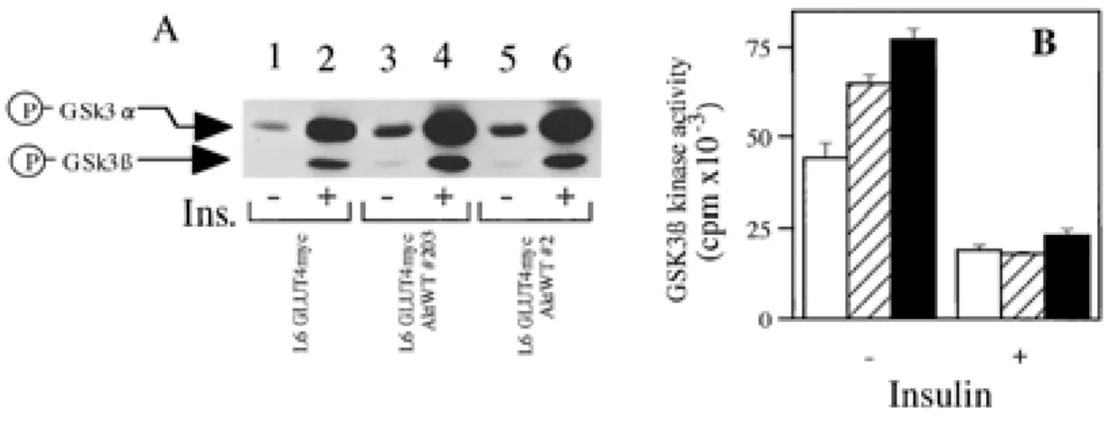
WT Akt1 results in about a 1.5~1.7 fold increase of basal GSK3β activity relative to that of parent cells (Fig.4B) (see“Discussion”). Insulin-stimulated GSK3β kinase activity was reduced about 43% in parental cells (L6GLUT4myc) and about 28% in the cells overexpressing WT Akt1 (#203, #2) (Fig. 4B). These results indicated that overexpression of WT Akt1 affected the insulin-induced phosphorylation and
the inactivation of GSK3β.
WT Akt1 overexpression did not affect the GLUT4 translocation or glucose uptake in L6 myotubes
Exogenously overexpressed Akt in L6 myotubes promoted insulin-stimulated Akt and p70S6K activities. Overexpression of WT Akt1 promoted
Fig. 3. Overexpression of WT Akt1 promoted the insulin-stimulated phosphorylation and kinase activity of p70S6K in L6 myotubes.
A : Phosphorylation of p70S6K in L6 myotubes. Cells were incubated in the presence or absence of 10-7M insulin for 10 min at 37℃,
lysed, and subjected to immnunoblotting with an anti-phospho-specific p70S6K antibody (BioLabs). By scanning densitometry, the relative intensity of the bands representing phosphorylated-p70S6K in lanes 2, 4, 6 was 569, 1577 and 1423, respectively.
B : Cells were incubated in the presence or absence of 10-7M insulin for 10 min at 37℃, lysed, immunoprecipitated with anti-p70S6K
antibody, and subjected to p70S6 kinase assay. Values represent means±S.E. of three determinations.
Fig. 4. Effect of WT Akt1 overexpression on the phosphorylations and activities of GSK 3β in L6 myotubes. A : Phosphorylation of GSK3α and GSK3β in L6 myotubes. Cells were incubated in the presence or absence of 10-7M insulin for 10 min
at 37℃, lysed, and subjected to immnunoblotting with an anti-phospho-specific GSK3α/β antibody. By scanning densitometry, the relative intensity of the bands representing phosphorylated-GSK3α in lanes 2, 4, 6 was 156, 225 and 224, and phospholyrated-GSK3β in lanes 2, 4, 6 was 59, 89 and 86, respectively.
B : Cells were incubated in the presence or absence of 10-7M insulin for 10 min at 37℃, lysed, immunoprecipitated with anti-GSK3β
antibody, and subjected to GSK3 kinase assay. L6-GLUT4myc (open bars) ; L6-GLUT4myc Akt1WT#203 (diagonally striped bars) ; L6-GLUT4myc Akt1WT#2 (closed bars). Values represent means±S.E. of three determinations.
51 The Journal of Medical Investigation Vol. 47 2000 51 The Journal of Medical Investigation Vol. 47 2000
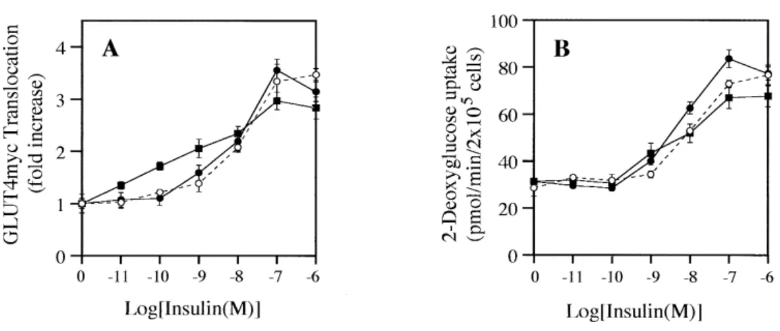
insulin-stimulated the phosphorylation (Ser 9) and reduced the activity of GSK3β. We therefore exam-ined insulin-stimulated GLUT4 translocation and glucose uptake in L6 myotubes overexpressing WT Akt1. Despite that exogenously overexpressed WT Akt1 promoted insulin-stimulated Akt activity, promoted p70S6K activity and reduced insulin-stimulated GSK3β activity, no significant differences of insulin-stimulated GLUT4 translocation or insulin-stimulated glucose uptake were found between L6GLUT4myc myotubes overexpressing WT Akt1 and parent L6GLUT4myc myotubes (Fig. 5A, B).
DISCUSSION
Overexpression of a constitutively active mutant of Akt promoted GLUT4 translocation and glucose uptake (21, 23, 24), while overexpression of a dominant negative mutant of Akt inhibited the translocation and uptake (23, 36). However, Kitamura et al. reported that overexpression of a constitutively active mutant of Akt and dominant negative mutant of Akt using an adenovirus expression system did not affect insulin-stimulated GLUT4 translocation or glucose uptake (27). Therefore, the relationship between Akt and insulin-stimulated GLUT4 translocation and glucose uptake is still controversial. Further-more, the effects of the dominant negative mutant of Akt, in which at two phosphorylation sites (Thr308
and Ser473
) the residue was changed to alanine, are
also controversial (25, 27, 36). We established stable CHO cells which overexpressed the mutant Akt at ten- fold the level of endogenous Akt, but a 10- fold overexpression was not sufficient to inhibit endog-enous insulin-stimulated Akt activation (data not shown). Stable overexpression of functional dominant negative mutant Akt might be difficult because Akt is a mediator of cell survival which prevents apoptosis (37). Therefore, we overexpressed wild type Akt in this study. Although Akt negatively regu-lated the activity of GSK3β by the phosphorylation of Ser 9, the stable overexpression of WT Akt1 increased basal activity of GSK3β about 1.7-fold compared to parent L6GLUT4myc cells (Fig. 4B). The same results were obtained in CHO clones (data not shown). The inconsistency of these results may be explained by the functions of Akt. There are several effectors of Akt and the regulation of GSK3β activity might be quite complicated. We found that overexpression of WT Akt1 in L6GLUT4myc affected downstream effectors, such as p70S6 kinase and GSK3β, but not GLUT4 translocation or glucose uptake. These results, however, do not indicate wheth-er Akt1 is a mediator of insulin-stimulated GLUT4 translocation and glucose uptake or not. Exogenous expression of Akt1 does not affect the insulin-stimulated GLUT4 translocation or glucose uptake, because the level of endogenous Akt1 is sufficient mediate this effect. This is not necessarily the case, however. An approximately 2-fold increase in Akt activity was observed in L6GLUT4myc cells after stimulation
Fig. 5. Effect of WT Akt1 overexpression on the insulin-stimulated GLUT4 translocation and glucose uptake in L6 myotubes.
L6 myoblasts in 24-well plates were differentiated into L6 myotubes by incubating with low serum, as described under MATERIALS AND METHODS, and incubated with various concentrations of insulin for 10 min at 37℃. The GLUT4myc translocation (A) and glucose uptake (B) were measured as described under MATERIALS AND METHODS. ○, parent L6-GLUT4myc cells ; ■, L6-GLUT4myc Akt1 WT #203 ; ●, L6-GLUT4myc Akt1 WT #2. Values represent means±S.E. of six determinations.
S. Noda et al. Effect of Akt1 on GLUT4 translocation
52 S. Noda et al. Effect of Akt1 on GLUT4 translocation
by insulin (Fig.2 B). The basal Akt activities in the cells overexpressing WT Akt1 were two-fold higher than the basal Akt activity of the parent cells (L6 GLUT4myc) (Fig.2B). However, there was no effect on the basal level of GLUT4 translocation or glucose uptake (Fig.5 B).
Our results indicated that Akt is not a main signaling molecule to transmit between PI3-kinase and insulin-stimulated GLUT 4 translocation.
ACKNOWLEDGMENT
We thank Dr. A. Bellacosa for kindly providing plasmids, as well as Y. Mitsumoto and A. Klip for kindly providing L6 cells.
REFERENCES
1. Birnbaum MJ : Identification of a novel gene encoding an insulin-responsive glucose trans-porter protein. Cell 57 : 305-315, 1989
2. James DE, Strube M, Mueckler M : Molecular cloning and characterization of an insulin-regulatable glucose transporter. Nature 338 : 83-87, 1989
3. Cushman SW, Wardzala LJ : Potential mechanism of insulin action on glucose transport in the isolated rat adipose cell. Apparent translocation of intracellular transport systems to the plasma membrane. J Biol Chem 255 : 4758-4762, 1980 4. Suzuki K, Kono T : Evidence that insulin causes
translocation of glucose transport activity to the plasma membrane from an intracellular storage site. Proc Natl Acad Sci USA 77 : 2542-2545, 1980
5. Kanai F, Nishioka Y, Hayashi H, Kamohara S, Todaka M, Ebina Y : Direct demonstration of insulin-induced GLUT4 translocation to the surface of intact cells by insertion of a c-myc epitope into an exofacial GLUT4 domain. J Biol Chem 268 : 14523-14526, 1993
6. Kamohara S, Hayashi H, Todaka M, Kanai F, Ishii K, Imanaka T, Escobedo JA, Williams LT, Ebina Y : Platelet-derived growth factor triggers translocation of the insulin-regulatable glucose transporter (type 4) predominantly through phosphatidylinositol 3-kinase binding sites on the receptor. Proc Natl Acad Sci USA 92 : 1077-1081, 1995
7. Kanai F, Ito K, Todaka M, Hayashi H, Kamohara
S, Ishii K, Okada T, Hazeki O, Ui M, Ebina Y : Insulin-stimulated GLUT 4 translocation is rel-evant to the phosphorylation of IRS-1 and the activity of PI3-kinase. Biochem Biophys Res Commun 195 : 762-768, 1993
8. Ishii K, Kamohara S, Hayashi H, Todaka M, Kanai F, Imanaka T, Ebina Y : Epidermal growth factor triggers the translocation of insulin-responsive glucose transporter (GLUT4). Biochem Biophys Res Commun 205 : 857-863, 1994
9. Kohn AD, Kovacina KS, Roth RA : Insulin stimulates the kinase activity of RAC-PK, a pleckstrin homology domain containing ser/ thr kinase. Embo J 14 : 4288-4295, 1995 10. Burgering BM, Coffer PJ : Protein kinase B
(c-Akt) in phosphatidylinositol-3-OH kinase signal transduction [see comments]. Nature 376 : 599-602, 1995
11. Franke TF, Yang SI, Chan TO, Datta K, Kazlauskas A, Morrison DK, Kaplan DR, Tsichlis PN: The protein kinase encoded by the Akt proto-oncogene is a target of the PDGF-activated phosphatidylinositol 3-kinase. Cell 81 : 727-736, 1995
12. Bellacosa A, Testa JR, Staal SP, Tsichlis PN : A retroviral oncogene, akt, encoding a serine-threonine kinase containing an SH2-like region. Science 254 : 274-277, 1991
13. Stephens L, Anderson K, Stokoe D, Erdjument-Bromage H, Painter GF, Holmes AB, Gaffney PR, Reese CB, McCormick F, Tempst P, Coadwell J, Hawkins PT : Protein kinase B kinases that mediate phosphatidylinositol 3,4,5- trisphosphate-dependent activation of protein kinase B [see comments]. Science 279 : 710-714, 1998 14. Jones PF, Jakubowicz T, Pitossi FJ, Maurer F,
Hemmings BA : Molecular cloning and iden-tification of a serine/threonine protein kinase of the second-messenger subfamily. Proc Natl Acad Sci USA 88 : 4171-4175, 1991
15. Cheng JQ, Godwin AK, Bellacosa A, Taguchi T, Franke TF, Hamilton TC, Tsichlis PN, Testa JR : AKT2, a putative oncogene encoding a member of a subfamily of protein-serine/threonine kinases, is amplified in human ovarian carcinomas. Proc Natl Acad Sci USA 89 : 9267-9271, 1992 16. Konishi H, Kuroda S, Tanaka M, Matsuzaki
H, Ono Y, Kameyama K, Haga T, Kikkawa U : Molecular cloning and characterization of a new member of the RAC protein kinase family : association of the pleckstrin homology domain of three types of RAC protein kinase with
53 The Journal of Medical Investigation Vol. 47 2000 53 The Journal of Medical Investigation Vol. 47 2000
protein kinase C subspecies and beta gamma subunits of G proteins. Biochem Biophys Res Commun 216 : 526-534, 1995
17. Cross DA, Alessi DR, Cohen P, Andjelkovich M, Hemmings BA : Inhibition of glycogen synthase kinase-3 by insulin mediated by protein kinase B. Nature 378 : 785-789, 1995
18. Moule SK, Welsh GI, Edgell NJ, Foulstone EJ, Proud CG, Denton RM : Regulation of protein kinase B and glycogen synthase kinase-3 by insulin and beta-adrenergic agonists in rat epididymal fat cells. Activation of protein kinase B by wortmannin-sensitive and-insensitive mechanisms. J Biol Chem 272 : 7713-7719, 1997 19. Alessi DR, Andjelkovic M, Caudwell B, Cron P, Morrice N, Cohen P, Hemmings BA : Mecha-nism of activation of protein kinase B by insulin and IGF-1. Embo J 15 : 6541-6551, 1996 20. Alessi DR, James SR, Downes CP, Holmes AB,
Gaffney PR, Reese CB, Cohen P : Characteriza-tion of a 3-phosphoinositide-dependent protein kinase which phosphorylates and activates protein kinase Balpha. Curr Biol 7 : 261-269, 1997
21. Tanti JF, Grillo S, Gremeaux T, Coffer PJ, Van Obberghen E, Le Marchand-Brustel Y : Potential role of protein kinase B in glucose transporter 4 translocation in adipocytes. Endocrinology 138 : 2005-2010, 1997
22. Ueki K, Yamamoto-Honda R, Kaburagi Y, Yamauchi T, Tobe K, Burgering BM, Coffer PJ, Komuro I, Akanuma Y, Yazaki Y, Kadowaki T : Potential role of protein kinase B in insulin-induced glucose transport, glycogen synthesis, and protein synthesis. J Biol Chem 273 : 5315-5322, 1998
23. Cong LN, Chen H, Li Y, Zhou L, McGibbon MA, Taylor SI, Quon MJ : Physiological role of Akt in insulin-stimulated translocation of GLUT4 in transfected rat adipose cells. Mol Endocrinol 11 : 1881-1890, 1997
24. Kohn AD, Summers SA, Birnbaum MJ, Roth RA : Expression of a constitutively active Akt Ser/Thr kinase in 3T3-L1 adipocytes stimulates glucose uptake and glucose transporter 4 trans-location. J Biol Chem 271 : 31372-31378, 1996 25. Hajduch E, Alessi DR, Hemmings BA, Hundal
HS : Constitutive activation of protein kinase B alpha by membrane targeting promotes glucose and system A amino acid transport, protein synthesis, and inactivation of glycogen synthase kinase 3 in L6 muscle cells. Diabetes
47 : 1006 -1013, 1998
26. Kohn AD, Takeuchi F, Roth RA : Akt, a pleckstrin homology domain containing kinase, is acti-vated primarily by phosphorylation. J Biol Chem 271 : 21920-21926, 1996
27. Kitamura T, Ogawa W, Sakaue H, Hino Y, Kuroda S, Takata M, Matsumoto M, Maeda T, Konishi H, Kikkawa U, Kasuga M : Requirement for activation of the serine-threonine kinase Akt (protein kinase B) in insulin stimulation of protein synthesis but not of glucose transport. Mol Cell Biol 18 : 3708 -3717, 1998
28. Mitsumoto Y, Burdett E, Grant A, Klip A : Dif-ferential expression of the GLUT1 and GLUT4 glucose transporters during differentiation of L6 muscle cells. Biochem Biophys Res Commun 175 : 652-659, 1991
29. Kishi K, Muromoto N, Nakaya Y, Miyata I, Hagi A, Hayashi H, Ebina Y : Bradykinin directly triggers GLUT4 translocation via an insulin-independent pathway [published erratum appears in Diabetes 1998 Jul ; 47 (7) : 1170]. Diabetes 47 : 550-558, 1998
30. Bellacosa A, Franke TF, Gonzalez-Portal ME, Datta K, Taguchi T, Gardner J, Cheng JQ, Testa JR, Tsichlis PN : Structure, expression and chromosomal mapping of c-akt : relationship to v-akt and its implications. Oncogene 8 : 745-754, 1993
31. Niwa H, Yamamura K, Miyazaki J : Efficient selection for high-expression transfectants with a novel eukaryotic vector. Gene 108 : 193-199, 1991
32. Sugden B, Marsh K, Yates J : A vector that replicates as a plasmid and can be efficiently selected in B-lymphoblasts transformed by Epstein-Barr virus. Mol Cell Biol 5 : 410-413, 1985
33. Hayashi H, Kamohara S, Nishioka Y, Kanai F, Miyake N, Fukui Y, Shibasaki F, Takenawa T, Ebina Y : Insulin treatment stimulates the tyrosine phosphorylation of the alpha- type 85-kDa subunit of phosphatidylinositol 3-kinase in vivo. J Biol Chem 267 : 22575-22580, 1992 34. Imanaka T, Hayashi H, Kishi K, Wang L, Ishii K,
Hazeki O, Katada T, Ebina Y : Reconstitution of insulin signaling pathways in rat 3Y1 cells lacking insulin receptor and insulin receptor substrate-1. Evidence that activation of Akt is insufficient for insulin-stimulated glycogen synthesis or glucose uptake in rat 3Y1 cells. J Biol Chem 273 : 25347-25355, 1998
S. Noda et al. Effect of Akt1 on GLUT4 translocation
54 S. Noda et al. Effect of Akt1 on GLUT4 translocation
35. Shaw M, Cohen P, Alessi DR : Further evi-dence that the inhibition of glycogen synthase kinase-3beta by IGF-1 is mediated by PDK1/ PKB-induced phosphorylation of Ser-9 and not by dephosphorylation of Tyr-216. FEBS Lett 416 : 307-311, 1997
36. Wang Q, Somwar R, Bilan PJ, Liu Z, Jin J, Woodgett JR, Klip A : Protein kinase B/Akt
participates in GLUT4 translocation by insulin in L6 myoblasts. Mol Cell Biol 19 : 4008 -4018, 1999
37. Dudek H, Datta SR, Franke TF, Birnbaum MJ, Yao R, Cooper GM, Segal RA, Kaplan DR, Greenberg ME : Regulation of neuronal survival by the serine-threonine protein kinase Akt [see comments]. Science 275 : 661-665, 1997
55 The Journal of Medical Investigation Vol. 47 2000 55 The Journal of Medical Investigation Vol. 47 2000