INTRODUCTION
Gastrointestinal stromal tumors (GISTs) are rela-tively rare and represent!3% of all gastrointestinal neoplasms, however, they are the most common mesenchymal tumor of the gastrointestinal tract (1). Approximately 70% of all GISTs are found in the stomach, 20% originate from the small intestine, and 10% are found elsewhere (2).
We retrospectively evaluated18 F-fluorodeoxyglu-cose (FDG) uptake and pathological risk category.
We also evaluated response to therapy with imatinib mesylate. We investigated the possibility of deter-mining the pathological risk category by18F-FDG positron emission tomography/computed tomogra-phy (18F-FDG PET/CT).
PATIENTS AND METHODS
This retrospective study was approved by the Ethics Committee of the Tokushima University Hospital. We retrospectively reviewed the results of 29 consecutive18F-FDG PET/CT studies in 20 patients (14 male, 6 female, 44-85 years old) with GISTs between October 2005 and December 2009. 18F-FDG was synthesized by the nucleophilic sub-stitution method using an18F - FDG - synthesizing
ORIGINAL
Relationship between FDG uptake and the pathological
risk category in gastrointestinal stromal tumors
Yoichi Otomi, Hideki Otsuka, Naomi Morita, Kaori Terazawa, Kaori Furutani,
Masafumi Harada, and Hiromu Nishitani
Department of Radiology, Institute of Health Biosciences, the University of Tokushima Graduate School, Tokushima, Japan
Abstract : Purpose. To evaluate18F-fluorodeoxyglucose (FDG) uptake and the pathologi-cal risk category of gastrointestinal stromal tumors (GISTs), and to investigate the pos-sibility of determining the pathological risk category by positron emission tomography/ computed tomography (PET/CT). Patients and Methods. We undertook 29 PET/CT studies in 20 patients with GISTs. Eleven of the 20 patients underwent PET/CT prior to therapy, with three of these also undergoing follow-up PET/CT after operation or imatinib ther-apy. Results. All eleven lesions imaged before treatment were FDG-positive on PET/CT. Seven of these eleven primary lesions were categorized as high risk and the other four primary lesions were categorized as low or intermediate risk. There was a significant difference between the maximum standardized uptake value (SUVmax) of the primary lesions categorized as high risk (11.8 3.15) and that of the primary lesions categorized as low and intermediate risk (2.88 0.47) (p 0.001). Recurrent tumors were also shown as FDG-positive. Conclusion. Primary GISTs and recurrent tumors can be detected by PET/ CT. Our study suggests that the degree of FDG uptake is a useful indicator of risk cate-gory. In addition, PET/CT is probably useful for follow-up examinations of GIST after op-eration or imatinib therapy. J. Med. Invest. 57 : 270-274, August, 2010
Keywords : GIST, Risk category, PET/CT
Received for publication December 28, 2009 ; accepted May 30, 2010.
Address correspondence and reprint requests to Yoichi Otomi, Department of Radiology, Institute of Health Biosciences, the University of Tokushima Graduate School, Kuramoto - cho, Tokushima 770 - 8503, Japan and Fax : + 81 - 88 - 633 - 7174.
instrument F100 (Sumitomo Heavy Industries, Ltd., Tokyo, Japan) and a cyclotron CYPRIS (Sumitomo Heavy Industries, Ltd.) in our institution. All patients were examined with PET/CT scanner (Aquiduo, Toshiba Medical Systems Corporation, Tochigi, Japan) 1 hour after FDG injection (3.7 MBq/kg). They were imaged from the top of the head to the middle of thigh. The attenuation - corrected PET image, non-attenuation-corrected PET image and CT image were reviewed, and the attenuation-cor-rected PET and CT image were co-registered us-ing AquariusNET viewer (TeraRecon, Inc.). Eleven of the 20 patients underwent PET/CT prior to ther-apy. Three of these eleven patients also underwent follow-up PET/CT after operation or imatinib ther-apy. Nine of the 20 patients underwent PET/CT only after operation. Three of these nine patients
had recurrent GIST. Of the eleven patients who underwent PET/CT prior to therapy, the primary tumor was located in the stomach in six patients, in the duodenum in two, and the small intestine in three.
The risk category was determined pathologi-cally by tumor size and the number of mitotic cells (Table 1) (3).
RESULTS
Tumor size, primary site, maximum standard-ized uptake value (SUVmax), and risk category of 20 patients who underwent PET/CT are presented in Table 2. All eleven lesions were FDG-positive upon pre-treatment PET/CT (SUVmax 2.2-15.7,
Table 2. Primary site, tumor size, SUVmax, risk category and treatment outcome of 20 GIST patients
Pt. no.
Age (y)/
gender Primary site Size (cm)
SUVmax
Risk category Treatment Follow - up
period (mo) Outcome PET/CT Prior to therapy Follow - up PET/CT after operation or chemotherapy
1 62/M stomach 3.5x3 3.0 - low op 1 rec (-)
2 72/F stomach 5x3.5 3.0 abnormal uptake (-) low op 23 rec (-)
3 72/M small intestine 5x4 2.2 - low op 24 rec (-)
4 62/F stomach 5.4x5.2 3.3 abnormal uptake (-) intermediate op 38 rec (-) 5 54/M stomach 21x5.5 10.0 - high neoadjuvant+ op + adjuvant 27 rec (-) 6 65/M stomach 7x4 14.3 - high op + adjuvant chemo 12 rec (-)
7 49/M stomach 11x7.4 15.7 - high chemo 3 PR
8 50/M duodenum 11x10 8.0 !5.0!10.0 high chemo 32* PR!PD
9 66/M duodenum 14x7.5 10.7 - high op 1 rec (-)
10 57/M small intestine 12x6.5 8.8 - high op 29 rec (-) 11 71/M small intestine 7x4.5 15.0 - high op + adjuvant chemo 25* rec (+) 12 61/M stomach - - 8.7 (liver meta.) unknown op + adjuvant chemo 40 rec (+) 13 85/M duodenum - - 5.5 (abdominal mass) unknown op + adjuvant chemo 28 rec (+) 14 73/M rectum - - 2.5 (pelvic mass) high neoadjuvant+ op + adjuvant 66 rec (+) 15 59/M stomach - - abnormal uptake (-) unknown op + adjuvant chemo 67 rec (+)!CR 16 62/F stomach - - abnormal uptake (-) unknown op + adjuvant chemo 85 rec (+)!CR 17 79/F stomach - - abnormal uptake (-) high op + adjuvant chemo 68 rec (-) 18 58/M stomach - - abnormal uptake (-) unknown op + adjuvant chemo 175 CR!rec (+) 19 44/F sigmoid colon - - abnormal uptake (-) high op 50 rec (-) 20 52/F unknown - - abnormal uptake (-) unknown op + adjuvant chemo 84 rec (+)!CR *Patient died.
Pt. no. 10 underwent PET/CT once after therapy. Pt. nos. 8, 17 underwent PET/CT twice after therapy. Pt. nos. 4, 19 underwent PET/CT three times after therapy.
SUVmax : maximum standardized uptake value GIST : gastrointestinal stromal tumor
PET/CT : positron emission tomography/computed tomography
Table 1. Risk category
Tumor size (cm)
Number of mitotic cells per 50 high- power fields
Very low !2 !5 Low 2 - 5 !5 Intermediate !5 5-10 6 - 10 !5 High "5 "10 Any size "5 Any mitotic rate
average 8.5). Seven of eleven primary lesions were categorized as high risk, and showed intense FDG uptake with SUVmax of 8.0, 8.8, 10.0, 10.7, 14.3, 15.0 and 15.7. Four of eleven primary lesions were categorized as low and intermediate risk and showed much lower FDG uptake (low risk : SUVmax 2.2, 3.0 and 3.0 ; intermediate risk : SUVmax 3.3). There was a significant difference between the SUVmax of the primary lesions categorized as high risk (11.8" 3.15) and that of the primary lesions categorized as low and intermediate risk (2.88"0.47) (p!0.001) (Fig. 1). All recurrent tumors showed high FDG uptake except for one case. The recurrent tumor of
this case, confirmed high-risk at initial diagnosis, showed relatively low uptake (SUVmax 2.5). Six of the nine patients who underwent PET/CT only af-ter operation had no abnormal FDG uptake on PET/CT.
CASE 1
A 72 - year - old woman had a mass behind the stomach, which measured 5.0
!
3.5 cm, and showed mild FDG uptake with an SUVmax of 3.0 on PET/ CT (Fig. 2). The abdominal mass was resected andFig. 1. Comparison of SUVmax (mean"SD) of the primary lesions categorized as high risk and that of the primary lesions cate-gorized as low and intermediate risk. The SUVmax of the primary lesions catecate-gorized as high risk was significantly higher than that of the primary lesions categorized as low and intermediate risk (11.8"3.15 vs 2.88"0.47 ; p!0.01).
Fig. 2. Transaxial CT (left), FDG - PET (middle), and fusion (right) images of the abdomen in a 72 - year - old woman with GIST cate-gorized as low risk. The tumor was behind the stomach, measured 5.0
!
3.5 cm, and showed mild FDG uptake with an SUVmax of 3.0 (arrow).confirmed pathologically as GIST of the stomach, and categorized as low risk. No recurrence and me-tastasis were detected on follow-up PET/CT per-formed 3.5 months after the operation.
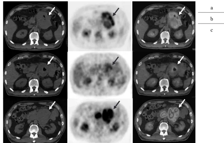
CASE 2
A 50-year-old man had an abdominal mass near the pancreatic body, which showed intense FDG uptake (SUVmax 8.0) (Fig. 3a). Tumor with ulcer formation was detected in the horizontal part of the duodenum at endoscopy. For biopsy, the mass was confirmed pathologically as GIST of the duodenum and categorized as high risk. Systemic chemother-apy with imatinib mesylate was started as neoadju-vant therapy. Follow-up PET/CT was performed 4 months after chemotherapy showed a reduction in tumor size and decreased FDG uptake (SUVmax 5.0) (Fig. 3b). However, follow-up PET/CT at 10 months after chemotherapy showed tumor enlarge-ment and increased FDG uptake (SUVmax 10.0) (Fig. 3c).
DISCUSSION
All primary GISTs and recurrent tumors can be detected by18F-FDG PET/CT. GIST can be shown to be FDG-positive, with a varying degree of uptake. Isis G et al. reported the sensitivity of GISTs includ-ing metastatic lesions by FDG-PET was 86% (1). Kamiyama et al. reported that the sensitivity of GISTs by FDG-PET was 100% (4). FDG uptake and pathological risk category may have some relation-ship. It has been reported that a high rate of FDG uptake in GIST indicates high metabolism and ma-lignant potential (4). There have been many reports that FDG accumulation and grade of malignancy are correlated with each other (4, 5). Kamiyama et al. reported that there was a significant difference be-tween the SUV of the primary lesions categorized as high and intermediate risk (6.52!1.15) and that of the primary lesions categorized as low risk (2.18! 0.06) (p!0.01) (4, 5). In our study, all the primary high - risk GISTs showed intense FDG uptake (SUVmax 11.8!3.15). The low- and intermediate-risk GISTs also showed FDG uptake (SUVmax
aa
b b
cc
Fig. 3. Transaxial CT (left), FDG - PET (middle), and fusion (right) images of the abdomen in a 50 - year - old man with GIST cate-gorized as high risk. The abdominal tumor showed intense FDG uptake (SUVmax 8.0) (a, arrow). Reduction in tumor size and lower FDG uptake (SUVmax 5.0) were observed on a PET/CT image (b, arrow) performed 4 months after chemotherapy with imatinib mesylate, but tumor enlargement and increased FDG uptake (SUVmax 10.0) were observed on a PET/CT image (c, arrow) performed 10 months after chemotherapy.
2.88!0.47). The SUVmax of the primary lesions categorized as high risk was significantly higher than that of the primary lesions categorized as low and intermediate risk. This observation suggested that the degree of FDG uptake may be a useful in-dicator of risk category. The size of the recurrent tumor in patient No.14 was small (about 1 cm). FDG uptake depends on spatial resolution of the PET/CT system and a partial volume effect may decrease FDG uptake, which leads to low SUV in a small tumor (6). Therefore, this recurrent tumor showed relatively low uptake (SUVmax 2.5) in spite of being high-risk.
The patients of GIST post operation showing no abnormal FDG uptake on PET/CT are clinically considered as no recurrence and no metastasis. PET/CT is useful for follow-up examinations of GIST post operation.
In case 2, follow-up PET/CT performed 4 months after chemotherapy showed a reduction in tumor size and lower FDG uptake. Imatinib mesylate was considered to be effective, but follow-up PET/CT performed 10 months after chemotherapy showed tumor enlargement and increased FDG uptake. This was considered to be indicative of resistance to imatinib mesylate (7). It has been reported that many GISTs may not change significantly in size during early response to imatinib mesylate admini-stration (8). PET has been shown to be highly sen-sitive in detecting early response to imatinib me-sylate therapy (8, 9). It has been reported that den-sity reduction on CT reflects necrotic changes in the tumor and is a predictor of early response to imatinib mesylate therapy. However, it is relatively difficult to evaluate early response to imatinib me-sylate therapy by CT alone (8, 10). If a tumor tem-porarily grew larger as an early response to imatinib mesylate therapy, it might be evaluated as show-ing a lack of response to imatinib mesylate therapy (10). By means of PET/CT, we can evaluate re-sponse to imatinib mesylate therapy before tumor shrinkage.
It may be that GISTs in pathologically high-risk patients have high glucose metabolism (4). Since 18F-FDG PET/CT can evaluate glucose metabolism, it may be a useful tool for determining GIST risk category.
REFERENCES
1. Isis G, Thuan V, Revathy I, Marcella J, Homer M, Nancy S, Donald P : The role of18F-FDG PET in staging and early prediction of response to therapy of recurrent gastrointestinal stromal tumors. J Nucl Med 45 : 17-21, 2004
2. Gerald A, Jorg K, Sebastian B, Hilmar K, Katrin RK, Jochen S, Andreas B, Jorg FD, Lutz SF : Comparison of PET, CT, and dual-modality PET/CT imaging for monitoring of imatinib (STI571) therapy in patients with gastrointes-tinal stromal tumors. J Nucl Med 45 : 357-365, 2004
3. Fletcher CD, Berman JJ, Corless C, Gorstein F, Lasota J, Longley BJ : Diagnosis of gastro-intestinal stromal tumors : A consensus ap-proach. Human Pathol 33 : 459-465, 2002 4. Kamiyama Y, Aihara R, Nakabayashi T, Mochiki
E, Asao T, Kuwano H, Oriuchi N, Endo K :18 F-fluorodeoxyglucose positron emission tomogra-phy : useful technique for predicting malignant potential of gastrointestinal stromal tumors. World J Surg 29 : 1429-1435, 2005
5. Kamiyama Y, Mochiki E, Kuwano H : The di-agnosis of gastrointestinal stromal tumors us-ing PET. Surgery 68 : 678-683, 2006
6. Tateishi U, Miyake M, Maeda T, Arai Y : Clini-cal radiology of GIST. GastroenterologiClini-cal Sur-gery 29 : 167-176, 2006
7. Nishida T : Evidence-based chemotherapy for gastrointestinal stromal tumor. Surgery 67 : 412-418, 2005
8. Komatsu Y, Yuki S, Asaka M : FDG-PET for prediction of response in GIST with imatinib mesylate (GIST). Gastroenterology 40 : 633-638, 2005
9. Banzo I, Quirce R, Martinez-Rodriguez I, Jimenez-Bonilla JF, Sainz-Esteban A, Barragan J, Portilla-Quattrociocchi H, Carril JM :18F-FDG PET/CT in response evaluation of gastrointesti-nal stromal tumors treated with imatinib. Rev Esp Med Nucl 27 : 168-175, 2008
10. Choi H, Charnsangavej C, de Castro Faria S, Tamm EP, Benjamin RS, Johnson MM, Macapinlac HA, Podoloff DA : CT evaluation of the response of gastrointestinal stromal tumors after imatinib mesylate treatment : a quantitative analysis correlated with FDG PET findings. AJR Am J Roentgenol 183 : 1619-1628, 2004