Anus,Rectum
andColon
http://journal-arc.jpReview Article
Current Status and Prospects of Endoscopic Resection Technique for
Colorectal Tumors
Keigo Suzuki1), Shoichi Saito1)and Yosuke Fukunaga2)
1) Department of Gastroenterological Medicine, Cancer Institute Hospital of Japanese Foundation for Cancer Research, Tokyo, Japan
2) Department of Gastroenterological Surgery, Cancer Institute Hospital of Japanese Foundation for Cancer Research, Tokyo, Japan
Abstract
Currently, endoscopic submucosal dissection (ESD) is a well-established and common treatment for intra-mucosal colorectal cancer in Japan. However, colorectal ESD is technically more difficult to perform than esophageal and gastric ESD, and some lesions, such as fibrotic lesions, are difficult to dissect by endo-scopy. Several techniques, such as the pocket-creation method and laparoscopically assisted endoscopic polypectomy, have been utilized for challenging targets. In recent years, endoscopic full-thickness resection (EFTR) using full-thickness resection devices have mainly been performed in Western countries. We have used laparoscopy and endoscopy cooperative surgery for colorectal tumors (LECS-CR) since 2011 for the challenging treatment of colorectal ESD. Improvements in ESD techniques have resulted in an increase in the literature on EFTR, and LECS-CR may be considered an effective endoscopic technique for colorectal ESD in the future.
Keywords
endoscopic submucosal dissection, laparoscopy and endoscopy cooperative surgery, combined of endo-scopic and laparoendo-scopic surgery, endoendo-scopic full-thickness resection
J Anus Rectum Colon 2021; 5(2): 121-128
Introduction
Colorectal cancer (CRC) is the second most common cause of mortality due to cancer in Japan[1]. Moreover, more than one million patients are diagnosed annually with CRC worldwide, with more than 600,000 deaths reported globally[2]. Meanwhile, most cases of CRC are sporadic and develop slowly over several years through the adenoma-carcinoma sequence[2]. Therefore, regular endoscopic screening every few years is useful, and resection at the ade-noma or intramucosal cancer stage has a very good five-year survival rate with little risk of metastasis.
In Japan, endoscopic treatment is recommended for
intra-mucosal carcinoma or carcinoma with slight subintra-mucosal in-vasion. Moreover, endoscopic submucosal dissection (ESD) is performed on lesions >2 cm in size[3]. Currently, colorec-tal ESD is a common procedure that is performed along with endoscopic mucosal resection and has become widely used since it was approved for under the national health in-surance for early stage colorectal neoplasia in 2012. How-ever, lesions with severe fibrosis, such as the presence of re-sidual or local recurrent lesions after endoscopic treatment, or tumors involving the diverticulum or appendix, are chal-lenging targets within the digestive tract. Endoscopic resec-tion has scarcely been utilized in the treatment of these col-orectal tumors due to technical difficulties and the
possibil-Corresponding author: Shoichi Saito, shoichi.saito@jfcr.or.jp Received: November 1, 2020, Accepted: December 16, 2020 CopyrightⒸ 2021 The Japan Society of Coloproctology
ity of perforation and failure associated with en bloc resec-tion[4-7].
Although there are reports regarding the utility of traction devices or the performance of pocket-creation methods for challenging targets[8-10], the technical challenges and risks of perforation still persist. In contrast, although more inva-sive than endoscopic treatment and particularly used for eld-erly patients or patients with poor general condition, surgical resection is the treatment of choice for patients undergoing procedures other than endoscopic treatment.
To overcome these problems, EFTR and other laparo-scopically assisted endoscopic treatments have been widely reported. In 2011, we developed a full-thickness resection method for colorectal tumors using laparoscopy and endo-scopy, which we called laparoscopy and endoscopy coopera-tive surgery for colorectal tumors (LECS-CR), which has been previously reported[11,12].
LECS-CR was developed based on a novel procedure for gastric submucosal tumors (SMTs) called laparoscopy and endoscopy cooperative surgery (LECS), which was reported in 2008 by Hiki et al.[13]. LECS was approved for imple-mentation under national health insurance for gastric SMT in 2014 and is now performed at several healthcare centers. In addition to the classical LECS procedure, several LECS procedures have been developed, such as the combination of laparoscopic and endoscopic approaches for neoplasia with nonexposure technique, nonexposed endoscopic wall-inversion surgery, and closed LECS[14-16].
LECS-CR achieves good results in the form of high en bloc resection rates and improved short-term out-comes[11,12]; LECS-CR differs from other EFTRs in that the resection line is endoscopically confirmed, and full-thickness resection along with the previously made resection line is performed both laparoscopically and endoscopically. This article presents the methods and treatment outcomes of LECS-CR and reviews the current situation of other meth-ods.
Patients and Methods
In total, 22 lesions were treated with LECS-CR at the Cancer Institute Hospital of the Japanese Foundation for Cancer Research from November 2011 to August 2020. LECS-CR was indicated in the following cases: (1) adenoma or intramucosal cancer with severe fibrosis; (2) adenoma or intramucosal cancer involving the diverticulum or appendix; and (3) SMT.
In all cases, the lesion was evaluated by magnified image-enhanced endoscopy prior to surgery, and for lesions sus-pected to have deep invasion into the submucosal layer, LECS-CR was not indicated owing to the risk of lymph node metastasis. If the lesion involving the appendix could be resected with a sufficient safety margin using a linear
sta-pler alone and ileocecal valve preservation was possible, we did not perform LECS-CR; instead, we performed laparo-scopic sleeve resection-assisted endolaparo-scopically and com-bined endoscopic and laparoscopic surgery (CELS). When-ever a surgeon approximated a laparoscopic linear stapler to the cecum, an endoscopist would ensure the appropriate margin from the lesion via the endoscopic direct vision. This approach can also be categorized as CELS[17,18].
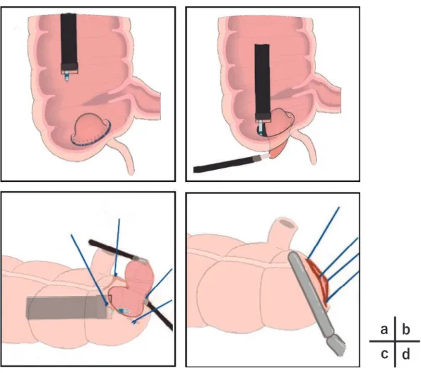
The methods of LECS-CR at our hospital are described below. Figure 1 shows a simple graphical illustration of the technique.
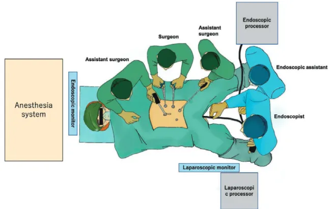
This procedure is performed under general anesthesia in the operating room, and the patient assumes a modified lithotomy position. Typically, a main operator and an assis-tant surgeon stand on the left side of the patient, with an-other assistant surgeon standing on the right side of the pa-tient; however, when approaching the cecum, all surgeons are positioned on the left side of the patient (Figure 2a). An endoscopist stands between the patient’s legs and operates the endoscope, while the assistant operates the treatment de-vice from behind the endoscopist.
First, five trocars are placed on the abdomen, starting with 8 mmHg CO2 pneumoperitoneum. Following confirma-tion of the tumor locaconfirma-tion by endoscopy and laparoscopy, the colon wall at the site of the lesion is exposed. Subse-quently, an endoscopist performs an intraluminal wash out around the lesion with approximately 1,000 ml of saline so-lution and takes the last 10 mL of the irrigated saline solu-tion as a sample for urgent cytology examinasolu-tion. After con-firming absence of malignant cells in the sample, circumfer-ential markings surrounding the lesion were made using a needle knife or a Dual Knife JⓇ (Olympus Medical, Tokyo, Japan).
The mucosal incision and submucosal dissection were performed endoscopically with an appropriate safety margin. To prevent the leaking of intraintestinal fluid in the abdomi-nal cavity, full-thickness resection was performed using the crown method reported by Nunobe et al.[19] in the treat-ment of gastric LECS. Complete full-thickness dissection and excision is performed by combining the endoscopic and laparoscopic procedures.
The specimen is retrieved in the intraluminal route via the anus and confirmed by macroscopic en bloc resection with a safety margin. After the resection, the colon wall was se-quentially closed using laparoscopic linear staplers.
All endoscopic procedures were performed using a single-channel endoscope with a water-jet system (PCF-Q260J, Olympus Optical Co., Ltd., Tokyo, Japan) with a large open-ing end attachment (D-201-11804; Olympus Medical, To-kyo, Japan).
A mixture of concentrate glyceride with small amounts of indigo carmine and epinephrine was used as a submucosal injection solution.
Figure 1. The procedure of laparoscopy and endoscopy cooperative surgery for colorectal tumors. a: The marking surrounds the lesion and mucosal incision with the endoscopic technique.
b: The submucosal dissection and seromuscular layer resection.
c: The lesion is lifted by the “crown method.” Laparoscopic ultrasound-activating scissors are also used for seromuscular layer resection and for the endoscopic collection of lesions.
d: Finally, the colon is closed with a laparoscopic linear stapler.
D E
F G
The mucosal incision was performed with a needle knife or a Dual Knife JⓇ, and seromuscular layer resection was performed with a Hook knife (Olympus Medical, Tokyo, Ja-pan), ITknife 2Ⓡ (Olympus Medical, Tokyo, Japan), or Dual KnifeⓇ. In normal colorectal ESD, ITknife2Ⓡ is significantly easier to use than ITknife nanoⓇ (Olympus Medical, Tokyo, Japan).
As an electrosurgical unit for the endoscopic procedure, we used VIO 300 D (ERBE Elektromedizin GmbH, Tübin-gen, Germany) under the following settings: EndoCut I mode (effect: 2, duration: 2, interval: 2) for the mucosal in-cision and resection of most of the seromuscular layer and SWIFT coagulation mode (effect: 3, 45 W) for numerous blood vessels. As a laparoscopic linear stapler, Endo GIA (Covidien, Dublin, Ireland) or Powered ECHELON (Ethicon Inc., Bridgewater Township, New Jersey, United States) was used.
Results
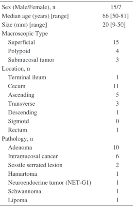
The number of lesions resected using the LECS-CR was 22. Patient demographics and lesion characteristics are shown in Table 1. With the most common histological diag-nosis being adenoma, followed by adenocarcinoma, in which all cases presented with invasion of the intramucosal layer, most of the lesions treated using LECS-CR were ob-served in the cecum and involved the appendix.
In the two histologically diagnosed cases of sessile ser-rated lesion (SSL), both cases presented with invasion that extended into the appendiceal orifice. These two cases had been previously diagnosed endoscopically as SSL without dysplasia. Therefore, we suggested a follow-up observation; however, both were resected based on the patient’s request.
There were three cases of SMTs, one of which was pa-thologically diagnosed as a neuroendocrine tumor (NET) having initially been suspected as GIST by ultrasound
endo-Figure 2a. Suggested endoscope and laparoscope placement for cecal lesions.
Figure 2b. The image of LECS-CR.
scopy. Although total mesorectal excision with lymph node dissection is generally recommended for rectal NETs >2 cm in diameter, this patient declined additional surgical inter-vention despite providing all disease-related information.
The intraoperative and postoperative data are shown in Table 2. Both the complete one-piece resection rate and R0 resection rate were 100%. While there were no cases of conversion to open surgery, one patient conversely under-went laparoscopic ileocecal resection due to intraoperative injury of the terminal ileum. We speculated that the reason for the injury was the strong adhesion between the terminal ileum and the appendix due to previous asymptomatic ap-pendicitis. Postoperative complications of grade III or higher according to the Clavien-Dindo classification were not
ob-served in this series. All 17 patients who were followed up for at least one year were free of recurrence and metastasis.
Procedures and Current Status of
Other Techniques
Combined endoscopic and laparoscopic surgery (CELS)
CELS is widely used as a common term for a collabora-tive technique of laparoscopy and endoscopy, and several methods have been reported. Since Beck et al.[20] reported a laparoscopically assisted snaring technique for the colon in 1993, several studies have been published, mainly in West-ern countries. CELS for colorectal lesions can be divided into the following subcategories. One category is laparoscopy-assisted endoscopic resection (LAER), which is defined as a technique involving endoscopic polypectomy with visualization using laparoscopy; another category in-cludes endoscope-assisted laparoscopic resection (EALR), which is characterized by the resection of a lesion using la-paroscopic techniques, while the lesion is visualized by en-doscopy. Several papers on EALR reported the technical benefits of this method, especially relating to the co-lon[20-26]. The technique reported by Beck et al. is called laparoscopic-assisted polypectomy, which is a type of LAER and is the most widely adapted technique among the cur-rently performed colorectal CELS procedures.
Table 1. Baseline Characteristics of the Patients and Lesions Treated with Laparoscopy and Endos-copy Cooperative Surgery for Colorectal Tumors.
Sex (Male/Female), n 15/7
Median age (years) [range] 66 [50-81]
Size (mm) [range] 20 [9-50] Macroscopic Type Superficial 15 Polypoid 4 Submucosal tumor 3 Location, n Terminal ileum 1 Cecum 11 Ascending 5 Transverse 3 Descending 1 Sigmoid 0 Rectum 1 Pathology, n Adenoma 10 Intramucosal cancer 6
Sessile serrated lesion 2
Hamartoma 1
Neuroendocrine tumor (NET-G1) 1
Schwannoma 1
Lipoma 1
Table 2. Intraoperative and Postoperative Data.
Hemorrhage (ml) 5
Median procedure time (min) 174
En bloc resection (%) 100
R0 resection (%) 100
Intraoperative adverse event, n (%) 1 (4.5)
Conversion to other surgery, n (%) 1 (4.5)
Postoperative adverse event (CD Grade III or more), n (%) 0 (0)
Postoperative days (days) 6
be considered as a type of CELS, although few resections have been reported to be used in the colon, colonic EALR is mainly performed using laparoscopic wedge resection under endoscopic observation, which is performed with a linear stapler along the long axis of the intestinal tract[27,28].
According to the literature, in each procedure, patients are operated in the lithotomy position under general anesthesia, similar to the LECS-CR. After trocar insertion via the peri-toneum, carbon dioxide insufflation is applied followed by laparoscopy. Endoscopy is sequentially performed for assis-tance in the laparoscopic procedures. These series of flows are similar to our LECS-CR. CELS can be converted to la-paroscopic surgery when adverse events, such as perforation, are observed.
Other endoscopic full-thickness resection (EFTR) proce-dures
Although there are few institutions that perform the la-paroscopic combined procedure in the treatment of colorec-tal lesions as frequently as in the upper GI tract, in the up-per GI tract, LECS and several other combination tech-niques of EFTR with laparoscopy are widely used. Mean-while, the incidence of EFTR without laparoscopy has gradually increased in the treatment of colorectal lesions in recent years. Particularly in the past couple of years, an in-creased number of studies on EFTR using FTRDⓇ (Ovesco
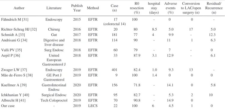
AG, Tübingen, Germany), which is an all-layer ablation de-vice based on the Over-The-Scope Clip system, have been reported. Table 3 shows some of the most recent studies on this subject.
A principal method of EFTR using FTRD is as follows: the attachment with a large suture clip is placed on the tip of the endoscope, then the colonoscope reaches the lesion, and the lesion is grasped and pulled enough into the attach-ment. The clip was then deployed, and the tissue above the clip was immediately resected with a snare. The lesions were collected via the perianal route.
This new technique is of interest to endoscopists in West-ern countries because it is easier to perform than ESD pro-cedure.
Discussion
Colonic partial resection is generally considered a chal-lenging target of ESD. However, in recent years, despite the efforts of laparoscopic and function-preserving surgeries having been attempted to achieve less invasive resections while ensuring radical cure, there is still a large difference in the degree of invasiveness between these techniques and endoscopic treatment. Although the technique of closing all layers, including the seromuscular layer, is still in its infancy and has not yet been standardized as a treatment method, in terms of invasiveness, endoscopy alone (pure EFTR) is the goal of endoscopists. To date, sufficient suturing techniques and instruments have been developed in the field of laparo-scopic surgery, and considering the current situation, where pure EFTR is not widely used, LECS-CR is a type of EFTR that can be performed at any institution.
Before the introduction of LECS-CR, conventional laparo-scopic colorectal resection was performed for challenging targets in ESD at our institution. Laparoscopic colorectal re-section is superior to open colon rere-section since it is less in-vasive and is associated with faster recovery of bowel func-tion[29]. In 2019, Suzuki et al.[12] reported that LECS-CR for challenging lesions in ESD resulted in a significantly shorter hospital stay compared with conventional laparo-scopic surgery for the same types of lesions. These results suggest that LECS-CR surgery should be preferred in these
Table 3. Literature of Colorectal EFTR.
Author Literature Publish
Year Method Case (n) R0 resection (%) hospital stay (days) Adverse events (%) Conversion to LAC/open surgery (n) Residual/ Recurrence (n)
Fähndrich M [31] Endoscopy 2015 EFTR 17
(colorectal 14)
100 - 0 0
-Richter-Schrag HJ [32] Chirurg 2016 EFTR 20 80 8.5 5.0 17 5.0
Schmidt A [33] Gut 2017 EFTR 181 77 4 9.9 - 12.3
Andrisani G [34] Digestive and liver disease
2018 EFTR 114 90 - 11 1 6.3
Valli PV [35] Surg Endosc 2018 EFTR 60 79 - 7 - 0
Aepil P [36] United
European Gastroenterol J
2018 EFTR 33 87.9 3.1 12.9 - 6.1
Zwager LW [37] Endoscopy 2019 EFTR 401 82.4 1.0 9.3 13
-Mão de-Ferro S [38] GE Port J Gastroenterol
2019 EFTR 9 100 1.4 0 0 0
Kuellmer A [39] GastroIntestinal Endosc
2020 EFTR 156 71.8 - 14.1 0 5.8
Ichkhanian Y [40] Surgical Endosc 2020 EFTR 95 82.7 - 5.3 2 10
Albrecht H [41] Tech Coloproctol 2019 EFTR 70 90.8 - 14.9 0
Our case - 2019 LECS 22 100 6 4.5 1 0
EFTR, endoscopic full-thickness resection; LAC, laparoscopic colectomy; LECS, laparoscopy and endoscopy cooperative surgery
cases owing to the degree of invasiveness of the method compared with conventional laparoscopic surgery.
CELS, EFTR with FTRD, and LECS-CR are still consid-ered minimally invasive treatment options that can be cur-rently performed. CELS could be considered a useful treat-ment because it has been developed for a relatively long time, and several studies have proven its utility; however, al-though LECS-CR is sometimes considered a part of CELS, we have emphasized on it as a different method. Its long-term results are also good, with only one case of local re-currence in a systematic review by Nakajima et al.[30]. The treatment outcomes of LECS-CR have been shown to be su-perior to those of CELS, especially in terms of the R0 re-section rate, which is higher than that of CELS[12]. LAER has a limitation in the size of tumors that can be resected en bloc owing to the snare size. It is considered that en bloc re-section would have better oncological results, such as lower local recurrence rate, than piecemeal resection, indicating the technical superiority of the LECS-CR over the LAER.
Recently, there have been an increasing number of reports on EFTR using FTRD. The number of multicenter studies is also increasing, and a prospective nonrandomized clinical trial (the WALL-RESECT study) was conducted at nine cen-ters in Germany and reported by Schmidt et al.[33]. EFTR with FTRD was not technically complex, and the success rate of all procedures was relatively good. However, the R0 resection rate varied among different reports, as shown in Table 3. Kuellmer et al.[39] reviewed 156 cases accumulated at a German multicenter and reported an R0 resection rate of 60.9%. Andrisani et al. reported results from 12 centers
in Italy and found that the R0 resection rate was similar to that reported by Ischkanian et al. regarding the treatment outcome at 12 centers in the USA, which was 82.7%[34]. A meta-analysis of nine studies showed a total of 469 patients with an R0 resection rate of 84.9%[42]. In the literature, the R0 resection rate of EFTR with FTRD was only 90% at best reported within two years of the accumulation of cases, in which the curative potential of EFTR with FTRD can be assured.
Where the EFTR with FTRD has shown better results in that it requires a shorter procedure time and can be com-pleted by endoscopy alone, the LECS-CR has an R0 resec-tion rate of 100% and is considered superior to CELS and EFTR with FTRD in terms of cure rate. However, EFTR with FTRD tends to have a lower R0 resection rate in cases of large tumors due to the characteristics of the device[40], which is one of the difficulties of EFTR with FTRD. Addi-tionally, the postoperative residuals rate is high[33].
It is remarkable that EFTR with FTRD has a higher inci-dence of adverse events than LECS-CR. The WALL-RESECT study shows a high complication rate, with a procedure-related adverse events rate of 9.9%[33]. Further-more, there have been several reports of acute appendicitis after EFTR with FTRD for lesions involving the appendix, which required an appendectomy[33]. While it has been considered that cases involving the appendix may be better candidates for LECS-CR, appendix resection in LECS-CR is performed in cases involving the appendix, which can pre-vent the subsequent development of appendicitis and unnec-essary surgical intervention. Other adverse events in FTRD
procedures include fistula formation, perforation, and bleed-ing[33].
The indication for LECS-CR is limited to intramucosal le-sions from the viewpoint of curative properties. Further-more, we have discussed the further adaptation for LECS-CR including the following: (1) deep submucosal invasive cancer after rapid intraoperative lymph node evaluation us-ing sentinel biopsy or one-step nucleic acid amplification; (2) treatment for metachronous cancer in patients with previ-ously operated CRC with a substantially shortened colon; and (3) treatment in elderly patients who may not be able to choose between routine surgery or no surgery at all.
Although colorectal ESD has been widely performed in Japan, it is not considered a popular technique in Western countries similar to other endoscopic procedures, with an-other problem with LECS-CR being the technical difficulty associated with the procedure. LECS-CR is based on ESD technology; therefore, it is more difficult than EFTR and CELS for endoscopists unfamiliar with ESD.
In LECS-CR, the crown method is used to prevent colo-nic content leakage; however, this method does carry the possible risk of peritoneal dissemination. Closed LECS-CR was attempted to be performed previously; nevertheless, we converted it to the conventional method due to technical dif-ficulties.
If it will be considered an indication for invasive cancers in the future, closed LECS should be adopted for use in gastric SMT[14-16]. At present, it is necessary to have a long-term follow-up to determine whether peritoneal dis-semination or multiple organ metastases occur.
Although this method has been mainly performed at our institution, there have been case reports from other institu-tions in recent years, and additional cases are expected to be reported in the future[43,44].
Conclusion
Minimally invasive treatments for colorectal tumors have merited attention and various techniques have been devel-oped, but to date their curability and safety have never been sufficient. In contrast, LECS-CR will be promised for a fea-sible treatment as a full-thickness resection near the future.
Acknowledgements
We would like to thank Dr. Mitsuaki Ishioka and Dr. Yuki Suzuki for providing illustrations.
Conflicts of Interest
There are no conflicts of interest.
References
1. Cancer Registry and Statistics. Cancer Information Service, Na-tional Cancer Center, Japan (Vital Statistics of Japan).
2. Brenner H, Kloor M, Pox CP. Colorectal cancer. Lancet. 2014 Apr; 383(9927): 1490-502.
3. Hashiguchi Y, Muro K, Saito Y, et al. Japanese Society for Cancer of the Colon and Rectum (JSCCR) guidelines 2019 for the treat-ment of colorectal cancer. Int J Clin Oncol. 2020 Jan; 25(1): 1-42. 4. Sato K, Ito S, Kitagawa T, et al. Factors affecting the technical difficulty and clinical outcome of endoscopic submucosal dissec-tion for colorectal tumors. Surg Endosc. 2014 Oct; 28(10): 2959-65.
5. Hayashi N, Tanaka S, Nishiyama S, et al. Predictors of incomplete resection and perforation associated with endoscopic submucosal dissection for colorectal tumors. Gastrointest Endosc. 2014 Mar; 79(3): 427-35.
6. Makino T, Kanmura S, Sasaki F, et al. Preoperative classification of submucosal fibrosis in colorectal laterally spreading tumors by endoscopic ultrasonography. Endosc Int Open. 2015 Aug; 3: E363-7.
7. Imai K, Hotta K, Yamaguchi Y, et al. Preoperative indicators of failure of en bloc resection or perforation in colorectal endoscopic submucosal dissection: implications for lesion stratification by technical difficulties during stepwise training. Gastrointest Endosc. 2016 May; 83(5): 954-62.
8. Faller J, Jacques J, Oung B, et al. Endoscopic submucosal dissec-tion with double clip and rubber band tracdissec-tion for residual or lo-cally recurrent colonic lesions after previous endoscopic mucosal resection. Endoscopy. 2020 May; 52(5): 383-8.
9. Oung B, Rivory J, Chabrun E, et al. ESD with double clips and rubber band traction of neoplastic lesions developed in the appen-diceal orifice is effective and safe. Endosc Int Open. 2020 Mar; 8 (3): E388-95.
10. Yoshida N, Naito Y, Yasuda R, et al. The efficacy of the pocket-creation method for cases with severe fibrosis in colorectal endo-scopic submucosal dissection. Endosc Int Open. 2018 Aug; 6(8): E 975-83.
11. Tamegai Y, Fukunaga Y, Suzuki S, et al. Laparoscopic and endo-scopic cooperative surgery (LECS) to overcome the limitations of endoscopic resection for colorectal tumors. Endosc Int Open. 2018 Dec; 6(12): E1477-85.
12. Suzuki S, Fukunaga Y, Tamegai Y, et al. The short-term outcomes of laparoscopic-endoscopic cooperative surgery for colorectal tu-mors (LECS-CR) in cases involving endoscopically unresectable colorectal tumors. Surgery Today. 2019 Dec; 49(12): 1051-7. 13. Hiki N, Yamamoto Y, Fukunaga T, et al. Laparoscopic and
endo-scopic cooperative surgery for gastrointestinal stromal tumor dis-section. Surg Endosc. 2008 Jul; 22(7): 1729-35.
14. Inoue H. Endoscopic mucosal resection, endoscopic submucosal dissection, and beyond: full-layer resection for gastric cancer with nonexposure technique (CLEAN-NET). Surg Oncol Clin N Am. 2012 Jan; 21(1): 129-40.
15. Goto O. NEWS: application and procedure. Gastroenterol Endosc. 2015; 57(8): 1632-40.
16. Kikuchi S, Nishizaki M, Kuroda S, et al. Nonexposure laparo-scopic and endolaparo-scopic cooperative surgery (closed laparolaparo-scopic and endoscopic cooperative surgery) for gastric submucosal tumor. Gastric Cancer. 2017 May; 20(3): 553-7.
17. Garrett KA. Combined endoscopic and laparoscopic surgery. Clin Colon Rectal Surg. 2015 Sep; 28(3): 140-5.
18. Noren ER, Wickham C, Lee SW. Current techniques for combined endoscopic and laparoscopic surgery (CELS). Ann Laparosc
En-dosc Surg. 2019 Aug; 4: 77.
19. Nunobe S, Hiki N, Gotoda T, et al. Successful application of la-paroscopic and endoscopic cooperative surgery (LECS) for a lateral-spreading mucosal gastric cancer. Gastric Cancer. 2012 Jul; 15(3): 338-42.
20. Beck DE, Karulf RE. Laparoscopic-assisted full-thickness endo-scopic polypectomy. Dis Colon Rectum. 1993 Jul; 36(7): 693-5. 21. Franklin ME Jr, Díaz-E JA, Abrego D, et al. Laparoscopic-assisted
colonoscopic polypectomy: the Texas Endosurgery Institute experi-ence. Dis Colon Rectum. 2000 Sep; 43(9): 1246-9.
22. Ommer A, Limmer J, Möllenberg H, et al. Laparoscopic-assisted colonoscopic polypectomy―indications and results [in German]. Zentralbl Chir. 2003 Mar; 128(3): 195-8.
23. Wilhelm D, von Delius S, Weber L, et al. Combined laparoscopic-endoscopic resections of colorectal polyps: 10-year experience and follow-up. Surg Endosc. 2009 Apr; 23(4): 688-93.
24. Lee SW, Garrett KA, Shin JH, et al. Dynamic article: long-term outcomes of patients undergoing combined endolaparoscopic sur-gery for benign colon polyps. Dis Colon Rectum. 2013 Jul; 56(7): 869-73.
25. Franklin ME Jr, Leyva-Alvizo A, Abrego-Medina D, et al. Laparo-scopically monitored colonoscopic polypectomy: an established form of endoluminal therapy for colorectal polyps. Surg Endosc. 2007 Sep; 21(9): 1650-3.
26. Franklin ME Jr, Portillo G. Laparoscopic monitored colonoscopic polypectomy: long-term follow-up. World J Surg. 2009 Jun; 33(6): 1306-9.
27. Lin AY, O’Mahoney PR, Milsom JW, et al. Full-thickness excision for benign colon polyps using combined endoscopic laparoscopic surgery. Dis Colon Rectum. 2016 Jan; 59(1): 16-21.
28. Leicher LW, De Vos tot Nederveen Cappel WH, van Westreenen HL. Limited endoscopic-assisted wedge resection for excision of colon polyps. Dis Colon Rectum. 2017 Mar; 60(3): 299-302. 29. Clinical Outcomes of Surgical Therapy Study Group, Nelson H,
Sargent DJ, et al. A comparison of laparoscopically assisted and open colectomy for colon cancer. N Engl J Med. 2004 May; 350 (20): 2050-9.
30. Nakajima K, Sharma SK, Lee SW, et al. Avoiding colorectal re-section for polyps: is CELS the best method? Surg Endosc. 2016 Mar; 30(3): 807-18.
31. Fähndrich M, Sandmann M. Endoscopic full-thickness resection for gastrointestinal lesions using the over-the-scope clip system: a case series. Endoscopy. 2015 Jan; 47(1): 76-9.
32. Richter-Schrag HJ, Walker C, Thimme R, et al. Full thickness re-section device (FTRD): experience and outcome for benign neo-plasms of the rectum and colon. Chirurg. 2016 Apr; 87(4):
316-25.
33. Schmidt A, Beyna T, Schumacher B. Colonoscopic full-thickness resection using an over-the-scope device: a prospective multicentre study in various indications. Gut. 2018 Jul; 67(7): 1280-9. 34. Andrisania G, Sorianib P, Manno M, et al. Colo-rectal endoscopic
full-thickness resection (EFTR) with the over-the-scope device (FTRDⓇ): a multicenter Italian experience. Dig Liver Dis. 2019
Mar; 51(3): 375-81.
35. Valli PV, Mertens J, Bauerfeind P, et al. Safe and successful resec-tion of difficult GI lesions using a novel single-step full-thickness resection device (FTRD). Surg Endosc. 2018 Jan; 32(1): 289-99. 36. Aepli P, Criblez D, Baumeler S. Endoscopic full thickness
tion (EFTR) of colorectal neoplasms with the full thickness resec-tion device (FTRD): clinical experience from two tertiary referral centers in Switzerland. United European Gastroenterol J. 2018 Apr; 6(3): 463-70.
37. Zwager LW, Bastiaansen BAJ, Bronzwaer MES, et al. Endoscopic full-thickness resection (eFTR) of colorectal lesions: results from the Dutch colorectal eFTR registry. Endoscopy. 2020 Jun. 38. Mão de-Ferro S, Castela J, Pereira D, et al. Endoscopic
full-thickness resection of colorectal lesions with the new FTRD sys-tem: single-center experience. GE Port J Gastroenterol. 2019; 26 (4): 235-41.
39. Kuellmer A, Mueller J, Caca K, et al. Endoscopic full-thickness resection for early colorectal cancer. Gastrointest Endosc. 2019 Jun; 89(6): 1180-9.
40. Ichkhanian Y, Vosoughi K, Diehl DL, et al. A large multicenter co-hort on the use of full-thickness resection device for difficult colo-nic lesions. Surg Endosc. 2020 Mar.
41. Albrecht H, Raithel M, Braun A, et al. Endoscopic full-thickness resection (EFTR) in the lower gastrointestinal tract. Tech Colo-proctol. 2019 Oct; 23(10): 957-63.
42. Li P, Ma B, Gong S, et al. Efficacy and safety of endoscopic full-thickness resection in the colon and rectum using an over-the-scope device: a meta-analysis. Surg Endosc. 2020 Jan.
43. Hiyoshi Y, Yamasaki A, Shono T, et al. Laparoscopic and endo-scopic cooperative surgery for rectal GI stromal tumor. Dis Colon Rectum. 2020 Jan; 63(1): 116.
44. Shi Q, Xu MD, Zhong YS, et al. The laparoscopic-endoscopic co-operative surgery for the colonic calcifying fibrous tumor: one case report. J Laparoendosc Adv Surg Tech A. 2012 Dec; 22(10): 996-8.
Journal of the Anus, Rectum and Colon is an Open Access journal distributed under the Creative Commons Attribution-NonCommercial-NoDerivatives 4.0 In-ternational License. To view the details of this license, please visit (https://creativ ecommons.org/licenses/by-nc-nd/4.0/).