Amount of Green Fluorescent Protein in the Anterior Chamber
after Intravitreal Injection of Triple-Mutated Self-Complementary AAV2 Vectors
is Not Affected by Previous Vitrectomy Surgery
Kazuhisa Takahashi
1,2, Tsutomu Igarashi
1,2,4, Koichi Miyake
1, Maika Kobayashi
1,2,
Yuko Katakai
3, Hiromi Hayashita-Kinoh
1,5, Chiaki Fujimoto
2, Shuhei Kameya
4,
Hiroshi Takahashi
2and Takashi Okada
1,51
Department of Biochemistry and Molecular Biology, Nippon Medical School, Tokyo, Japan 2
Department of Ophthalmology, Nippon Medical School, Tokyo, Japan 3
The Corporation for Production and Research of Laboratory Primates, Ibaraki, Japan 4Department of Ophthalmology, Nippon Medical School Chiba Hokusoh Hospital, Chiba, Japan 5
Division of Molecular and Medical Genetics, The Institute of Medical Science, The University of Tokyo, Tokyo, Japan Background: The adeno-associated virus (AAV) vector is a promising vector for ocular gene therapy.
Surgical internal limiting membrane peeling before AAV vector administration is useful for efficient reti-nal transduction. However, no report has investigated localization of AAV vectors after administration into a post-vitrectomy eye. This study investigated the effects of vitrectomy surgery on intravitreal-injected AAV vector-mediated gene expression in the anterior segment and examined the presence of neutralizing antibodies (NAbs) in serum before and after AAV vector administration.
Methods: Of six eyes from three female cynomolgus monkeys, four were vitrectomized (Group VIT)
and two were non-vitrectomized (Group IV). All eyes were injected with 50 μL of triple-mutated
self-complementary AAV2 vector (1.9 × 10¹³
v.g./mL) encoding green fluorescent protein (GFP). NAbs in the serum were examined before administration and at 2 and 6 weeks after administration. GFP expres-sion was analyzed at 19 weeks after administration.
Results: Immunohistological analysis showed no GFP expression in the trabecular meshwork in any
eye. The GFP genome copy in two slices of the anterior segment was 2.417 (vector genome copies/dip-loid genome) in Group VIT and 4.316 (vector genome copies/dipcopies/dip-loid genome) in group IV. The NAb titer was 1:15.9 (geometric mean) before administration, 1:310.7 at 2 weeks after administration, and 1: 669.4 at 6 weeks after administration.
Conclusion: Previous vitrectomy surgery did not affect gene expression in the anterior segment after
in-travitreal injection of AAV vectors. (J Nippon Med Sch 2021; 88: 103―108)
Key words: AAV vector, intravitreal, injection, vitrectomy, neutralizing antibodies (NAbs), trabecular
meshwork
Introduction
The adeno-associated virus (AAV) vector is a promising vector for ocular gene therapy. Gene therapy for eye dis-eases was initially proposed, in 2008, in a clinical study
of Leber’s congenital amaurosis by Bainbridge et al.1
.
Many ongoing clinical trials include patients with Leber’s
congenital amaurosis2―5
, age-related macular degenera-tion6
, and retinoschisis7
. The two strategies used to de-liver the vector to the retina are subretinal and in-travitreal injection. Because the efficiency of transduction
Correspondence to Tsutomu Igarashi, MD, PhD, Department of Ophthalmology, Nippon Medical School, 1―1―5 Sendagi, Bunkyo-ku, Tokyo 113―8602, Japan
E-mail: tutomu@nms.ac.jp
https://doi.org/10.1272/jnms.JNMS.2021_88-203 Journal Website (https://www.nms.ac.jp/sh/jnms/)
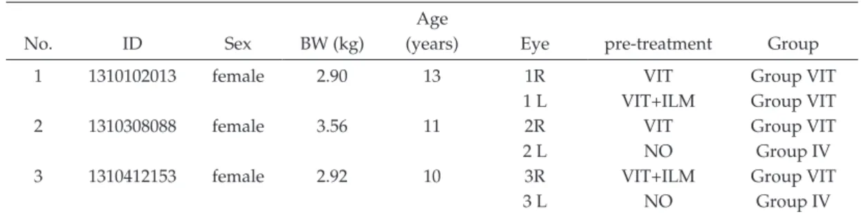
Table 1 Animal characteristics and surgical treatment Age
No. ID Sex BW (kg) (years) Eye pre-treatment Group
1 1310102013 female 2.90 13 1R VIT Group VIT
1 L VIT+ILM Group VIT
2 1310308088 female 3.56 11 2R VIT Group VIT
2 L NO Group IV
3 1310412153 female 2.92 10 3R VIT+ILM Group VIT
3 L NO Group IV
VIT: vitrectomy, ILM: internal limiting membrane peeling, NO: no operation, IV: intravitreal injection, BW: body weight
by injection of subretinal AAV vectors has been previ-ously demonstrated, we performed AAV experiments by
using subretinal AAV vector injections8―10
. However, subretinal injections require creation of iatrogenic retinal
detachment, which could lead to retinal dysfunction11
. In contrast, intravitreal injections are safe and advantageous for gene transduction to the inner retina. The transduc-tion efficiency by intravitreal injectransduc-tion of AAV vectors is high in rodents12,13
. In contrast, the transduction efficiency for simple intravitreal injection of AAV vectors remains low when used in non-human primate retinas, such as
those of monkeys14
. The internal limiting membrane (ILM) may act as a barrier against gene transduction by
intravitreal injection15
. Boye et al. reported that although sub-ILM injection of the AAV vector improved transduc-tion efficiency16
, pressure from sub-ILM injection caused retinal damage; thus, adopting this method for glaucoma would be difficult. Takahashi et al. previously reported that surgical ILM peeling before intravitreal administra-tion of AAV vectors was safe and useful for efficient
transduction of non-human primate retina17
. Lebherz et al. reported that intravitreal AAV7 vector injection was
able to transduce anterior segment structures18
. In addi-tion, because the vitreous is a gel-like collagen, it may prevent widespread exposure of a vector solution in the retina19
. Vitrectomy can cause spreading of AAV vectors, which are injected intravitreally, to the anterior chamber. However, no study has investigated localization of AAV vectors when administered into the vitreous of a vitrec-tomy eye. In the present study, we compared transduc-tion efficiency in the anterior segment of vitrectomized and non-vitrectomized eyes.
Intravitreal administration of AAV vectors generates neutralizing antibodies (NAbs) against the AAV capsid that block AAV vector-mediated gene expression upon re-administration via an intravitreal route into the other eye20
. Kotterman et al. reported that NAb levels in serum
are correlated with those in vitreal fluid21
. Moreover, af-ter intravitreal administration of AAV, they found that the presence of pre-existing NAb titers in serum from monkeys was associated with weak, decaying, or no
transgene expression21
. These previous findings suggest that when performing intravitreal administration of AAV vectors, monitoring NAb titers in the serum may be im-portant. Thus, this study compared serum NAb titers be-fore, and at 2 and 6 weeks after, intravitreal administra-tion of AAV vectors.
Materials and Methods Animals
Three adult female cynomolgus monkeys from the Tsukuba Primate Research Center were used for this study. Each animal weighed approximately 3 kg and was 10-13 years of age (Table 1). All animal procedures were conducted in accordance with the Association for Re-search in Vision and Ophthalmology (ARVO) statement for the Use of Animals in Ophthalmic and Vision Re-search and the Animal Experimental Ethical Review Committee of Nippon Medical School (approval number 27-055). The Animal Welfare and Animal Care Committee of the National Institute of Biomedical Innovation ap-proved the protocols and experimental procedures.
Production and purification of AAV vectors
Dr. Arun Srivastava of the University of Florida kindly provided an AAV packaging plasmid (pACG2-3M) gen-erated by introducing a triple tyrosine-to-phenylalanine (Y444+500+730F) mutation into the VP3 region of the AAV serotype 2 capsid and a self-complementary AAV vector plasmid carrying cDNA encoding enhanced green
fluorescent protein (EGFP) (pdsCBA-GFP)22
. A recombi-nant scAAV vector (tm-scAAV2/GFP) was produced
with a previously described method 17
. We determined the genome titers of the AAV vector by using quantita-tive polymerase chain reaction (qPCR) analysis with a
7500 Fast Real-Time PCR Instrument (Applied Biosys-tems, Carlsbad, CA, USA) and the following primers for EGFP: forward, 5’-AGCAGCACGACTTCTTCAAGTCC-3’ and reverse, 5’-TGTAGTTGTACTCCAGCTTGTGCC-3’.
The total vector genome (v.g.) number for tm-scAAV2/
EGFP was 1.9 × 10¹³
v.g./mL.
Surgical procedures
We used mixed anesthesia (ketamine and xylazine) to anesthetize all animals. Of the six eyes from three mon-keys, four received a standard 3-port vitrectomy (VIT)
before the AAV injection, as previously described17
(Group VIT). Two eyes in the Group VIT underwent ILM
peeling in addition to VIT, for use in another study17
. No pretreatment was performed for the remaining two eyes
(Group IV)17
(Table 1). The VIT and ILM peeling were
performed as described previously17
.
Intravitreal injection
At 1 month after pretreatment, all six eyes were in-jected with AAV. We chose 1 month for the injection time to avoid any adverse effects of pretreatment. All animals were anesthetized with mixed anesthesia (ketamine and xylazine), after which we penetrated the vitreous at the pars plana and administered 50 μL of tm-scAAV2/GFP
with a 30-gauge needle, as previously described17
.
Histological analysis
At 19 weeks after the AAV vector injection, animals were euthanized for histological analysis, as previously
described17
. Eyes were enucleated and fixed with 4% paraformaldehyde in phosphate-buffered saline (PBS)
overnight at 4̊C. The cornea and lens were then
col-lected at the pars plana, followed by removal of the lens. The posterior segments, which contained the retina and optic nerve, were examined and GFP expression was
confirmed, as previously described17
. The anterior seg-ments, which contained the ciliary body and iris, were again fixed in 4% paraformaldehyde in PBS overnight at
4̊C. The eyes were subsequently fixed in 10% sucrose
for 4 h, in 20% sucrose overnight, and in 30% sucrose overnight, after which they were frozen in O.C.T. com-pound (Tissue-Tek, Sakura Finetechnical Co., Ltd., Tokyo, Japan) on dry ice/ethanol. We cut 10-μm-thick sections with a CM1950 cryostat (Leica Microsystems, Wetzlar, Germany).
Immunohistochemistry
The sections from each eye were
immunohistochemi-cally analyzed, as previously described10,17
. The primary antibody was a rabbit anti-GFP IgG antibody (1:1,000; In-vitrogen, Carlsbad, CA, USA) and the secondary anti-body was Alexa 488-conjugated goat anti-rabbit IgG (1:
500; Invitrogen). The stained sections were washed with PBS with Triton X (PBST), mounted using medium
con-taining 4,6-diamidino-2-phenylindole (DAPI) (Vector
Laboratories, Burlingame, CA, USA), and examined un-der a fluorescence microscope (DP-70, Olympus Fluores-cence Microscope, Tokyo, Japan). We examined three slices per eye.
qPCR
Using the DNeasy Blood & Tissue Kit (Qiagen, Hilden, Germany), we extracted DNA from two 10-μm slices cut from the frozen block of the anterior segment. We used vector plasmid (pdsAAV-CB-EGFP) as the standard. The GFP titer was determined with qPCR using a 7500 Fast Real-Time PCR Instrument (Applied Biosystems) and the
following primers for EGFP : forward, 5 ’
-AGCAGCACGACTTCTTCAAGTCC-3’ and reverse, 5’-TGTAGTTGTACTCCAGCTTGTGCC-3’. PCR conditions
were as follows: 95̊C for 10 s, followed by 40 cycles of
95̊C for 5 s and 60 ̊C for 34 s.
NAb assay
To detect NAbs to AAV, human embryonic kidney (HEK) 293 cells were used for the assay. Cells were
plated at a density of 5 × 10⁴
cells in each well of a
96-well plate and incubated for 6 h at 37̊C in 5% CO2.
Se-rum samples were diluted in 50 μL of culture medium at ratios of 1:4, 1:10, 1:20, 1:50, 1:100, 1:300, 1:1,000, 1:2,500, 1:5,000, or 1:10,000. The diluted serum samples were then mixed with an equal volume of culture medium
contain-ing tmAAV2-GFP (2 × 10⁹
v.g./well). A mixture of the se-rum sample and tmAAV2-GFP was added to each well of the HEK 293 cells. After 72 h of additional incubation, cells were washed three times with PBS, followed by the addition of 100 μL of PBS to each well. GFP expression was measured with a fluorescence plate reader at 485 nm/535 nm (Wallac 1420 ARVO; PerkinElmer). Serum NAbs were scored as positive if the fluorescence intensity
was !50% of that observed when tmAAV-GFP-infected
HEK 293 cells were pre-incubated without sera23
. This test was performed once in duplicate.
Results
GFP expression in the anterior chamber
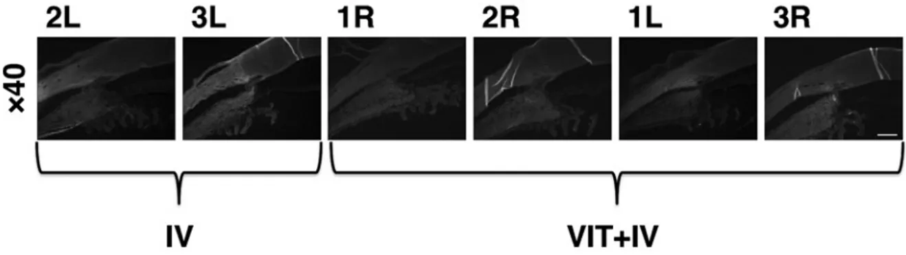
Transduction efficiency was assessed immunohisto-chemically with an anti-GFP antibody at 19 weeks after intravitreal injection of 50 μL of tm-scAAV2/GFP vector. No GFP expression was detected in Group VIT or Group IV (Fig. 1).
AAV genome copy number in the anterior chamber
As-Fig. 1 Histological analysis for the detection of GFP
Sections from each eye were immunohistochemically analyzed. The primary antibody was rabbit GFP IgG anti-body and the secondary antianti-body was Alexa 488-conjugated goat anti-rabbit IgG.
GFP expression was not detected in group IV or group VIT+IV.
IV: The AAV vector was injected intravitreally without first performing a vitrectomy. VIT+IV: Vitrectomy was performed before the AAV vector was injected intravitreally. (magnification ×40; scale bar, 500 μm)
Fig. 2 Results of qPCR analysis
DNA was extracted from two 10-μm slices cut from a frozen block. The GFP titer was determined with qPCR.
a) Results of qPCR for GFP in each eye.
b) PCR analysis revealed the presence of the vector genome in the anterior chamber in the VIT group (2.417 vector genome copies (g.c.) /diploid genome (d.g.) ). The amount did not exceed that in group IV (4.316 g.c./d.g.).
suming 6.181 pg genomic DNA per diploid genome (d. g.), qPCR results indicated that the vector genome copies (g.c.) of GFP were 2.417 g.c./d.g. in Group VIT and 4.316 g.c./d.g. in Group IV (Fig. 2). Only a small amount of GFP was detected with qPCR, and GFP detected in the VIT group did not exceed that in the IV group.
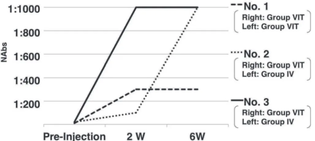
AAV NAbs in the serum
We collected serum samples from each monkey before, and at 2 and 6 weeks after, AAV vector injection. The NAb titers were 1:10, 1:20, and 1:20 (Geometric mean; 15.9) before administration; 1:100, 1:300, and 1:1,000 (310.7) at 2 weeks post-administration; and 1:300, 1:1,000, and 1:1,000 (669.4) at 6 weeks post-administration (Fig.
Fig. 3 AAV neutralizing antibodies (NAbs) in the serum
To detect NAbs to AAV, human embryonic kidney (HEK) 293 cells were used for the as-say. Serum NAbs were scored as positive if fluorescence intensity was ≤50% of that ob-served when tmAAV-GFP-infected HEK 293 cells were pre-incubated without sera. This test was performed once in duplicate.
The NAb titers were 1:10, 1:20, and 1:20 (geometric mean, 15.9) before administration; 1:100, 1:300, and 1:1,000 (310.7) at 2 weeks post-administration; and 1:300, 1:1,000, and 1:1,000 (669.4) at 6 weeks post-administration.
3).
Discussion
In our study, immunohistochemical analysis showed no GFP in the anterior chamber after vitreous administration of AAV vectors. PCR showed a miniscule amount of GFP in the anterior chamber; however, performing vitrectomy in advance did not increase the amount of GFP in the an-terior chamber. Intravitreal administration of AAV vec-tors resulted in increased NAbs in serum.
The two strategies for delivering vectors to the retina are subretinal injection and intravitreal injection. The in-travitreal route is considered safer, as it does not cause
retinal detachment11
. However, transduction efficiency in
the primate retina is low24
. Takahashi et al. previously re-ported that ILM peeling improved intravitreal AAV-mediated inner retinal gene transduction in cynomolgus
monkeys17
. When ILM peeling is performed, vitrectomy is required. Because the vitreous is a gel-like collagen, re-searchers have hypothesized that it can prevent
wide-spread exposure of a vector solution to the retina19
. After vitrectomy, AAV vectors injected intravitreally can spread to the anterior chamber. However, localization of AAV vectors after intravitreal injection has not been reported. In the present report, we compared transduction effi-ciency in the anterior segment in vitrectomized and non-vitrectomized eyes. Vitrectomy did not affect the amount of vector that was transduced in the anterior chamber,
which suggests that vitrectomy is not a risk factor for un-intended gene transfer into the anterior chamber when performing gene therapy in the retina by vitreous ad-ministration. The present study aids our understanding of the local effect in the eye when gene therapy in the retina-for diseases such as retinitis pigmentosa,
age-related macular degeneration, retinoschisis, and
glaucoma-is performed with ILM peeling17
.
Because of ocular immune privilege, researchers have believed that the entire body has little effect after travitreal administration. However, we confirmed that in-travitreal administration of AAV vectors generated NAbs against the AAV vector in monkeys. After intravitreal ad-ministration, the level of NAbs in the serum increased 40-fold. This result is similar to those reported by Li et al.
for mice20
and by Korrterman et al. for non-human
pri-mates (2-50-fold)21
.
It was reported that presence of the AAV vector in the anterior chamber, Schlemm’s canal, or collector channels
could result in systemic exposure25
. In our study, PCR de-tected a miniscule amount of GFP in the anterior cham-ber in all eyes. This may be associated with systemic ex-posure and increased NAbs in serum. Clinical application of retinal AAV-mediated gene therapy will require moni-toring of NAbs and consideration of possible systemic exposure. Future experiments should examine how AAV vectors are systemically exposed and how NAbs are gen-erated.
Acknowledgments: We thank Dr. Arun Srivastava at the Uni-versity of Florida for providing the pACG2-3M (pAAV2-Y730 +500+444F) packaging plasmids and the pdsCBA-GFP vector plasmid. This study was conducted as part of the Cooperative Research Program in the Tsukuba Primate Research Center, National Institutes of Biomedical Innovation, Health and Nu-trition. This work was supported in part by a JRPS Research Grant from the Japanese Retinitis Pigmentosa Society.
Conflict of Interest: None.
References
1.Bainbridge JW, Smith AJ, Barker SS, et al. Effect of gene therapy on visual function in Leber’s congenital amauro-sis. N Engl J Med. 2008;358:2231―9.
2.Bainbridge JW, Mehat MS, Sundaram V, et al. Long-term effect of gene therapy on Leber’s congenital amaurosis. N Engl J Med. 2015;372:1887―97.
3.Cideciyan AV, Aleman TS, Boye SL, et al. Human gene therapy for RPE65 isomerase deficiency activates the reti-noid cycle of vision but with slow rod kinetics. Proc Natl Acad Sci U S A. 105:15112―7.
4.Maguire AM, High KA, Auricchio A, et al. Age-dependent effects of RPE65 gene therapy for Leber’s con-genital amaurosis: a phase 1 dose-escalation trial. Lancet. 2009;374:1597―605.
5.Maguire AM, Simonelli F, Pierce EA, et al. Safety and effi-cacy of gene transfer for Leber’s congenital amaurosis. N Engl J Med. 2008;358:2240―8.
6.Rakoczy EP, Lai CM, Magno AL, et al. Gene therapy with recombinant adeno-associated vectors for neovascular age-related macular degeneration: 1 year follow-up of a phase 1 randomised clinical trial. Lancet. 2015;386:2395― 403.
7.Cukras C, Wiley HE, Jeffrey BG, et al. Retinal AAV8-RS1 gene therapy for X-linked retinoschisis: Initial findings from a phase I/IIa trial by intravitreal delivery. Mol Ther. 2018;26:2282―94.
8.Igarashi T, Miyake K, Kato K, et al. Lentivirus-mediated expression of angiostatin efficiently inhibits neovasculari-zation in a murine proliferative retinopathy model. Gene Ther. 2003;10:219―26.
9.Igarashi T, Miyake K, Masuda I, Takahashi H, Shimada T. Adeno-associated vector (type 8)-mediated expression of soluble Flt-1 efficiently inhibits neovascularization in a murine choroidal neovascularization model. Hum Gene Ther. 2010;21:631―7.
10.Igarashi T, Miyake N, Fujimoto C, et al. Adeno-associated virus type 8 vector-mediated expression of siRNA target-ing vascular endothelial growth factor efficiently inhibits neovascularization in a murine choroidal neovasculariza-tion model. Mol Vis. 2014;20:488―96.
11.Igarashi T, Miyake K, Asakawa N, Miyake N, Shimada T, Takahashi H. Direct comparison of administration routes for AAV8-mediated ocular gene therapy. Curr Eye Res. 2013;38:569―77.
12.Igarashi T, Miyake K, Kobayashi M, et al. Tyrosine triple
mutated AAV2-BDNF gene therapy in a rat model of transient IOP elevation. Mol Vis. 2016;22:816―26.
13.Day TP, Byrne LC, Schaffer DV, Flannery JG. Advances in AAV vector development for gene therapy in the retina. Adv Exp Med Biol. 2014;801:687―93.
14.Da Costa R, Röger C, Segelken J, Barben M, Grimm C, Neidhardt J. A novel method combining vitreous aspira-tion and intravitreal AAV2/8 injecaspira-tion results in retina-wide transduction in adult mice. Invest Ophthalmol Vis Sci. 2016;57:5326―34.
15.Dalkara D, Kolstad KD, Caporale N, et al. Inner limiting membrane barriers to AAV-mediated retinal transduction from the vitreous. Mol Ther. 2009;17:2096―102.
16.Boye SE, Alexander JJ, Witherspoon CD, et al. Highly effi-cient delivery of adeno-associated viral vectors to the pri-mate retina. Hum Gene Ther. 2016;27:580―97.
17.Takahashi K, Igarashi T, Miyake K, et al. Improved in-travitreal AAV-mediated inner retinal gene transduction after surgical internal limiting membrane peeling in cyno-molgus monkeys. Mol Ther. 2017;25:296―302.
18.Lebherz C, Maguire A, Tang W, Bennett J, Wilson JM. Novel AAV serotypes for improved ocular gene transfer. J Gene Med. 2008;10:375―82.
19.Boyd RF, Boye SL, Conlon TJ, et al. Reduced retinal trans-duction and enhanced transgene-directed immunogenicity with intravitreal delivery of rAAV following posterior vitrectomy in dogs. Gene Ther. 2016;23:548―56.
20.Li Q, Miller R, Han PY, et al. Intraocular route of AAV2 vector administration defines humoral immune response and therapeutic potential. Mol Vis. 2008;14:1760―9. 21.Kotterman MA, Yin L, Strazzeri JM, Flannery JG, Merigan
WH, Schaffer DV. Antibody neutralization poses a barrier to intravitreal adeno-associated viral vector gene delivery to non-human primates. Gene Ther. 2015;22:116―26. 22.Li M, Jayandharan GR, Li B, et al. High-efficiency
trans-duction of fibroblasts and mesenchymal stem cells by tyrosine-mutant AAV2 vectors for their potential use in cellular therapy. Hum Gene Ther. 2010;21:1527―43. 23.Kay MA, Manno CS, Ragni MV, et al. Evidence for gene
transfer and expression of factor IX in haemophilia B pa-tients treated with an AAV vector. Nat Genet. 2000;24: 257―61.
24.Yin L, Greenberg K, Hunter JJ, et al. Intravitreal injection of AAV2 transduces macaque inner retina. Invest Oph-thalmol Vis Sci. 2011;52:2775―83.
25.Dang Y, Loewen R, Parikh HA, Roy P. Loewen NA. Gene transfer to the outflow tract. Exp Eye Res. 2017;158:73―84.
(Received, (Accepted, December February 16, 2019) 26, 2020)
Journal of Nippon Medical School has adopted the Creative Com-mons Attribution-NonCommercial-NoDerivatives 4.0 International License (https://creativecommons.org/licenses/by-nc-nd/4.0/) for this article. The Medical Association of Nippon Medical School re-mains the copyright holder of all articles. Anyone may download, reuse, copy, reprint, or distribute articles for non-profit purposes under this license, on condition that the authors of the articles are properly credited.