R E S E A R C H
Open Access
Optimal vitamin D spurs serotonin:
1,25-dihydroxyvitamin D represses
serotonin reuptake transport (
SERT) and
degradation (
MAO-A) gene expression in
cultured rat serotonergic neuronal cell lines
Marya S. Sabir
1, Mark R. Haussler
2*, Sanchita Mallick
1, Ichiro Kaneko
1,2, Daniel A. Lucas
1, Carol A. Haussler
2,
G. Kerr Whitfield
2and Peter W. Jurutka
1,2Abstract
Background: Diminished brain levels of two neurohormones, 5-hydroxytryptamine (5-HT; serotonin) and 1,25-dihydroxyvitamin D3(1,25D; active vitamin D metabolite), are proposed to play a role in the atypical social behaviors
associated with psychological conditions including autism spectrum disorders and depression. We reported previously that 1,25D induces expression of tryptophan hydroxylase-2 (TPH2), the initial and rate-limiting enzyme in the biosynthetic pathway to 5-HT, in cultured rat serotonergic neuronal cells. However, other enzymes and transporters in the pathway of tryptophan metabolism had yet to be examined with respect to the actions of vitamin D. Herein, we probed the response of neuronal cells to 1,25D by quantifying mRNA expression of serotonin synthesis isozymes, TPH1 and TPH2, as well as expression of the serotonin reuptake transporter (SERT), and the enzyme responsible for serotonin catabolism, monoamine oxidase-A (MAO-A). We also assessed the direct production of serotonin in cell culture in response to 1,25D. Results: Employing quantitative real-time PCR, we demonstrate thatTPH-1/-2 mRNAs are 28- to 33-fold induced by 10 nM 1,25D treatment of cultured rat serotonergic neuronal cells (RN46A-B14), and the enhancement ofTPH2 mRNA by 1,25D is dependent on the degree of neuron-like character of the cells. In contrast, examination ofSERT, the gene product of which is a target for the SSRI-class of antidepressants, andMAO-A, which encodes the predominant catabolic enzyme in the serotonin pathway, reveals that their mRNAs are 51–59% repressed by 10 nM 1,25D treatment of RN46A-B14 cells. Finally, serotonin concentrations are significantly enhanced (2.9-fold) by 10 nM 1,25D in this system.
Conclusions: These results are consistent with the concept that vitamin D maintains extracellular fluid serotonin concentrations in the brain, thereby offering an explanation for how vitamin D could influence the trajectory and development of neuropsychiatric disorders. Given the profile of gene regulation in cultured RN46A-B14 serotonergic neurons, we conclude that 1,25D acts not only to induce serotonin synthesis, but also functions at an indirect, molecular-genomic stage to mimic SSRIs and MAO inhibitors, likely elevating serotonin in the CNS. These data suggest that optimal vitamin D status may contribute to improving behavioral pathophysiologies resulting from dysregulation of serotonergic neurotransmission.
Keywords: Autism spectrum disorders, Depression, Neuropsychiatric disorders, Social behavior, Tryptophan metabolism, Vitamin D receptor, 5-Hydroxytryptamine
* Correspondence:[email protected]
2Department of Basic Medical Sciences, University of Arizona College of
Medicine, Phoenix, AZ, USA
Full list of author information is available at the end of the article
© The Author(s). 2018 Open Access This article is distributed under the terms of the Creative Commons Attribution 4.0 International License (http://creativecommons.org/licenses/by/4.0/), which permits unrestricted use, distribution, and reproduction in any medium, provided you give appropriate credit to the original author(s) and the source, provide a link to the Creative Commons license, and indicate if changes were made. The Creative Commons Public Domain Dedication waiver (http://creativecommons.org/publicdomain/zero/1.0/) applies to the data made available in this article, unless otherwise stated.
Background
Serotonin, a neurotransmitter derived from the nutri-tionally essential amino acid tryptophan, executes critical functions in the brain such as control of appetite, energy expenditure, sleep, temperature, mood, and social cogni-tion [1]. Serotonergic neurons innervate vast areas of the brain, with projections arising from cell bodies in the dor-sal and median raphe and neighboring nuclei of the lower brain stem. These projections radiate to the hippocampus, amygdala, hypothalamus, nucleus accumbens, and lateral prefrontal cortex, as well as to a widespread array of cor-tical areas, wherein serotonin modulates a broad range of behavioral actions via metabotropic G protein-coupled and ionotropic, ligand-gated ion channel receptors [2].
In what constitutes the first step of serotonin produc-tion, tryptophan in the CNS is hydroxylated to 5-hydroxytryptophan by the enzyme tryptophan hydroxy-lase type 2 (TPH2), the rate-limiting step in brain sero-tonin synthesis. This is followed by subsequent decarboxylation of 5-hydroxytryptophan, catalyzed by aromatic amino acid decarboxylase, to serotonin (5-hy-droxytryptamine; 5-HT). Serotonin is then packaged by vesicularization through the action of monoamine trans-porter isoform 2 in raphe neurons. After the release of 5-HT-containing vesicles from axon terminals, and sub-sequent reuptake of serotonin from the synapse via a serotonin reuptake transporter (SERT), degradation of serotonin is catalyzed by monoamine oxidase-A (MAO-A) and aldehyde dehydrogenase to the predomin-ant serotonin metabolite, 5-hydroxyindoleacetic acid [2]. Therefore, three important enzymes/transport proteins that determine serotonin concentration and dynamics in the CNS are TPH2, SERT, and MAO-A. It is generally accepted that the amount and activity of these three pro-teins governs serotonin levels in the CNS [3].
In a recent publication [4], we proposed that serotonin concentrations are controlled in part by 1,25-dihydroxy-vitamin D3 (1,25D), the active, hormonal metabolite of
vitamin D. Specifically, we demonstrated that 1,25D dra-matically inducesTPH2 gene expression in differentiated serotonergic rat raphe RN46A-B14 cells [4], a finding that is reinforced by in vivo experiments conducted by Jiang et al. [5] revealing a significant induction of TPH2 mRNA in the prefrontal cortex of rats after chronic 1,25D administration. Central among the plethora of serotonin bioeffects are those that influence executive function, sensory gating, and prosocial behavior; import-antly, serotonin levels are often suboptimal in disorders of these actions. Patrick and Ames [6] have reviewed the observed association between aberrant serotonin con-centrations, both during development and in the adult, in a broad range of behavioral illnesses including ADHD, autism, bipolar disorder, depression, and schizophrenia, as well as anti-social, obsessive-compulsive, and suicidal
behaviors. They hypothesized that an association be-tween vitamin D insufficiency and low central serotonin concentrations represents a common denominator in a myriad of neuropsychiatric disorders. While it is con-ceivable that the ability of the vitamin D hormone, 1,25D, to upregulateTPH2 expression is a key factor in maintaining serotonin output by serotonergic neurons, other avenues of serotonin metabolism have yet to be in-vestigated with respect to the effects of vitamin D. In the present communication, we examined the influence of a range of 1,25D concentrations on the expression of SERT, MAO-A/B, and TPH-1/2 to elucidate the mechan-ism of action underlying the regulation of serotonin re-uptake, degradation, and synthesis by vitamin D in serotonergic neurons.
Results
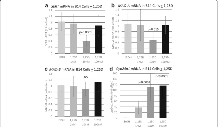
To determine if vitamin D affects the major route of serotonin reuptake and degradation, the influence of 1,25D on the expression of SERT and MAO-A was assessed by quantitative real-time PCR in differentiated serotonergic rat raphe RN46A-B14 cells. Cells in culture were challenged with 1, 10, and 100 nM 1,25D, and mRNA was isolated, reverse transcribed to cDNA and probed forSERT and MAO-A expression. Figure1a illus-trates that SERT is unaffected by 1 and 100 nM 1,25D-treatment, but a concentration of 10 nM 1,25D significantly (p = 0.0001) represses SERT mRNA expres-sion by 59%. Thus, the response of SERT to 1,25D is biphasic, reminiscent of the U-shaped curves generated when plotting circulating 25-hydroxyvitamin D levels versus disease and death in humans [7]. Strikingly, MAO-A displays an identical regulatory pattern of expression when challenged with a range of 1,25D con-centrations in differentiated serotonergic rat raphe RN46A-B14 cells (Fig. 1b). MAO-A is similarly un-affected by 1 and 100 nM 1,25D-treatment, but a con-centration of 10 nM 1,25D significantly (p = 0.015) repressesMAO-A mRNA expression by 51%. Therefore, two neurally expressed genes exhibit a similar profile of regulation by 1,25D, and the data suggest that certain levels of the vitamin D hormone appear to be capable of suppressing serotonin reuptake and catabolism.
As a negative-control neural gene, MAO-B was evalu-ated for modulation of expression by 1,25D in RN46A-B14 cells (Fig. 1c). Interestingly, MAO-B was not significantly repressed by 1,25D at any of the tested dosages, indicating that the influence of 1,25D on SERT and MAO-A expression may be selective for biologically relevant serotonin metabolism genes. Furthermore, as a positive control to confirm endogenous expression of a sufficient copy number of biologically functional nuclear vitamin D receptors (VDRs) to mediate transcriptional regulation by 1,25D in RN46A-B14 cells, we examined
Cyp24a1 mRNA enhancement in response to the vita-min D hormone. As depicted in Fig.1d,Cyp24a1 expres-sion is dramatically (38- to 116-fold) augmented by exposure of the cells to 1–100 nM 1,25D, with the pertin-ent observation that this gene displayed the traditional dose-response curve for induction. Cyp24a1 serves not only as a control for positive modulation of gene expres-sion via VDR in RN46A-B14 cells, but because the RNA probed was isolated from the same treated cells forSERT and MAO-A mRNA assessment, any systematic error in the technology can be eliminated from consideration as an explanation for the unique dose-response results for serotonin catabolic gene expression.
Next, we determined if 1,25D exerted actions on the expression of SERT, MAO-A, MAO-B, and CYP24A1 in the human glioblastoma cell line, U-87 MG (U87), which does not exhibit serotonergic neuronal characteristics. In contrast to the results in RN46A-B14 cells, the data for U87 cells reveal that 1,25D does not significantly alter the expression of eitherSERT or MAO-A in glioblastoma cells (Fig. 2a, b). This observation suggests that the repressive modulation of these two serotonin-degradative
reactions by 1,25D may be specific for serotonergic neu-rons. Further, as depicted in Fig.2c, unlike in RN46A-B14 cells, 1,25D significantly represses (25%, p = 0.005) MAO-B mRNA levels in U87 cells, pointing to a potential selectivity of 1,25D action on the MAO-B isozyme in regions of the CNS separate from serotonergic neuro-transmission. Interestingly, the same biphasic pattern of U-shaped dose-responsiveness to 1,25D obtained forSERT and MAO-A repression in RN46A-B14 cells occurs for MAO-B in U87 cells in terms of the optimal repressive dose of 1,25D being 10 nM. That this finding is valid is supported by the classic dose-response relationship occur-ring for 1,25D induction ofCYP24A1 in U87 cells (Fig.2d), which serves as a positive control.
In order to investigate the influence of serotonergic neuronal cell state on the control of serotonin synthesis by 1,25D, we next studied RN46A-B14 cells cultured in three different media formulations designed to generate varying degrees of neuronal morphology. The three media, designated media 1, 2, and 3, are described in de-tail in the“Methods” section. Figure3apictorially repre-sents RN46A-B14 cells grown in media 1, illustrating
b
a
d
c
Fig. 1 Effect of 1,25D on selected serotonin degrading pathway and control mRNAs in a serotonergic neuronal cell line.P values listed at the top of bars represent statistical significance for comparison between 1,25D treatment and ethanol vehicle control group in each and all cases. Statistical analyses were performed using ANOVA with a post hoc adjustment ofP values via the Dunnett test that compares every mean to a control mean and takes into account the scatter of all the groups. a Expression ofSERT in serotonergic rat raphe RN46A-B14 cells treated with 1,25D. Each mRNA value is the average of nine biological replicates ± std. dev. b Expression ofMAO-A in serotonergic rat raphe RN46A-B14 cells treated with 1,25D. Each mRNA value is the average of nine biological replicates ± std. dev. c Expression ofMAO-B in serotonergic rat raphe RN46A-B14 cells treated with 1,25D. Each mRNA value is the average of 18 biological replicates ± std. dev. d Expression ofCyp24a1 in serotonergic rat raphe RN46A-B14 cells treated with 1,25D. Each mRNA value is the average of nine biological replicates ± std. dev
their fusiform stellate morphology and neurite intercon-nections; cells maintained in media 1 displayed the most neuronal-like morphology and exhibited the slowest growth rate among the tested media. Figure 3b shows RN46A-B14 cells grown in media 2, with the cells retaining generally neuronal morphology, but acquiring some fibroblast-like character with increased growth rate and confluency. RN46A-B14 cells maintained in media 3 (Fig. 3c) displayed mostly fibroblast morphology during proliferation, with the fastest growth rate, likely induced by components of fetal bovine serum (FBS) and add-itional glucose, both present in the media. Therefore, media 1 elicited the most neuron-like character in RN46A-B14 cells, and the cells assumed a more fibroblast-like morphology and proliferated more rapidly as the media progressed from 1 to 3. Figure 3d depicts the results ofTPH2 mRNA induction by treatment with 10 nM 1,25D for 24 h of RN46A-B14 cells maintained in media 1–3. TPH2 was significantly (32-fold) induced by 1,25D only in media 1, revealing that 1,25D regulation of tryptophan metabolizing gene products occurs in cells most resembling serotonergic neurons.
Next, the concentration-dependence of 1,25D-regula-tion of bothTPH1 and TPH2 mRNAs was evaluated in RN46A-B14 cells maintained in media 1. As illustrated in Fig.4a, the profile ofTPH1 mRNA response to 1,25D as a function of concentration of the vitamin D hormone is analogous to the results obtained for SERT and MAO-A but inverted, in that treatment of the cells with 1 or 100 nM 1,25D is without significant effect whereas exposure of the cells to 10 nM 1,25D induces TPH1 28-fold. Again, this biphasic induction over a range of hormone concentrations is not the consequence of ex-perimental error because when assayed in RNA from the same biological replicate cells,Cyp24a1 mRNA exhibits the traditional dose-response curve for induction follow-ing treatment of the cells with 1,25D (Fig. 4c). This non-traditional inverted U-shaped dose response is ad-dressed in the “Discussion” section, but at present the
authors postulate that specific, CNS-expressed genes may be modulated in their expression differently than are typical vitamin D target genes in peripheral tissues such as bone, intestine, and kidney. Consistent with this interpretation is the 1,25D concentration-dependence of
a
b
c
d
Fig. 2 Effect of 1,25D on serotonin degrading pathway and control mRNAs in U87 glioblastoma cells.P values listed at the top of bars represent statistical significance for comparison between 1,25D-treatment and ethanol vehicle control group in each and all cases. Statistical analyses were performed using ANOVA with a post hoc adjustment ofP values via the Dunnett test that compares every mean to a control mean and takes into account the scatter of all the groups. a Expression ofSERT in U87 human glioblastoma cells treated with 1,25D. Each mRNA value is the average of seven biological replicates ± std. dev. b Expression ofMAO-A in U87 human glioblastoma cells treated with 1,25D. Each mRNA value is the average of 11 biological replicates ± std. dev. c Expression ofMAO-B in U87 glioblastoma cells treated with 1,25D. Each mRNA value is the average of 27 biological replicates ± std. dev. d Expression ofCYP24A1 in U87 glioblastoma cells treated with 1,25D. Each mRNA value represents the average of seven biological replicates ± std. dev
regulation ofTPH2 mRNA in RN46A-B14 cells (Fig.4b). TPH2 is documented to be the isoform that is respon-sible for catalyzing brain serotonin synthesis in the adult [8]. In non-neuronal serotonergic cells, TPH1 mRNA is the primary isoform expressed, whereas serotonergic neurons express TPH2 mRNA predominantly [9]. As depicted in Fig.4b, the profile ofTPH2 mRNA response to 1,25D as a function of concentration of the vitamin D hormone mimics the pattern of regulation of TPH1, in that treatment of the cells with 1 or 100 nM 1,25D pro-duces no significant effect onTPH2 mRNA, whereas ex-posure of the cells to 10 nM 1,25D induces TPH2 33-fold (p < 0.0001). What could be important with
respect to serotonin dynamics is that the inverted U-shaped curve forTPH2 induction by 1,25D as a func-tion of concentrafunc-tion of hormone (Fig. 4b) is comple-mentary to the U-shaped curves for SERT (Fig. 1a) and MAO-A (Fig. 1b) mRNA repression in response to in-creasing 1,25D concentration.
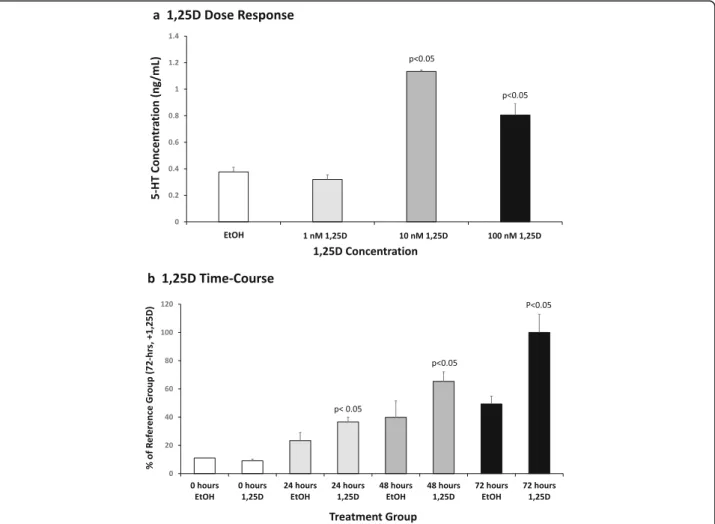
In a final set of in vitro experiments, RN46A-B14 cells were exposed to 1, 10, or 100 nM 1,25D for 72 h and serotonin concentrations were quantified by 5-HT ELISA of the cell culture medium. Interestingly, 1 nM 1,25D exerted a slight (15%), non-significant suppression of serotonin compared to ethanol vehicle control, which contrasted to the 2.9-fold increase (p < 0.05) in serotonin
a b c
d
Fig. 3 Morphology of serotonergic rat raphe RN46A-B14 cells grown in a media 1, b media 2, and c media 3, see the“Methods” section for
descriptions of the media formulations. d RN46A-B14 cells grown in media 1–3 and treated with 10 nM 1,25D for 24 h, followed by TPH2 mRNA quantification, expressed as fold relative to EtOH vehicle control (not shown)
c b
a
Fig. 4 Effect of 1,25D onTPH1, TPH2 and Cyp24a1 mRNAs in a serotonergic neuronal cell line. P values listed at the top of bars represent statistical significance for comparison between 1,25D-treatment and ethanol vehicle control group in each and all cases. Statistical analyses were performed using ANOVA with a post hoc adjustment ofP values via the Dunnett test that compares every mean to a control mean and takes into account the scatter of all the groups. a Expression ofTPH1 in serotonergic rat raphe RN46A-B14 cells treated with 1,25D. The exact fold-effect values for 1 and 100 nM 1,25D are too small to be perceived in the figure as scaled; they are 0.41 ± 0.22 and 0.73 ± 0.17, respectively. Each mRNA value is the average of nine biological replicates ± std. dev. b Expression ofTPH2 in serotonergic rat raphe RN46A-B14 cells treated with 1,25D. The exact fold-effect values for 1 and 100 nM 1,25D are too small to be perceived in the figure as scaled; they are 0.36 ± 0.23 and 0.65 ± 0.31, respectively. Each mRNA value is the average of 30 biological replicates ± std. dev. c Expression ofCyp24a1 in serotonergic rat raphe RN46A-B14 cells treated with 1,25D. Each mRNA value is the average of 20 biological replicates ± std. dev
induced by 10 nM 1,25D (Fig.5a). Remarkably, consider-ing that this assay is cumulative, Fig. 5a also illustrates that 100 nM 1,25D treatment of RN46A-B14 cells pro-duced a diminished induction of serotonin to 2.0-fold, generating a biphasic dose-response curve for total sero-tonin that reflects the biphasic dose-response curves for the induction of TPH2 as well as for the repression of SERT and MAO-A. Taken together, this stimulatory ef-fect of 10 nM 1,25D on serotonin synthesis, coupled with suppressive actions on serotonin reuptake and metabolic elimination, resulting in almost a tripling of total free serotonin, vividly integrates what a significant effect an optimal concentration of 1,25D (10 nM) exerts to bolster extracellular serotonin. Figure5b displays the time-course of serotonin enhancement by 10 nM 1,25D in RN46A-B14 cell culture. Augmented serotonin is ap-parent at 24 h (1.7-fold; p < 0.05), levels off at 48 h
(1.6-fold; p < 0.05), and reaches 2.0-fold (p < 0.05) by 72 h of exposure to 1,25D versus control ethanol vehicle. Such a time-course is consistent with induction/repres-sion but does not distinguish between primary and sec-ondary mechanisms.
Discussion
Although recent research has highlighted the possible significance of the relationship between vitamin D and the tryptophan-derived neurotransmitter, serotonin [4], previous studies focused solely on serotonin synthesis. In the present communication, we have investigated, for the first time, the effects of 1,25D on the regulation of serotonin reuptake via a serotonin reuptake transporter and on the first step of serotonin degradation by monoamine oxidase-A in serotoninergic neuronal cell lines. SERT (SLC6A4, 5HTT), the 5-HT transporter,
Fig. 5 Effect of 1,25D on total 5-HT levels in rat serotonergic neuron cell culture medium. a A dose-response experiment was performed in serotonergic rat raphe RN46A-B14 cells treated with the indicated concentrations of 1,25D or ethanol (EtOH) vehicle. A 200μL sample of medium was removed from each plate after 72 h of treatment and 5-HT quantitated by ELISA as detailed in the“Methods” section. Values are the average
of three biological replicates (n = 3) ± SD, with duplicate or triplicate samples averaged in each group. b A time-course experiment was carried out in RN46A-B14 cells treated with the optimal concentration of 1,25D (10 nM) for 0, 24, 48, and 72 h followed by removal of a 200μL sample of medium from each plate and serotonin assayed by 5-HT ELISA. Values are the average of three biological replicates (n = 3) ± SD, with duplicate or triplicate sample groups averaged in each case
plays a crucial role in the control of serotonergic neuro-transmission by determining the magnitude and dur-ation of the 5-HT synaptic signal and has been an attractive target in rational neuropsychiatric drug design [10]. Fluoxetine (Prozac), citalopram (Celexa), paroxetine (Paxil), and sertraline (Zoloft) represent selective inhibi-tors of SERT. In the presence of these selective serotonin reuptake inhibitors (SSRIs), 5-HT remains in the extra-cellular space for an extended duration, allowing pro-longed 5-HT receptor activation in the postsynaptic neuron. Thus, monoamine transporters such as SERT constitute the primary mechanism for terminating neurotransmission through removal of the released transmitter from the synaptic cleft. These transporters, including SERT, function as oligomeric complexes in the cell membrane, and are regulated primarily through phosphorylation, protein-protein interactions, and traf-ficking events [11]. Limited data are available on the control ofSERT at the transcriptional level, although the genes encoding mammalian forms have been cloned [12]. The 5-prime flanking region of the SERT (5HTT) human gene contains a cyclic AMP-responsive element (CRE) at − 99 bp plus binding sites for AP-1, AP-2, and SP1, all just upstream of a TATA-like motif. Sequences within − 1.4 kb of the transcription start site confer cell-specific expression, and the basal promoter responds to induction by cAMP [12]. A functional polymorphism (5HTT-LPR) has been identified in the human 5HTT promoter [13], and evidence has accumulated for an as-sociation between alleles of this variation and anxiety traits, affective disorders, as well as autism. Notably, three ChIP-seq databases generated in human immune cells for 1,25D-dependent occupation of vitamin D-responsive elements (VDREs) by VDR yield appar-ently functional sites at − 64.5 and + 118 kb in relation to the transcription start site of the SERT gene [14–16]. Thus, 1,25D could potentially modulate human SERT expression in a primary fashion via remote VDREs.
Examining the influence of 1,25D onSERT mRNA ex-pression in rat raphe RN46A-B14 cells, we observed a repression of SERT mRNA by 59% when the cells were treated with 10 nM 1,25D (Fig.1a). This repression was selective for serotonergic neuronal cells in that it was not found in U87 human glioblastoma cells (Fig. 2a). The initial conclusion is that 1,25D, analogous to SSRIs, suppresses serotonin reuptake, possibly potentiating se-rotonergic neurotransmission. However, this conclusion must be reached with caution, because neither 1 nor 100 nM 1,25D treatment of RN46A-B14 cells signifi-cantly repressesSERT mRNA expression (Fig.1a), gener-ating an unusual, biphasic dose-response curve. Nevertheless, this dose response is reminiscent of the U-shaped curves generated when plotting circulating 25-hydroxyvitamin D levels versus disease in humans,
most recently reported in a study of autism risk in 3-year-old Chinese children as a function of circulating 25-hydroxyvitamin D levels at birth [17]. In other words, there appears to exist an optimal level of circulating vita-min D for health, and this may correspond empirically to 10 nM 1,25D in the case of RN46A-B14 cell treatment.
However, the cellular and molecular mechanism that accounts for the observed effects of various concentra-tions of 1,25D in RN46A-B14 cell treatment experiments requires careful consideration. The authors favor the concept that limited, but more physiologically relevant, concentrations of 1,25D signal biologic actions such as cell proliferation, whereas higher levels of the hormone cause the opposing responses, namely inhibition of cell growth and apoptosis. We first identified this phenomenon when studying osteosarcoma cells in cul-ture, observing that low concentrations of 1,25D stimu-lated cell growth, whereas higher levels drastically curbed proliferation [18]. Therefore, we propose that the data in Fig. 1a can be explained if one assumes that the minimum effective concentration to stimulate prolifera-tion (and other effects such as altered transcripprolifera-tion fac-tor profile and epigenetic modifications) in RN46A-B14 cells is 10 nM 1,25D, conditioning the neuronal cells to exhibit a repression ofSERT expression, then the lack of response to treatment with 1 nM is appropriate. If one then assumes that 100 nM 1,25D, analogous to the ac-tion of this level in many cell types, promulgates an in-hibition of cell proliferation with attending alteration of transcription factor profile and epigenetic modifications, then the lack of responsiveness of SERT expression to this higher dose of 1,25D of these growth-inhibited RN46A-B14 cells is also readily explainable. Neverthe-less, the observations, in vitro, that higher concentra-tions of 1,25D are less effective than 10 nM hormone must be viewed with some skepticism and it will ultim-ately need to be determined if data from cell lines can be directly equated to the U-shaped dose-response curves, in vivo, because cell growth aspects and cell cul-ture conditions (i.e., interaction with other media com-ponents) could be responsible for the results with high 1,25D concentrations. However, very recent data ob-tained in autistic children suggest that maintaining cir-culating 25-hydroxyvitamin D levels in an optimal range elicits maximal therapeutic efficacy in vitamin D-responsive children [19]. Regardless of the mechanis-tic nuances, we provide herein more support for the vitamin D-serotonin hypothesis in the pathophysiology of neuropsychiatric disease that has been professed by Patrick and Ames [1, 6]. Consistent with the relevance of SERT modulation by 1,25D is a report [20] of in-creased concentrations of SERT protein (monitored by positron emission tomography (PET) brain scans) in
patients with seasonal affective disorder (SAD). SERT levels in SAD patients were 5% higher in winter, when vitamin D generated by sunlight is deficient, than in summer when sunlight is at a maximum; control subjects without SAD manifest no change in SERT levels with season. It is accepted that sunlight is mood-elevating, and the 1,25D effect to repress SERT reported herein may represent a facet of this benefit.
When examining MAO-A modulation by 1,25D in rat raphe RN46A-B14 cells, we observed a repression of MAO-A mRNA by 51% when the cells were treated with 10 nM 1,25D (Fig.1b). The 1,25D dose-response profile was equivalent for MAO-A and SERT repression, and the effect onMAO-A was also selective for serotonergic neuronal cells as it was not found in U87 human glio-blastoma cells (Fig. 2b). Thus, the same arguments can be applied in discussing the control ofMAO-A by 1,25D that were elaborated above for SERT repression. How-ever, there exists a caveat to the observed repression of MAO-A by 1,25D in cultured rat serotonergic cells, in that Jiang et al. [5] demonstrated a significant induction of MAO-A mRNA in the prefrontal cortex of rats after chronic 1,25D administration. This conflict between the in vitro and in vivo results with respect to MAO-A could be because Jiang et al. [5] analyzed the entire pre-frontal cortex, whereas the cell culture data are selective to serotonergic neuronal cells. Notably, MAO-A also catalyzes the degradation of dopamine and norepineph-rine, as well as serotonin, implying a broader array of functions beyond serotonin catabolism in the cortex, perhaps explaining the differences between cell culture results and those obtained in rodent models.
MAO-A is a gene of interest that has been given the moniker “warrior gene” as it has been linked to aggres-sion in observational and survey-based studies [21]. In addition, MAO-A knockout mice are more aggressive than their wild-type littermates or mice null forMAO-B, which degrades a different set of neurotransmitters [22]. The transcriptional regulation of MAO-A has not been fully characterized but, in a recent computational and experimental analysis ofMao-A expression, Gupta et al. [23] identified critical roles for a conserved proximal promoter domain (− 71/− 40 bp) in cis, and transcription factors Sp1/Gata2/Tbp intrans, to govern mouse Mao-A gene basal expression in a coordinated manner. We have located, in silico, a near-consensus candidate VDRE, consisting of a direct repeat with a spacer of three nucle-otides (DR3), CGGACActgAGGTCA (antisense), at + 776 in relation to the start of transcription of the human MAO-A gene. Furthermore, Tuoresmaki et al. [16] have reported two VDR ChIP-seq target regions (roughly 200 bp in size) in the human MAO-A gene, the first of which overlaps the candidate DR3 VDRE at + 776 bp cited above and the second, located approximately
350 bp 3-prime to the first, which contains two add-itional candidate (antisense)VDREs: GGTGCAggaGGTT CT and GGTGCAggaGGTGCT. It is not yet known whether these putative VDREs are involved in MAO-A control by 1,25D, but their proximity to the core regula-tory module at − 71/− 40 bp is suggestive of a possible cis regulatory module spanning the proximal promoter and first exon of the MAO-A gene. If this concept is verified, yet another avenue may exist whereby 1,25D could not only benefit patients with behavioral patho-physiologies resulting from dysregulation of serotonergic neurotransmission, but also impact other behavioral traits and neurological disorders, including Alzheimer’s dementia, aggression, and attention deficit hyperactivity disorder [23].
The present results support the vitamin D-serotonin hypothesis both experimentally and mechanistically. In fact, when we assessed the direct production of sero-tonin in neuronal cell culture in response to treatment with 10 nM 1,25D for 72 h, serotonin concentrations were significantly enhanced (two- to threefold) in the medium (Fig.5), consistent with the concerted induction of TPH2 (Fig. 4b) and repression of SERT (Fig. 1a) and MAO-A (Fig. 1b) by the empirically optimum level of 1,25D in cultured serotonergic neurons. Taking all of these results together, it seems highly likely that vitamin D can amplify serotonin in the central nervous system, making vitamin D a candidate for the treatment of neuropsychiatric disorders in which vitamin D and/or serotonin are implicated. Indeed, vitamin D supplemen-tation has been reported to improve inattention, hyper-activity, and impulsivity in children and adults with ADHD [24].
Conclusions
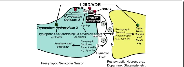
The results reported herein, combined with our previous work and that of Patrick and Ames [1,6], lead us to con-clude that a set of tryptophan metabolism pathway en-zymes/proteins relevant to CNS levels of serotonin are transcriptionally regulated by 1,25D in differentiated se-rotonergic rat raphe RN46A-B14 cells in such a fashion as to suggest that optimum levels of the vitamin D hor-mone represent a natural effector in spurring serotonin. Strikingly, the modulation of the tryptophan metabolism pathway by 1,25D at the transcriptional level is con-served to the degree that it is even prominent in a verte-brate only distantly related to mammals, namely developing zebrafish [25]. By potentially fine-tuning serotonin concentrations in the synaptic cleft, as depicted schematically in the model in Fig.6, 1,25D may be able to steer neurological control of such processes as social behavior to prevent autism spectrum disorders and depression. In summary, as illustrated in hypothet-ical form in Fig. 6, we conclude that the vitamin D
hormone may be capable of governing serotonin con-centrations in relevant regions of the brain where both VDR and TPH2, as well as SERT and MAO-A, are expressed, placing this nutrient at the forefront of nutri-genomic efforts to guide gene expression in serotonergic neurons toward ameliorating neuropsychiatric disease by spurring the actions of serotonin.
Methods
Mammalian cell culture
Two mammalian cell lines were employed in this study: embryonic rat medullary raphe (RN46A-B14; obtained from Dr. Robert J. Handa/Dr. John Neumaier) cultured with neurobasal A (Gibco/Life Technologies, Grand Island, NY) medium with B-27 supplement (Invitrogen Corporation) andL-glutamine (Gibco) and human brain glioblastoma/astrocytoma (U-87 MG, U87; obtained from the American Type Culture Collection) grown in DMEM/ high glucose + L-glutamine + pyruvate (Hyclone, Logan, Utah). All cells were passaged in the above-indicated media supplemented with 10% FBS and penicillin/strepto-mycin (pen-strep) under a humidified atmosphere of 5% CO2in air at 37 °C. RN46A-B14 cells in culture were
uti-lized in their native state, i.e., not transfected with any expression vector. However, U87 cells were transfected with a VDR overexpression plasmid to ensure that they consistently expressed biologically functional levels of the nuclear receptor. The PolyJET transfection reagent (Signa-Gen, Rockville, MD) was utilized to transfect U87 cells according to the manufacturer’s protocol. Briefly, each well was transfected with 15 μL PolyJET reagent and 500 ng of pSG5-hVDR (plasmid-expressing human VDR).
The clonal cell line, RN46A-B14, was isolated following transfection of the gene encoding rat brain-derived neuro-trophic factor (BDNF) into RN46A cells [26]. RN46A-B14 cells synthesize and secrete biologically active BDNF in vitro and synthesize serotonin following partial mem-brane depolarization. To optimize the growth conditions and neuronal morphology of adherent, rat serotonergic B14 cells in culture, three different media formulations were examined: media 1: neurobasal A medium, B-27 sup-plement, L-glutamine, and pen-strep (serum free); media 2: media 1 plus 10% FBS; media 3: DMEM/high glucose +
L-glutamine + pyruvate, 10% FBS, and pen-strep.
Quantitative real-time PCR
U87 and RN46A-B14 cells were plated at 500,000– 800,000 cells per well in a 6-well plate, and dosed with 1–100 nM 1,25D (see Figs. 1,2,3 and 4) or ethanol ve-hicle control for 24 h. Total RNA was isolated from each well using an Aurum Total RNA Mini kit (Bio-Rad, Hercules, CA) according to the manufacturer’s instruc-tions. The RNA quantity and quality were assessed using A260/280 spectrophotometry. DNase-treated RNA (1 μg)
was reverse transcribed using an iScript cDNA Synthesis kit (Bio-Rad) to produce 20μL of first-strand cDNA. For real-time PCR, 2μL of cDNA was used in a 10 μL PCR reaction containing 5 μL FastStart Universal SYBR Green Master Mix + Rox (Roche Applied Science, In-dianapolis, IN) and forward and reverse primers. Reac-tions were performed in 96-well PCR plates in a Bio-Rad CFX96 instrument using a standard 40-cycle profile. Data were analyzed using the comparativeΔΔCt method as means of relative quantitation, normalized to an en-dogenous reference (GAPDH) and relative to a calibrator
Fig. 6 Actions of 1,25D/VDR on serotonin dynamics in the CNS at the serotonin synapse. This model depicts the fine-tuning of serotonin levels by 1,25D via transcriptional regulation ofTPH2, SERT, and MAO-A; the result of which is the potentiation of serotonergic nerve transmission. Initially, (left) 1,25D inducesTPH2 to enhance serotonin output and, because circled pathway 3 (reuptake and degradation) is attenuated by 1,25D via repression ofSERT and MAO-A, serotonin action is spurred at both presynaptic (circled pathway 2) and postsynaptic (circled pathway 1) sites through type 1A and 2A serotonin receptors, respectively
(normalized Ct value from vehicle-treated cells) and expressed as 2-ΔΔCt according to Applied Biosystems’ User Bulletin 2: Rev. B, “Relative Quantitation of Gene Expression.” Primer sets for the PCR were as follows:
RatGAPDH (forward 5′-AGGTCGGTGTGAACGGATT TG-3′, reverse 5′-CATTCTCAGCCTTGACTGTGC-3′)
Rat Cyp24a1 (forward 5’-AACGAAGCCTACGGGTT GATG-3′, reverse 5’-AGAAAGTCAGCCAAGACCTCA-3′) Rat SERT [27] (forward 5′-TCTGAAAAGCCCCACT
GGACT-3′, reverse 5′-TAGGACCGTGTCTTCATCAG GC-3′)
Rat MAOA [28] (forward 5′-CCACAGCCAGAGCGT
TCAG-3′, reverse 5′-TGAGAGCCTTTGCCCAGATT G-3′)
Rat MAOB [28] (forward 5′-CAGTGGAAGCAGAGG
AGAG-3′, reverse 5′-TGCTGCCATACCTGAGATG-3′) Rat TPH1 [29] (forward 5′-GCTGAACAAACTCT
ACCCAAC-3′, reverse 5′-TTCCCGATAGCCACAGTA TT-3′)
Rat TPH2 [30] (forward 5′-CTCCAAGCTTCGCA TCACAG-3′, reverse 5′-AGCACTTCAGGAAGCGTAC C-3′)
Human GAPDH (forward 5′-ACAACTTTGGTATC GTGGAAGGAC-3′, reverse 5′-CAGGGATGATGTTC TGGAGAGC-3′)
Human CYP24A1 (forward 5′-CAGCGAACTGAACA AATGGTCG-3′, reverse 5′-TCTCTTCTCATACAACA CGAGGCAG-3′)
Human SERT [31] (forward 5′-TTCTCCCCTCCAAG
TGAATG-3′, reverse 5′-GGGGAATCCATGGAAATAA G-3′)
Human MAOA [32] (forward 5′-TAAATGGTCTC
GGGAAGGTG-3′, reverse 5′-CCCAGGGCAGTTAC TGATGT-3′)
Human MAOB [33] (forward 5′-GCTCTCTGGTT
CCTGTGGTATGTG-3′, reverse 5′-TCCGCTCACTCA CTTGACCAGATC-3′)
Serotonin ELISA
To measure serotonin (5-HT) concentrations, colorimet-ric competitive 5-HT enzyme-linked immunosorbent assay (ELISA) experiments were performed utilizing the Serotonin Research ELISA kit (Rocky Mountain Diag-nostics, Inc., Colorado Springs, CO). RN46A-B14 were plated at 700,000 cells per well in a six-well plate in media 2 (see above for details) and incubated in a hu-midified atmosphere of 5% CO2at 37 °C overnight. The
following day, media 2 was aspirated and the cells were gently washed with PBS to remove residual serum. Media 1 (see above for details) was added, and the cells were incubated overnight to generate a serum-deprived cell state. Next, the cells were treated with either ethanol vehicle control or 10 nM 1,25D for 0–72 h in media 1. Samples of media/culture supernatant (200 μL) were
removed from each well (without disrupting the adher-ent cells) at 0, 24, 48, or 72 h and assayed for 5-HT con-tent in accordance with the manufacturer’s protocol. Briefly, the diluent, wash buffer, six standards, and two positive controls were prepared at the time of the assay, and 100 μL of the diluted standards, controls, and ex-perimental samples were acylated for 30 min at room temperature. Next, 100 μL of the acylated standards, controls, and experimental samples were placed into the appropriate wells of the serotonin/5-HIAA microtiter strips, and 25μL of the 5-HT antiserum were added to each well and incubated for 15–20 h at 4 °C. The wells were washed, and 100 μL of the enzyme conjugate were added and allowed to incubate for 30 min followed by additional washes and incubation with 100 μL of the substrate. After the final incubation, 100μL of the stop solution were added and the absorbance was read within 10 min using a microplate reader set to 450 nm and a reference wavelength between 620 and 650 nm. The cali-bration curve was obtained by plotting the absorbance readings measured for the standards (linear, y axis) against the corresponding standard concentrations (lin-ear,x axis, ng/mL). A logarithmic trendline was fitted to the data for the standards and the corresponding equa-tion was utilized to determine the serotonin concentra-tions in the experimental samples. The mean of three biological replicates, each with duplicate treatment groups, was utilized in the analysis.
Statistical analysis
Data are expressed as means ± SD. All data are presented as fold-effects, with transcription of the gene in question (i.e., mRNA level) in the absence of 1,25D (i.e., ethanol ve-hicle) set at 1.0-fold. Thus, all results are normalized to basal transcription and presented graphically and numer-ically as fold-effect of the single tested variable, i.e., 1,25D. Because the design of the experiments was a simple motif in which the fold-ability of 1,25D to enhance transcription over basal (i.e., ethanol vehicle) was assessed, statistical differences between two groups were determined initially by a two-tailed Student’s t test. However, because variance was high in several experiments, further statistical analyses were performed using GraphPad Prism 7 to generate ANOVA data, followed by a post hoc Dunnett test that compares every mean to a control mean and takes into ac-count the scatter of all the groups. In each figure illustrat-ing the fold-effect of 1,25D, exact P values (unless < 0.0001) are listed above the bar for concentrations of 1,25D versus EtOH control, except when the difference from the vehicle control is not significant (NS); aP value less than 0.05 is considered significant.
Abbreviations
1,25D:1,25-Dihydroxyvitamin D3; 5-HT: 5-Hydroxytryptamine; 5-HTT: 5-HT
MAO-A: Monoamine oxidase-A; MAO-B: Monoamine oxidase-B; PET: Positron emission tomography; SAD: Seasonal affective disorder; SERT: Serotonin reuptake transporter; SSRI: Selective serotonin reuptake inhibitor; TPH1: Tryptophan hydroxylase-1; TPH2: Tryptophan hydroxylase-2; VDR: Vitamin D receptor; VDRE: Vitamin D-responsive element Funding
This research was supported in part by NIH grants DK033351 to MRH and CA140285 to PWJ and a grant from the University of Arizona, Department of Basic Medical Sciences to GKW.
Authors’ contributions
MSS contributed to the study design, data collection, data analysis/
interpretation, and developing a preliminary version of the manuscript in poster format. MRH, the corresponding author, contributed via conception/design of the work and wrote the initial version, as well as the final revisions of the manuscript. SM, IK, and DAL contributed to the data collection/analysis and to the revision of this manuscript. CAH and GKW contributed the critical experimental insight and interpretations, as well as to the independent revision of the article. PWJ, the senior investigator directing the study, contributed via conception/design of work, data interpretation, and critical revision of the article. All authors read and approved the final manuscript.
Competing interests
The authors declare that they have no competing interests. Author details
1School of Mathematical and Natural Sciences, Arizona State University,
Phoenix, AZ, USA.2Department of Basic Medical Sciences, University of Arizona College of Medicine, Phoenix, AZ, USA.
Received: 15 February 2018 Accepted: 8 June 2018
References
1. Patrick RP, Ames BN. Vitamin D hormone regulates serotonin synthesis. Part 1: relevance for autism. FASEB J. 2014;28(6):2398–413.
2. Jenkins TA, Nguyen JC, Polglaze KE, Bertrand PP. Influence of tryptophan and serotonin on mood and cognition with a possible role of the gut-brain Axis. Nutrients. 2016;8(1):56.
3. Andrews PW, Bharwani A, Lee KR, Fox M, Thomson JA Jr. Is serotonin an upper or a downer? The evolution of the serotonergic system and its role in depression and the antidepressant response. Neurosci Biobehav Rev. 2015;51:164–88.
4. Kaneko I, Sabir MS, Dussik CM, Whitfield GK, Karrys A, Hsieh JC, Haussler MR, Meyer MB, Pike JW, Jurutka PW. 1,25-Dihydroxyvitamin D regulates expression of the tryptophan hydroxylase 2 and leptin genes: implication for behavioral influences of vitamin D. FASEB J. 2015;29(9):4023–35. 5. Jiang P, Zhang LH, Cai HL, Li HD, Liu YP, Tang MM, Dang RL, Zhu WY, Xue
Y, He X. Neurochemical effects of chronic administration of calcitriol in rats. Nutrients. 2014;6(12):6048–59.
6. Patrick RP, Ames BN. Vitamin D and the omega-3 fatty acids control serotonin synthesis and action, part 2: relevance for ADHD, bipolar disorder, schizophrenia, and impulsive behavior. FASEB J. 2015;29(6):2207–22. 7. Keisala T, Minasyan A, Lou YR, Zou J, Kalueff AV, Pyykko I, Tuohimaa P.
Premature aging in vitamin D receptor mutant mice. J Steroid Biochem Mol Biol. 2009;115(3–5):91–7.
8. Zhang X, Beaulieu JM, Sotnikova TD, Gainetdinov RR, Caron MG. Tryptophan hydroxylase-2 controls brain serotonin synthesis. Science. 2004;305(5681):217. 9. Gutknecht L, Kriegebaum C, Waider J, Schmitt A, Lesch KP. Spatio-temporal
expression of tryptophan hydroxylase isoforms in murine and human brain: convergent data from Tph2 knockout mice. Eur Neuropsychopharmacol. 2009;19(4):266–82.
10. Linden DR, White SL, Brooks EM, Mawe GM. Novel promoter and alternate transcription start site of the human serotonin reuptake transporter in intestinal mucosa. Neurogastroenterol Motil. 2009;21(5):534–41. e510-531 11. Torres GE, Gainetdinov RR, Caron MG. Plasma membrane monoamine
transporters: structure, regulation and function. Nat Rev Neurosci. 2003;4(1):13–25. 12. Heils A, Teufel A, Petri S, Seemann M, Bengel D, Balling U, Riederer P, Lesch KP.
Functional promoter and polyadenylation site mapping of the human serotonin (5-HT) transporter gene. J Neural Transm Gen Sect. 1995;102(3):247–54.
13. Flattem NL, Blakely RD. Modified structure of the human serotonin transporter promoter. Mol Psychiatry. 2000;5(1):110–5.
14. Handel AE, Sandve GK, Disanto G, Berlanga-Taylor AJ, Gallone G, Hanwell H, Drablos F, Giovannoni G, Ebers GC, Ramagopalan SV. Vitamin D receptor ChIP-seq in primary CD4+ cells: relationship to serum 25-hydroxyvitamin D levels and autoimmune disease. BMC Med. 2013;11:163.
15. Heikkinen S, Vaisanen S, Pehkonen P, Seuter S, Benes V, Carlberg C. Nuclear hormone 1alpha,25-dihydroxyvitamin D3 elicits a genome-wide shift in the locations of VDR chromatin occupancy. Nucleic Acids Res. 2011;39(21):9181–93. 16. Tuoresmaki P, Vaisanen S, Neme A, Heikkinen S, Carlberg C. Patterns of
genome-wide VDR locations. PLoS One. 2014;9(4):e96105.
17. Wu D-M, Wen X, Han X-R, Wang S, Wang Y-J, Shen M, Fan S-H, Zhuang J, Li M-Q, Hu B, et al. Relationship between neonatal vitamin D at birth and risk of autism spectrum disorders: the NBSIB study. J Bone Miner Res. 2018;33(3): 458–66.
18. Dokoh S, Donaldson CA, Haussler MR. Influence of 1,25-dihydroxyvitamin D3
on cultured osteogenic sarcoma cells: correlation with the 1,25-dihydroxyvitamin D3receptor. Cancer Res. 1984;44:2103–9.
19. Jia F, Shan L, Wang B, Li H, Feng J, Xu Z, Saad K. Fluctuations in clinical symptoms with changes in serum 25(OH)vitamin D levels in autistic children: three cases report. Nutr Neurosci. 2018; Epub ahead of print April 8 20. Mc Mahon B, Andersen SB, Madsen MK, Hjordt LV, Hageman I, Dam H,
Svarer C, da Cunha-Bang S, Baare W, Madsen J, et al. Seasonal difference in brain serotonin transporter binding predicts symptom severity in patients with seasonal affective disorder. Brain. 2016;139(Pt 5):1605–14.
21. McDermott R, Tingley D, Cowden J, Frazzetto G, Johnson DD. Monoamine oxidase A gene (MAOA) predicts behavioral aggression following provocation. Proc Natl Acad Sci U S A. 2009;106(7):2118–23. 22. Shih JC, Chen K. MAO-A and -B gene knock-out mice exhibit distinctly
different behavior. Neurobiology (Bp). 1999;7(2):235–46.
23. Gupta V, Khan AA, Sasi BK, Mahapatra NR. Molecular mechanism of monoamine oxidase A gene regulation under inflammation and ischemia-like conditions: key roles of the transcription factors GATA2, Sp1 and TBP. J Neurochem. 2015;134(1):21–38.
24. Rucklidge JJ, Frampton CM, Gorman B, Boggis A. Vitamin-mineral treatment of attention-deficit hyperactivity disorder in adults: double-blind randomised placebo-controlled trial. Br J Psychiatry. 2014;204:306–15. 25. Craig TA, Zhang Y, McNulty MS, Middha S, Ketha H, Singh RJ, Magis AT, Funk C,
Price ND, Ekker SC, et al. Research resource: whole transcriptome RNA sequencing detects multiple 1alpha,25-dihydroxyvitamin D(3)-sensitive metabolic pathways in developing zebrafish. Mol Endocrinol. 2012;26(9):1630–42. 26. Eaton MJ, Whittemore SR. Autocrine BDNF secretion enhances the survival
and serotonergic differentiation of raphe neuronal precursor cells grafted into the adult rat CNS. Exp Neurol. 1996;140(2):105–14.
27. Abumaria N, Rygula R, Havemann-Reinecke U, Ruther E, Bodemer W, Roos C, Flugge G. Identification of genes regulated by chronic social stress in the rat dorsal raphe nucleus. Cell Mol Neurobiol. 2006;26(2):145–62.
28. Birgner C, Kindlundh-Hogberg AM, Oreland L, Alsio J, Lindblom J, Schioth HB, Bergstrom L. Reduced activity of monoamine oxidase in the rat brain following repeated nandrolone decanoate administration. Brain Res. 2008; 1219:103–10.
29. Charoenphandhu J, Teerapornpuntakit J, Nuntapornsak A, Krishnamra N, Charoenphandhu N. Anxiety-like behaviors and expression of SERT and TPH in the dorsal raphe of estrogen- and fluoxetine-treated ovariectomized rats. Pharmacol Biochem Behav. 2011;98(4):503–10.
30. Castro B, Sanchez P, Torres JM, Ortega E. Effects of adult exposure to bisphenol a on genes involved in the physiopathology of rat prefrontal cortex. PLoS One. 2013;8(9):e73584.
31. Bernard R, Kerman IA, Meng F, Evans SJ, Amrein I, Jones EG, Bunney WE, Akil H, Watson SJ, Thompson RC. Gene expression profiling of neurochemically defined regions of the human brain by in situ hybridization-guided laser capture microdissection. J Neurosci Methods. 2009;178(1):46–54.
32. Zhao H, Nolley R, Chen Z, Reese SW, Peehl DM. Inhibition of monoamine oxidase a promotes secretory differentiation in basal prostatic epithelial cells. Differentiation. 2008;76(7):820–30.
33. Jiang H, Jiang Q, Liu W, Feng J. Parkin suppresses the expression of monoamine oxidases. J Biol Chem. 2006;281(13):8591–9.