Posted at the Institutional Resources for Unique Collection and Academic Archives at Tokyo Dental College, Available from http://ir.tdc.ac.jp/
Title
Perspectives on the role of the lateral pterygoid
muscle and the sphenomandibular ligament in
temporomandibular joint function
Author(s)
Abe, S; Ouchi, Y; Ide, Y; Yonezu, H
Journal
Cranio : the journal of craniomandibular practice,
15(3): 203-207
URL
http://hdl.handle.net/10130/1099
0886-9634/1503-203$03.00/0, THE JOURNAL OF CRANIOMANDIBULAR PRACTICE, Copyright © 1997 by CHROMA, Inc.
ABSTRACT: The lateral pterygoid muscle plays an important role in the movement of the mandible and has been studied from several points of view, including structural and functional anatomy. What matters clinically is the relative position of the muscle fibers attached medially to the mandibular condyle. In the following study, we observed not only the attachment of the lateral pterygoid muscle fibers to the articular disk, but also the relative position of the mandibular condyle to a base line set up on the mandibular condyle . According to our observations, the lateral pterygoid muscle fibers attach to the articular disk at the inner point of the medial pole. Based on this finding, we can say that the muscle fibers can both draw the articular disk anteriorly and balance it by supporting it posteriorly. That is to say, the lateral pterygoid muscle has two actions: to elevate the articular disk anteriorly and to support the articular disk. Furthermore, the sphenomandibular ligament has continuity with the articular disk tissue medially. This relationship suggests that the ligament fibers attached to the articular disk draw the disk posteriorly in its course of mandibular closing, thus enabling the articular disk to move smoothly.
Dr. Shinichi Abe received his D.D.S. degree from Tokyo Dental College in 1989 and his Ph.D. degree in 1993 from the same college. Dr. Abe is currently an instructor in the Department of Anatomy at Tokyo Dental College.
Dr. Yoshihito Ouchi received his D.D.S. degree from Tokyo Dental College in 1992 and his Ph.D. from the same college in 1996. Dr. Ouchi is currently in resi-dency at the Tokyo Dental College in the Department of Anatomy.
T
he lateral pterygoid muscle plays an important role in the movement of the mandible and has been studied from several points of view, including biology and functional anatomy.1,2For the attachment ofthe lateral pterygoid muscle fiber to the temporo-mandibular joint, especially at the insertion, numerous studies have been done3,4but without consistent results. A
recent application of a different dissection approach has made it possible to fully observe the medial attachment of the lateral pterygoid muscle fibers to the articular disk.5,6
Since the superior and inferior heads of the lateral ptery-goid muscle approach the insertion combined and entan-gled, it is difficult to determine where these muscle fibers originate even using the superior approach. Even though we can observe on slices or by the superior approach, how the muscle fibers are attached to the articular disk, it is difficult to determine whether they are from the supe-rior head or the infesupe-rior head. The possibility of error exists when determining the attachment through the supe-rior observation approach method or on slices. Therefore, it is only possible to make a correct determination by tracing each individual thread of fiber under a micro-scope. Dr. Abe has pointed out limitations in the superior approach and reported the attachment of the lateral ptery-goid muscle fibers to the articular disk.7In describing the
Perspectives On the Role of the Lateral Pterygoid
Muscle and the Sphenomandibular Ligament in
Temporomandibular Joint Function
Shinichi Abe, D.D.S., Ph.D.; Yoshihito Ouchi, D.D.S., Ph.D.; Yoshinobu Ide, D.D.S., Ph.D.; Hakubun Yonezu, D.D.S., Ph.D.
Manuscript received August 19, 1996; revised manuscript received December 11, 1996; accepted March 31, 1997
Address for reprint requests: Dr. Shinichi Abe Tokyo Dental College Dept. of Anatomy 1-2-2 Masago, Mihama-ku Chiba-shi, Chiba 261 Japan Email: [email protected]
heads of origin of the fibers attached to the articular disk, what matters clinically is the relative position of the muscle fibers attached medially to the mandibular condyle. In this study, we observed not only the attach-ment of the lateral pterygoid muscle fibers to the articular disk, but also the relative position of the head of mandible to a base line set up on the head of mandible.
When we consider the role of the lateral pterygoid muscle in the temporomandibular joint, it is necessary to describe the lateral pterygoid muscle fibersÕ attachment to the articular disk. But we wondered what made the articular disk go back to the initial position during jaw-closing. When the bone surrounding the temporo-mandibular joint is normal, the articular disk may return to its initial position by being drawn with the posterior connective tissue of the temporomandibular joint in coor-dination with translocation of the mandibular condyle. However in practice, the bone and peripheral soft tissue surrounding the temporomandibular joint is subject to changes due to aging, occlusal collapse, or temporo-mandibular joint disease. Is there a tissue that draws back the articular disk posteriorly without being affected by these changes?
Materials and Methods
All subjects for this study had 20-29 teeth. The sub-jects ranged in age from 38 to 59 years.
Attachments of the Lateral Pterygoid Muscle to the Articular Disk
Studies of the attachment of the lateral pterygoid muscle to the articular disk were made on 20 samples from ten cadavers at the Department of Anatomy of the Tokyo Dental College. All specimens were free of damage to the temporomandibular joint.
The condylar neck was cut at its junction with the ramus of the mandible in order to remove the mandibular component of the TMJ along with the disk and lateral pterygoid muscle (Figure 1). With the articular disk viewed from the superior head, we marked the spots we deemed the lateral pole and the medial pole of the mandibular condyle. We then drew a line (the base line) joining the lateral and medial poles. This line corresponds to the long axis of the head of the mandible(Figure 2). With the muscle fibers still attached to the articular disk, all muscle fibers attached to the fossa of the lateral ptery-goid were cut, the mandible removed, and the articular disk observed from below.(Figure 3) We marked the most medial point of the muscle fibers attached to the articular disk, and drew a line joining that point and the lateral pole. The angle formed between this line and the
MUSCLE AND LIGAMENT FUNCTION IN THE TMJ ABE ET AL.
204 THE JOURNAL OF CRANIOMANDIBULAR PRACTICE JULY 1997, VOL. 15, NO. 3
Figure 1
The lateral pterygoid muscle, joint projection and articular disk were extracted as a unit. D: articular disk; U.LPM: upper head of the lateral pterygoid muscle; L.LPM: Lower head of the lateral pterygoid muscle.
Figure 2
The articular disk viewed from superior. M.P.: medial pole;
L.P.: lateral pole; B.L.: base line.
Figure 3
The articular disk observed from below. D: articular disk; B.L.: base line; L.P.: lateral pole; M.P.: medial pole; U.LPM: upper head of the lateral pterygoid muscle; L.LPM: lower head of the lateral pterygoid muscle; ❉: the most medial point of the muscle fiber attached to the articular disk.
base line was measured. The unit of angle is shown as a plus when the attachment end extends medially beyond the base line, and a minus when it does not extend beyond the base line. In the same manner, we marked the most medial point of the attachment area with the upper head muscle fibers and joined the most medial point and the lateral pole to create a line. We then measured the angle between this line and the baseline.
Existence of the Ligament which Draws the Articular Disk Posteriorly During Jaw-closing
Studies of the existence of the ligament that draws the articular disk posteriorly during jaw-closing were made on four samples from two cadavers in the collection of the Department of Anatomy of the Tokyo Dental College and were made on eight samples from four cadavers in the collection of the Institute for Anatomy of the Free University Berlin. All specimens were muscles with upper and lower heads, and they were all free of damage to the temporomandibular joint.
In our lateral approach, after resectioning the zygo-matic arch and masseter muscle, we severed the coronoid process of the mandible at its basal part and removed the temporalis muscle (Figure 4). In the medial approach, we cut the cadaver head in the sagittal section, removed the soft tissue surrounding the ramus of mandible, and finally removed the medial pterygoid muscle (Figure 5). Thus it was possible to observe the attachment of the spheno-mandibular ligament medially.
Results
Attachments of the Lateral Pterygoid Muscle to the Articular Disk
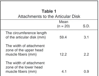
The attachment of fibers from both heads of the lateral pterygoid muscle to the articular disk was observed in all specimens. The circumference of the articular disks aver-aged 58.1 mm, the width of attachment zone of the supe-rior head muscle fibers averaged 11.9 mm, and the width of the attachment zone of the inferior head muscle fibers was 4.3 mm (Table 1). The angle formed between the tip of the attachment area of the upper head muscle fibers and the baseline averaged -4.5¡, and that between the tip of the attachment area of the lower head muscle fibers averaged 6.1¡ (Table 2).
Existence of the Ligament which Draws the Articular Disk Posteriorly During Jaw-closing
As a result of our detailed observation of the spheno-mandibular ligament at its attachment area with periph-eral bones of the temporomandibular joint, we found the ligament was firmly attached to the spine of sphenoid,
and under a stereoscope the sphenomandibular liga-mentÕs fibers extended toward the tympanosqua-mosal fissure. With all 12 sides of the specimens, we confirmed that these fibers were connected with the artic-ular disk (Figure 6).
Discussion
Studies of the insertions of the lateral pterygoid muscle from lateral or superior perspectives or reconstruction from sections alone are not optimal because of the limited perspective. What then is most important? The answer is simple: to determine whether the muscle fibers are from the superior head or inferior head by tracing the fibers under a stereoscopic microscope. This practice of identi-fying the muscle fibers with oneÕs own eyes, however simple it may seem, is really difficult because of the muscle fibersÕ positional relation and thus requires a high level of technical dexterity. After experimenting
Figure 4
Lateral approach. J.P.: joint projection; U.LPM: upper head of the lateral pterygoid muscle; L.LPM: lower head of the lateral pterygoid muscle.
Figure 5
Medial approach. LPM: lateral pterygoid muscle; S.L.: sphenoman-dibular ligament; M.B.: mansphenoman-dibular bone.
with various methods, Dr. Abe, one of the authors of this study, has established an observation method suitable for observing the peripheral tissues of articular disks.7
Contrary to previous studies4which had demonstrated
that there was very little attachment, if any, to the articu-lar disk, the method applied in this study proved the infe-rior head muscle fibers were firmly attached to the articular disk medially. In terms of the relative position with the mandibular condyle, the lower head muscle fibers extended medially beyond the baseline between the lateral and medial poles. Based on this information, we can say that the muscle fibers can both draw the articular disk anteriorly and balance it by supporting it posteriorly. Since most of the muscle fibers are attached to the anterior portion of the articular disk, they can cause the anterior transposition of the articular disk as previously reported.6,8In consideration of the medio-posterior
attachment of the muscle fibers, however, they can pull the articular disk posteriorly. Therefore, we are con-vinced that these fibers attach to the articular disk beyond the base line and play an important role in balancing,
maintaining, and stabilizing the complicated movements of the articular disk.
It has long been supposed that the major role of the sphenomandibular ligament is to suppress the movement of the mandible during jaw-opening.8,9After observing
12 specimens, we found that the sphenomandibular liga-ment has two major roles. We found two major bundles of fibers in the ligament. One bundle flows from the spine of the sphenoid and is attached to the tympanosquamosal fissure, and the other attaches to the articular disk.
The sphenomandibular ligament has two attachments to its fibers. The fibers attached to the spine of the sphe-noid bone were observed to be strained during the mandibleÕs opening motion. Therefore, we can say that the sphenomandibular ligament functions to limit the movements of the mandible, as previously demonstrated.9
Close observation of the sphenomandibular ligament during jaw-closing showed that the fibers attached to the spine of the sphenoid bone are relaxed, and the fibers attached to the articular disk are strained. These findings suggest the ligament fibers attached to the articular disk draw the disk posteriorly in its course of mandibular clos-ing, thus enabling the articular disk to move smoothly. As described earlier,9there are few accurate reports on
posterior supports for the articular disk. Although there have been many studies on the structure of tissues using slice images as Òthe posterior connective tissue of the articular disk,Ó they are not convincing as to tissue sup-porting the articular disk posteriorly. In this study, we obtained new knowledge not only by observing the tem-poromandibular joint, but by observing the whole ramus of the mandible medially and tracing the sphenomandibu-lar ligamentÕs fibers as they travel to the temporo-mandibular joint. These important sphenotemporo-mandibular ligament fibers are hidden in Òthe posterior connective tissue of the articular disk.Ó
MUSCLE AND LIGAMENT FUNCTION IN THE TMJ ABE ET AL.
206 THE JOURNAL OF CRANIOMANDIBULAR PRACTICE JULY 1997, VOL. 15, NO. 3
Table 1
Attachments to the Articular Disk
Mean
(n = 20) S.D. The circumference length
of the articular disk (mm) 59.4 3.1 The width of attachment
zone of the upper head
muscle fibers (mm) 12.2 2.2 The width of attachment
zone of the lower head
muscle fibers (mm) 4.1 0.9
Table 2
The Angle
Mean
(n = 20) S.D. The angle formed between
the tip of the attachment area -4.7 1.3 of the upper head muscle
fibers and the base line (˚) The angle formed between the tip of the attachment area
of the lower head muscle 6.2 2.1 fibers and the base line (˚)
Figure 6
Acknowledgements
The authors wish to thank Professors Bogusch Gottfried, Graf Renate, and Merker Hans Joachim, the Free University Berlin and the Institute for Anatomy, for supporting this study.
References
1. Toller M, Juniper RP: The embryologic development of the human lateral pterygoid muscle and its relationships with the temporomandibular joint disk and MeckelÕs cartilage. J Oral Maxillofac Surg 1993; 51:772-778 2. Schmolke C: The relationship between the temporomandibular joint capsule,
articular disk and jaw muscles. J Anat 1994; 184:335-345
3. Sicher H: Structual and functional basis for disorders of the temporo-mandibular articulation. J Oral Surg 1955; 13:275-279
4. Pinkert R: Die Beziehungen zwischen dem M. pterygoideus lateralis und dem discus articularis und deren Bedeutung fur die Bewegungen im Kiefergelenk. Zahn-. Mund-u. Kieferheilkd, 1984; 72:553-558
5. Troiano MF: New concept of the insertion of the lateral pterygoid muscle.
J Oral Surg 1967; 25:337-340
6. Porter MF: The attachment of the lateral pterygoid muscle to the meniscus.
J Prosthet Dent 1970; 24:555-562
7. Abe S, Takasaki I, Ichikawa K, Ide Y: Investigations of the run and the attach-ment of the lateral pterygoid muscle in Japanese. Bulletin, Tokyo Dent Coll 1993; 34: 135-139
8. Wilkinson TM: The relationship between the disk and the lateral pterygoid muscle in the human temporomandibular joint. J Prosthet Dent 1988; 60:715-724
9. Burch, J: Activity of the accessory ligaments of the temporomandibular joint.
J Prosthet Dent 1970; 24:621-628
Dr. Yoshinobu Ide received his D.D.S. degree from Tokyo Dental College in 1976 and his Ph.D. degree from the same college in 1980. Dr. Ide is currently a Professor in the Department of Anatomy at Tokyo Dental College.
Dr. Hakubun Yonezu received his D.D.S. degree from Tokyo Dental College in 1984 and his Ph.D. degree from the same college in 1988. Dr. Yonezu is currently an Instructor at the Tokyo Dental College in the Department of Oral and Maxillofacial Surgery.