Japan Advanced Institute of Science and Technology
JAIST Repository
https://dspace.jaist.ac.jp/Title
Assembly of Ag@Au Nanoparticles Using
Complementary Stranded DNA Molecules and Their Detection Using UV-Vis and Raman Spectroscopic Techniques
Author(s) Mott, Derrick; Nguyen, T. B. Thuy; Aoki, Yoshiya; Maenosono, Shinya
Citation Materials Research Society Symposium Proceedings, 1272: PP06-12
Issue Date 2010
Type Conference Paper
Text version publisher
URL http://hdl.handle.net/10119/9556
Rights
Copyright (C) 2010 Materials Research Society. It is posted here by permission of the Materials Research Society. Derrick Mott, Nguyen T. B. Thuy, Yoshiya Aoki, and Shinya Maenosono,
Materials Research Society Symposium Proceedings, 1272, 2010, PP06-12. http://www.mrs.org/
Assembly of Ag@Au Nanoparticles Using Complementary Stranded DNA Molecules and Their Detection Using UV-Vis and Raman Spectroscopic Techniques.
Derrick Mott, Nguyen T. B. Thuy, Yoshiya Aoki, and Shinya Maenosono.
School of Materials Science, Japan Advanced Institute of Science and Technology, 1-1 Asahidai, Nomi, Ishikawa 923-1292, Japan
ABSTRACT
Silver nanoparticles coated by a layer of gold (Ag@Au) have received much attention because of their potential application as ultra sensitive probes for the detection of biologically important molecules such as DNA, proteins, amino acids and many others. However, the ability to control the size, shape, and monodispersity of the Ag@Au structure has met with limited success. In our own research we have addressed this challenge by creating an aqueous wet chemical synthesis technique towards size and shape controllable Ag@Au nanoparticles. These materials are highly interesting because of the tunable silver core size, and the tunable gold shell thickness, opening many avenues to the modification of the particle properties in terms of bio-molecular sensing. The resulting nanoparticle probes were functionalized with two
complementary stranded DNA oligonucleotides. When combined, the complementary strands hybridized, causing the Ag@Au nanoparticles to assemble into large nano-structures. The presence of the oligonucleotide was confirmed through a series of techniques including UV-Vis and Raman spectroscopy, as well as TEM, XPS, DLS, and many others. The results reflect the role that the nanoparticle physical properties play in the detection of the bio-molecules, as well as elucidate the characteristics of the bio-molecule/nanoparticle interaction.
INTRODUCTION
To date there have been several attempts to detect DNA using nanoparticles as sensitive probes [1]. Such detection techniques often rely on assembly of complementary stranded NPs (Ag, Au or Ag@Au NPs) and their subsequent hybridization which is detected by colorimetric techniques or by labeling of the nanoparticles with a reporter label which can then be detected using Raman Spectroscopy [1]. These techniques open the doors to the ultra-sensitive detection of DNA using nanoparticle probes, but there is still much work to be done in understanding the nanoparticle probe properties themselves. Both silver and gold nanoparticles (NPs) have
received wide attention for their enhanced properties in a multitude of potential applications such as sensing, microelectronics, and catalysis, [2,3] because of the many desirable chemical and physical properties of the materials. In terms of bio-diagnostics and sensing, it is the optical properties that make Ag NPs exceptional, while for Au NPs it is the resistance to oxidation and enhanced thiol chemistry that is attractive [1,4]. The current trend in this area of research is the coupling of these two materials as a core@shell structure that takes advantage of the optical properties of silver and the stability/thiol chemistry of gold. These Ag@Au nanoparticles are expected to have use as bio-probes with unprecedented sensitivity and selectivity. Despite the excitement surrounding these materials though, there are still many challenges to address, including the ability to synthesize Ag NPs in aqueous phase with a desired size, shape, or monodispersity [5], and the ability to coat the silver particle with a uniform and controllable layer of gold. Finally, the detection of DNA using these nanoscale probes presents many unique challenges of its own including attachment of DNA to the nanoparticle probe surface, assembly of the NPs using a target strand of DNA, hybridization of the matched DNA strands, and finally
detection of the DNA [6]. Our approach to these unique challenges is a straightforward assembly of the Ag@Au nanoparticle probes using specific sequenced DNA strands through a photoligation reaction, and finally detection of the interaction using a dual buffer/reporter molecule with Raman spectroscopy.
EXPERIMENT
Chemicals. Silver nitrate, sodium acrylate, gold tetrachloroaurate trihydrate and common solvents were obtained from Aldrich. Water was purified with a Millipore Direct-Q system (18.2 MΩ). Dialysis membranes with molecular weight pore size of 10,000 daltons were obtained from Spectra/Por and were rinsed in pure water before use. Calcium chloride and sodium chloride were purchased from Wako Chemicals. Cacodylate buffer solution (0.2M) was purchased from Nacalai Tesque Inc., Japan. Nuclease P1 was purchased from Yamasa Corp., Japan. ODN sequences were synthesized using an Applied Biosystems 3400 DNA synthesizer. The reagents for the synthesis of DNA components was purchased from Glen Research, USA.
Synthesis of Ag NP Cores: Ag NPs were synthesized by first mixing 50ml of water with 1.25×10⁻⁵ moles of silver nitrate, and then adding 6.75x10⁻⁶ moles of sodium hydroxide, which results in a dilute yellow colored solution of silver hydroxide. This solution is purged with argon and is then brought to reflux. At reflux, 2.55×10⁻⁴ moles of sodium acrylate are added causing the solution to turn completely clear. The solution is refluxed for 1 hour, over this time the solution color changes from clear to green-yellow to yellow-orange.
Purification of as-synthesized Ag NPs: Prior to deposition of Au on the Ag NP seeds, the as-synthesized particles are purified to remove excess acrylate, silver, sodium, and other ions from the solution. Purification is performed by enveloping the particle solution inside of a cellulose dialysis membrane with pore size of 10,000 daltons and soaking in a distilled water bath. The water was changed every 12 hours for 48 hours.
Deposition of Au on the Ag Cores to form Ag@Au NPs: 50ml of the dialysized Ag particles are brought to reflux and 10ml of a gold tetrachloroaurate trihydrate solution (ranging from 6.25×10⁻⁷ to 3.13×10⁻⁶ moles according to the thickness of the Au shell desired) and 10ml of a sodium acrylate solution (from 5.10×10⁻⁵ to 2.55×10⁻⁴ moles) are added dropwise
simultaneously. The solution color changes depending on the amount of Au added. In general, as Au and sodium acrylate is added to the Ag particles, the color changes from yellow-amber to dark amber to grey to grey-purple and finally to purple.
Instrumentation and Measurements: An array of techniques including Transmission Electron Microscopy (TEM), High Resolution TEM and Energy Dispersive Spectroscopy (HR-TEM, EDS), X-Ray Photoelectron Spectroscopy (XPS) and Ultra-Violet Visible Spectroscopy (UV-Vis) were used to characterize the size, shape, composition and other properties of the materials. TEM analysis was performed on an Hitachi H-7100 transmission electron microscope operated at 100kV. HR-TEM and EDS analysis was performed on an Hitachi H-9000NAR transmission electron microscope operated at 300kV. TEM samples were prepared by dropping the suspended particles onto a carbon coated copper grid and drying in air overnight.
DISCUSSION
In general, our synthetic rout towards Ag@Au NPs consists of 2 main steps. First we synthesize the Ag cores in aqueous phase with an acrylate capping agent. In the first part of the
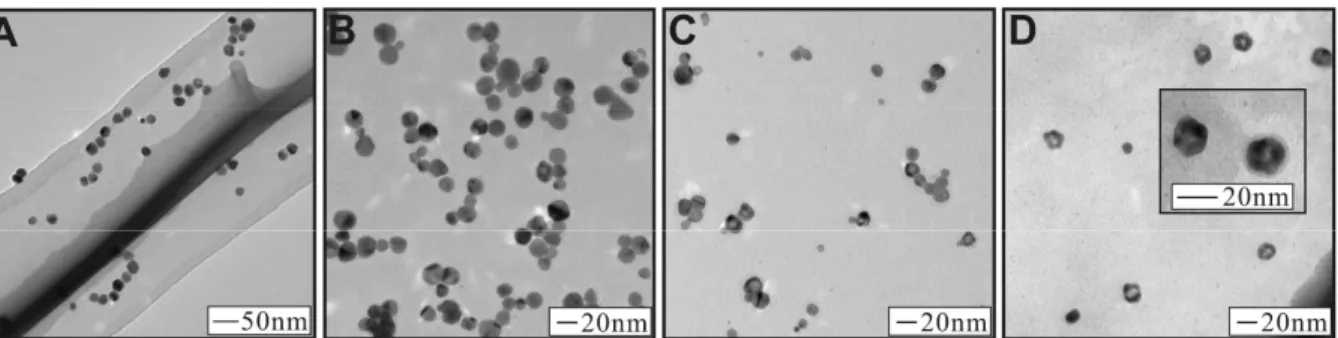
results and discussion section we show our synthetic approach to monodispersed Ag particles. The second step of this research is the coating of Au on the Ag particle surface. In the second part of the results and discussion section we illustrate the coating of the Ag particles by Au and discuss the ability to control the thickness of the Au layer and the resulting morphology of the nanostructures. The overall technique of our synthetic approach is shown in Scheme 1. In this approach, first we used silver nitrate and sodium hydroxide to create a dilute solution of silver hydroxide. This solution was brought to reflux, whereupon sodium acrylate was added, initiating the formation of Ag NPs. The Ag particles formed over the course of one hour as evidenced by the appearance of a yellow-amber solution. Figure 1A shows the TEM image of the
as-synthesized Ag NPs capped by the acrylate molecule. The particle size distribution is 20.5 ± 3.3 nm (16% deviation). Given the size of the particles and their total mass, we calculated the Ag NP concentration to be 7.28×10⁻¹¹ M and their extinction coefficient to be 2.11×10¹⁰ M−1cm−1.
After synthesis, the particles were purified by using dialysis to remove excess acrylate, silver ions, and other species. The coating of the silver NPs by gold is achieved in what is essentially a seeded growth reaction. Briefly, first the Ag NPs are brought to reflux, then dilute aqueous gold and sodium acrylate solutions are added simultaneously, dropwise, causing the Au to reduce at the Ag NP surface, causing a coating of Au to be formed. The primary challenge in this reaction is the propensity of Ag to be oxidized as Au is reduced during the coating. Such an occurrence can lead to either hollow Au particles or alloyed Au and Ag NPs. In our own
reaction, the alloying or etching of the Ag NPs can be minimized through control of the concentration of gold being added, the rate that it is added, and by addition of the sodium acrylate capping and reducing agent. Here, the role of sodium acrylate is to cause the Au to become reduced before the Ag can be oxidized.
Scheme 1: Reaction Rout for the Synthesis of Ag NPs and their Coating with Au.
Figure 1 shows a series of TEM images of Ag@Au NPs synthesized using the method described above. For each sample, the amount of gold and sodium acrylate added was
incrementally increased. Figure 1B shows a sample of Ag@Au NPs synthesized by adding 6.25×10⁻⁷ moles of Au and 1.38×10⁻⁴ moles of sodium acrylate (corresponding to 5% Au in terms of atomic composition). Inspection of the TEM image reveals particles with a uniform spherical morphology. The direct observation of a deposition of a layer of gold on the surface of the silver nanoparticles is difficult to ascertain in this image. The size distribution of these particles is 17.5 ± 3.7nm. Figure 1C shows a sample of Ag@Au NPs synthesized by adding 1.88×10⁻⁶ moles of Au and 1.38×10⁻⁴ moles of sodium acrylate (corresponding to 15% Au in terms of atomic composition). The TEM image shows several particles with a spherical morphology, but now several particles are observed that have a lighter spherical center and darker outside ring. The observation of this dark outside and light center is inconsistent among different particles in the sample, some particles display no darker ring and light center at all, or
some particles display simply a light spot near the periphery of the NP. We attribute this observation to the formation of an incomplete Au shell on the Ag particle surface. In effect, the round holes that are observed on the particles in the TEM image are a gap or hole in the Au shell, allowing us to see the Ag core inside the particle. The size distribution of these NPs is 16.3 ± 2.7 nm. In addition, a few NPs are observed in the TEM image with much smaller size than the parent Ag NPs (~9nm), which could be attributed to the non-specific formation of Au NPs without coating on the Ag surface. The presence of these small particles was not observed in techniques such as UV-Vis, probably because of their low concentration as compared to the coated particles. Finally, Figure 1D shows a sample of Ag@Au NPs synthesized by adding 3.13×10⁻⁶ moles of Au and 1.38×10⁻⁴ moles of sodium acrylate (corresponding to 25% Au in terms of atomic composition). The TEM image reveals many particles with a light center and thick dark outside. Now the particles seem to have adopted roughly hexagonal or pentagonal shapes, likely reflecting the tendency of Ag nanocrystals to be oriented in the twinned structure, templating the growth of Au at their surface. The size distribution of these particles is 17.5 ± 5.1nm. Among these three samples the particle sizes are generally smaller than the precursor Ag NP seeds (size of 20.5 ± 3.3 nm). We attribute this size decrease to a small degree of etching of the silver surface at the initial reaction stage as the gold layer is deposited. As the reaction progresses, the acrylate reducing agent plays a more significant role in reducing the gold as it is deposited on the particle surface, thereby preventing the entire silver core from being etched away.
Figure 1. TEM images for as-synthesized Ag NPs (A), and Ag@Au NPs with atomic feeding ratio of: 5% Au (B), 15% Au (C), and 25% Au (D).
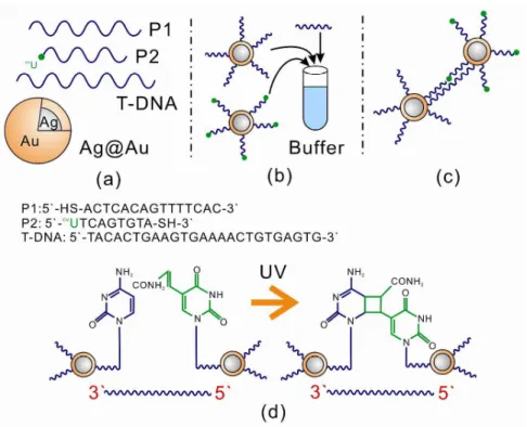
The nanoparticles synthesized above were used as probes for the detection of DNA (Ag@Au NPs synthesized with 5% Au in terms of atomic feeding ratio). Scheme 2 shows our approach to the detection of DNA. Scheme 2A shows the components of the detection. The DNA sensing technique exploits the photoligation reaction between oligodeoxynucleotides (ODNs) attached on the surfaces of NPs in the presence of target DNA (T-DNA). In this approach, two ODNs, a common probe (P1) and a specific discriminating probe (P2) are separately conjugated on the surface of the NPS (Scheme 2B). The conjugated NPs hybridize and form aggregates via photoligation between the P1 and P2 strands (Scheme 2B and C). Once aggregation has taken place the interparticle spacing between the NPs acts as a “hot spot”, causing a SERS signal from the Raman active buffer (cacodylic acid) used in the assembly.
Figure 2 shows Raman spectra for the functionalization of the Ag@Au NPs with the P1, P2 and target DNA strands. Figure 2a shows the Raman spectra of the sample before incubation, while Figure 2b shows the Raman spectra after 6 hours incubation at 40 ºC. The successful functionalization of the DNA strands can be identified by the observation of the ring breathing modes for Adenine and Cytosine in the Raman spectrum. Photoligation of the materials and subsequent characterization of the detection of DNA is part of our ongoing work in this area of research.
Figure 2. Detection of the DNA hybridization using Raman Spectroscopy. Before hybridization (A) and after 6 hours hybridization (B).
CONCLUSIONS
We have demonstrated a straightforward technique for the synthesis of monodispersed silver nanoparticles and their coating by a shell of gold of variable thickness that can be
controlled by the amount of gold precursor used in the coating process. The use of acrylate as a dual reducing and encapsulating agent minimized the etching of metallic silver particles by the aqueous gold precursor. The use of these probes to detect DNA has been explored, the
preliminary results are promising and further study of the system is part of our ongoing work. ACKNOWLEDGMENTS
Derrick Mott gratefully acknowledges support by the Japan Society for the Promotion of Science (JSPS) fellowship. We thank Mr. Nobuaki Ito for assistance in the use of XPS
instrumentation and Mr. Koichi Higashimine for assistance in the use of TEM instrumentation.
REFERENCES
1. Y-W. Cao, R. Jin and C. A. Mirkin, J. Am. Chem. Soc. 123, 7961 (2001). 2. L. Lu, A. Kobayashi, K. Tawa and Y. Ozaki, Chem. Mater. 18, 4894 (2006). 3. Z. S. Pillai and P. V. Kamat, J. Phys. Chem. B 108, 945 (2004).
4. Y. Cui, B. Ren, J-L. Yao, R-A. Gu and Z-Q. Tian, J. Phys. Chem. B 110, 4002 (2006). 5. V. K. Sharma, R. A. Yngard and Y. Lin, Adv. Colloid Interf. Sci. 145, 83 (2009).
6. N. T. B. Thuy, R. Yokogawa, Y. Yoshimura, K. Fujimoto, M. Koyano and S. Maenosono, Analyst 135, 595 (2010).