Effect of moderate exercise intensities on the cortical activity in young adults
著者 橋本 直之
著者別表示 Hashimoto Naoyuki journal or
publication title
博士論文本文Full 学位授与番号 13301甲第4805号
学位名 博士(保健学)
学位授与年月日 2018‑09‑26
URL http://hdl.handle.net/2297/00053139
Original Article
Effect of moderate exercise intensities on the cortical activity in young adults
Naoyuki Hashimoto, RPT, MSc
1, 2)*, Masami Yokogawa, RPT, PhD
3),
Haruyuki Kojima, PhD
4), Shoji Tanaka, RPT, PhD
3), Takao Nakagawa, MD, PhD
3)1)
Section of Rehabilitation, Kanazawa University Hospital: 13-1 Takara-machi, Kanazawa, Ishikawa 920-8641, Japan
2)
School of Health Science, College of Medical, Pharmaceutical and Health Sciences, Kanazawa University, Japan
3)
Institute of Medical, Pharmaceutical and Health Sciences, Kanazawa University, Japan
4)
Institute of Human and Social Sciences, Kanazawa University, Japan
Abstract. [Purpose] To examine the influence of different exercise intensities on cortical activity. [Participants and Methods] Twenty-six healthy adults aged 20–30 years performed exercise at three intensities on a bicycle er- gometer as follows: (a) 15-minute exercise at 40% peak oxygen uptake, (b) same as (a) but at 60% peak oxygen uptake, and (c) 15 minutes of rest. The cognitive function of the participants was measured before and after exercise by the Paced Auditory Serial Addition Test (PASAT) under these three conditions. The cerebral blood flow in the left prefrontal and temporal cortices was measured using near-infrared spectroscopy during the PASAT. [Results]
The PASAT score was significantly higher after exercise under condition (b) than before exercise (41.4 ± 9.1 vs. 47.7
± 8.3). The cerebral blood flow in the prefrontal cortex under condition (b) was significantly increased compared to that under condition (c), as determined by the Tukey method (0.019 ± 0.030 vs. −0.008 ± 0.044). Significant differ- ences were not observed in the cerebral blood flow in the temporal cortex under these three conditions. [Conclusion]
Cortical activation of the frontal lobe increased after high-intensity aerobic exercise with no change in the cortical activity of the temporal lobe.
Key words: Exercise intensity, Aerobic exercise, Cortical activity
(This article was submitted May 26, 2018, and was accepted Jul. 20, 2018)
INTRODUCTION
Cognitive function is intellectual activity, including attention, execution, information processing, language, visual spatial skills, psychomotor ability, learning, and memory. Physical exercise not only improves the functional capabilities of the body, such as muscular strength and muscle endurance but also affects cognitive functions associated with structural changes in the brain
1). Studies in mice have indicated that physical activity can regulate hippocampal neurogenesis and improve spatial memory
2). Colcombe et al.
3)have reported that cognitive functions in elderly people, in particular their executive functions, improved after aerobic exercise for 6 months. The volume of their cerebral white matter and gray matter had also increased
4). These findings indicate that physical exercise plays an important role in the maintenance and improvement of cognitive function
5). However, several questions still need to be addressed with regard to the different effects that physical exercise has on the brain according to the conditions under which the exercise takes place.
In long-term intervention studies, examining the factors that affect the brain is difficult as daily lifestyle rhythms differ from person to person and the amount of activity that each individual takes part in also affects study results
6). For this reason,
J. Phys. Ther. Sci. 30: 1257–1261, 2018
*Corresponding author. Naoyuki Hashimoto (E-mail: nao_hashimot531@med.kanazawa-u.ac.jp)
©2018 The Society of Physical Therapy Science. Published by IPEC Inc.
This is an open-access article distributed under the terms of the Creative Commons Attribution Non-Commercial No Deriva-
The Journal of Physical Therapy Science
The Journal of Physical Therapy Science
transient exercise is used in studies that aim to examine the effects that physical exercise conditions have on the brain
7–9). Test batteries are used in these studies, which test aspects of cognitive function such as reaction time, attention, crystallized intel- ligence, executive function, and memory; the studies suggest that the results of each of these aspects of cognitive function will improve after exercise
8). From the perspective of exercise intensity, results improved as the intensity level increased from low to a specified high-intensity peak. However, in contrast, the results worsened if the intensity level was increased beyond this point, indicating an inverse U-curve relationship
10). Therefore, as a general rule, for exercise that aims at improving cognitive function, a moderate-intensity level is recommended
9).
As possible reasons for the relationship between exercise intensity and cognitive function exhibiting an inverted U-shaped curve, Kashihara et al.
11)cite the possibility that high-intensity exercise leads to the release of an excessive quantity of neurotransmitters, causing failure of the nerve network. In addition, they also cite the possibility of the effects of exhaustion and stress. Regarding changes in event-related potential, the P3 (or P300) amplitude after moderate-intensity exercise was larger than after high- and low-intensity exercise
12). It has been previously reported that the P3 amplitude is associated with attention allocation
13). Moreover, a study using near-infrared spectroscopy (NIRS) reported improved prefrontal cortex activ- ity following 15 minutes of exercise with a 50% VO
2peak. Specifically, cognitive function might be improved by creating an appropriate environment in which brain neural activity can function easily after moderate-intensity exercise
14).
Therefore, exercise is known to produce changes in neural activity in the brain resulting in changes in cognitive function.
However, as the cortical activity is examined under a uniform intensity, the differences in cortical activity according to exer- cise intensity remain unknown. Moreover, in terms of the contents of the cognitive aspects, auditory stimulation and visual stimulation are known, for example, to improve activity in the applicable cortical areas
15, 16). However, whether any changes occur in cortical activation other than in the prefrontal cortex after exercise remains unknown. Therefore, we aimed in this study to examine the influences of different exercise intensity on cortical activity. We used the Paced Auditory Addition Task (PASAT) to evaluate cognitive function. The PASAT is a task that is often used for checking attention functions and is known to affect the temporal cortex
8). In addition, fMRI was used to reveal the area activated during the task
17).
PARTICIPANTS AND METHODS
The study included 26 healthy, right-handed adults aged 20–30 years (14 males, 12 females). The mean age, height, and weight were 24.3 ± 3.5 years, 164.3 ± 7.8 cm, and 59.5 ± 9.5 kg, respectively. The exclusion criteria included history of neurological disorder or difficulty riding a bicycle. The purpose and procedures of the study were explained verbally and in writing, and informed consent was obtained from all participants. This study was approved by the Ethics Committee on Human and Social Sciences, Kanazawa University (approval number: 2013-2).
The experiments for the study took place over 4 days. On the first day, an exercise test was carried out to determine the appropriate exercise intensity level. The peak oxygen uptake (VO
2peak) during testing was measured using a respiratory metabolism device (AE-300S, Minato Medical Science Co. Ltd., Osaka, Japan). Using a bicycle ergometer (Rehcor500P, Lode, the Netherlands), a 3-minute warm-up was followed by a cycling exercise with a ramp protocol of 20 W/min with the pedal frequency maintained at 60 rpm, wherein the participants cycled until exhaustion. The exercise test was terminated if the participant chose to stop, if the pedaling rate dropped to 50 rpm, or if the participant’s condition corresponded to the criteria set by the American College of Sports Medicine
18). The exercise was followed by a 3-minute cool down. As a safety measure during testing, the participants’ heart rate was monitored by a medical telemeter (DS-2202, Fukuda Denshi Co., Ltd., Tokyo, Japan) and blood pressure was measured manually every 2 minutes.
Between the second and the fourth day of the study, the participants took part in 2 exercise groups and rested in a seated position (Control). For the exercise groups, each participant took part in a 15-minute exercise on the bicycle ergometer (EX90, Combi Corporation, Tokyo, Japan), at an intensity of 40% of the VO
2peak (G40) and 60% of the VO
2peak (G60), which was calculated from each participant’s VO
2peak level. In the Control condition, the participant sat on the bicycle ergometer and rested for 15 minutes. All participants underwent a cognitive function test before and after the exercise conditions and Control. After the exercise the participant was required to rest for 15 minutes to exclude the influence of the temporary increases in cerebral blood flow that occur after exercise, in accordance with Yanagisawa et al
14). Participants were instructed to minimize movements of the head during the NIRS measurement and the PASAT. During the cognitive function test, the cerebral blood flow of the frontal and temporal areas was measured using a NIRS, as described below. The order of individual measurements was randomly determined. Each measurement was taken with a 3–7-day interval.
We assessed cognitive function using the Paced Auditory Serial Addition Test (PASAT) that is often used to assess at- tention in neuropsychology. This test requires the participant to listen to a digital recording of digits presented one at a time and to mentally add the number they just heard with the number they heard before it. For example, if the numbers “3,” “1,”
and “2” were presented, the participant should answer “4” and then “3.” In adults, scores have been reported to decline with
age
19). As for the interval between each number, 1-second and 2-second intervals were tested in a preliminary experiment. For
the participants of this study, 1 second was decided to be more suitable as the level of difficulty. Therefore, the intervals were
set at 1 second for the test. For the cerebral blood flow measurements to fit into a block design, the PASAT tasks were carried
out for 15 seconds with 10 seconds of rest and were repeated 5 times. After completion of cerebral blood flow measurements,
all 60 questions of the PASAT tasks were answered in succession by the participants, and the number of correct responses
was recorded.
For the cerebral blood flow measurements, a NIRS (ETG-4000, Hitachi, Ltd., Tokyo, Japan) that uses 2 near-infrared lights (695 nm and 830 nm) was used. This device applies the optical data based on the modified Beer–Lambert Law
20)and can obtain data on oxygenated hemoglobin (oxygenated Hb), deoxygenated hemoglobin, total hemoglobin concentration, and changes in hemoglobin concentration (mM•mm)
21). The sampling frequency was set at 100 ms.
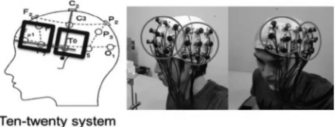
The NIRS probes were positioned so they covered the participant’s left frontal lobe and the temporal lobe. With 24 channels per 1 set (3 cm space), each set is composed of 8 irradiation probes and 8 light absorption probes. When the probes were installed onto the participant’s head, 2 multichannel probe holders (4 × 4) were used. During the PASAT tasks, the left prefrontal cortex, the outer premotor cortex, the supplementary motor area, the cingulate gyrus, the left parietal lobe, the superior temporal gyrus, the right temporal pole, and the visual association cortex were reported to be the brain’s activated regions
17). The probes are installed in accordance with the international 10–20 system. The left frontal lobe is joined with the probe by fastening the Fp1 and F7 to it in a straight line, ensuring that the F7 is covered when installed, and the temporal lobe is joined with the vertical row of probes by fastening the Cz and T3 to it in a straight line, ensuring that the T3 is covered when installed (Fig. 1).
This study used a block design comprising the 5 PASAT tasks and the 5 rest periods, and the cerebral blood flow was measured using the NIRS to determine brain activity. The oxygenated Hb levels were used for the evaluation; the resting scores were subtracted from the PASAT task scores, and the resulting calculations were averaged using the NIRS. Changes in the cerebral blood flow during the cognitive tasks were calculated. Next, the difference between the post-exercise and pre-exercise scores was calculated to determine the changes in cerebral blood flow according to each condition.
The number of correct responses from the PASAT before and after exercise was compared using a t-test for each group.
A one-way analysis of variance (ANOVA) was used for the number of correct responses from the PASAT in each before and after exercise, and a multiple comparison test was used if a significant difference was observed. After the one-way ANOVA, the cerebral blood flow among the groups was analyzed using a multiple comparison test. The significance level for all tests was set at <5%.
RESULTS
The number of the correct responses on the PASAT was significantly higher after exercise than before exercise only in G60 (p<0.05). As for the number of correct responses among the conditions, no significant difference was observed before exercise (F
2,75=0.073, p=0.92). However, the post-exercise results showed a significant difference (F
2,75=3.11, p<0.001).
Moreover, the score for G60 was significantly higher compared with G40 and Control (p<0.05) (Table 1). The cerebral blood flow of the frontal lobe showed a significant difference (F
2,75=4.11, p<0.05), and in Tukey’s method, the G60 was significantly increased compared with the Control (p<0.05). No significant difference was observed among the conditions for the cerebral blood flow in the temporal lobe (F
2,75=0.32, p=0.73) (Table 2).
DISCUSSION
In the present study, we examined the influence of different exercise intensities on cortical activity in the frontal and temporal lobes. An increase in cortical activity after exercise was observed only in the frontal lobe at G60.
Concerning the changes in cognitive function task scores as a result of exercise, the graph, which represents exercise intensity on the x-axis and cognitive task scores on the y-axis, is said to produce an inverse U curve. Therefore, the optimum exercise intensity level to improve cognitive function, namely, the top of the inverse U curve, should extend to the anaerobic and lactate threshold (LT) areas
12). The LT of adults who do not exercise regularly is 50–60% VO
2max
22, 23), and should reach 65–75% VO
2max
24)for athletes. As the participants of this study were young adults aged 20–30 years, the peak exercise intensity level that will improve cognitive function should be approximately 60%. Thus, we estimated that both G40 and G60 would improve cortical activity, with G60 providing the greatest amount of improvement. However, our results
Fig. 1. Positioning and installation of the probes.
indicated that the G60 cortical activity increased compared with the Control but was not significantly different from G40.
Also, there was no significant difference between G40 and the Control.
To control the effect of a temporary increase in blood flow during exercise
14), we measured the tasks after the participants had rested for 15 minutes following their exercise activity. Regarding why cognitive function improves, Kashihara et al.
11)state that cerebral blood flow, the quantity of brain-derived neurotrophic factor (BDNF) released in the blood, increase with exercise intensity. Moreover, blood glucose
25)and BDNF
26)have been found to return to the resting level once a certain amount of time passes after exercise. The blood concentration and quantity released into the blood of the substances affecting brain neural activity are thought to affect nerve activity and are expected to attenuate sooner with low-intensity exercise than with high-intensity exercise. Since it was confirmed that increased of cerebral blood flow caused by transient exercise reverts back to the resting level after 15 minutes
14), the cognitive function tasks in this study were implemented 15 minutes after exercise was finished. The improvement in cortical activity might not have been observed at G40 because cerebral blood flow had already returned to the resting level when PASAT was measured after exercise.
The results of this study, similar to previous studies
14, 27), showed that the cortical activity of the frontal lobe did increase after transient exercise. In the current study, the temporal lobe, as an area other than the frontal lobe, was measured. The NIRS measurement scores for the temporal lobe are thought to reflect the activity of the auditory association area. The results from this study thus suggest that the auditory association area bears no relationship with the improvement of cognitive function.
However, the adaptability of the auditory association area with regard to repetitive sounds is remarkable. Therefore, the reaction is also thought to almost disappear once the sound has been presented a few times
28). In the current study, a repetitive block design was used for the tasks and rest periods, so there is a possibility that it may have been influenced by the task conditions. We assume that the increased cortical activity in all areas is not necessarily directly related to improvement in function but, on the contrary, that a decline or lack of change in activity may be beneficial. We also think that this reflects the role difference in the domain.
The present study has several limitations. First, we did not assess the exercise habits of the participants. LT is well known to be higher in athletes
24); therefore, it is thought to be also affected by exercise habits. There is a possibility that other than exercise intensity, factors such as gender, age, exercise habits, and the interval between exercise and performing the tasks affect cognitive function. Therefore, further research on cognitive function after transient exercise is needed with regards to such factors. An improvement in cortical activation was demonstrated at 60% in young adults in this study. However, age-related differences were not taken into consideration. It is likely that the optimum level of exercise intensity is lower in the elderly because cognitive function
29)and physical fitness
30)decrease year by year. Thus, further studies of elderly people are necessary.
Conflict of interest None.
Table 2. Changes in cerebral blood flow (mM•mm)
Control G40 G60 Tukey’s method
Frontal lobe −0.008 ± 0.044 0.012 ± 0.029 0.019 ± 0.030 Control < G60***
Temporal lobe −0.003 ± 0.045 −0.004 ± 0.033 0.005 ± 0.046 Mean ± standard deviation.
***p<0.05: Comparison between groups.
Table 1. Number of correct responses (score) to Paced Auditory Serial Addition Test
Control G40 G60 Tukey’s method
Number of
correct responsesBefore exercise 42.1 ± 9.9 41.1 ± 9.6 41.4 ± 9.1
After exercise 40.8 ± 8.4 41.9 ± 8.5 47.7 ± 8.3* Control < G60**
G40 < G60**
Mean ± standard deviation.
*p<0.05: Comparison before and after exercise.
**p<0.05: Comparison between groups after exercise.
REFERENCES
1) Colcombe SJ, Erickson KI, Raz N, et al.: Aerobic fitness reduces brain tissue loss in aging humans. J Gerontol A Biol Sci Med Sci, 2003, 58: 176–180. [Medline]
[CrossRef]
2) van Praag H, Christie BR, Sejnowski TJ, et al.: Running enhances neurogenesis, learning, and long-term potentiation in mice. Proc Natl Acad Sci USA, 1999, 96: 13427–13431. [Medline] [CrossRef]
3) Colcombe S, Kramer AF: Fitness effects on the cognitive function of older adults: a meta-analytic study. Psychol Sci, 2003, 14: 125–130. [Medline] [CrossRef]
4) Colcombe SJ, Erickson KI, Scalf PE, et al.: Aerobic exercise training increases brain volume in aging humans. J Gerontol A Biol Sci Med Sci, 2006, 61:
1166–1170. [Medline] [CrossRef]
5) Barnes DE, Yaffe K: The projected effect of risk factor reduction on Alzheimer’s disease prevalence. Lancet Neurol, 2011, 10: 819–828. [Medline] [CrossRef]
6) Colcombe SJ, Kramer AF, Erickson KI, et al.: Cardiovascular fitness, cortical plasticity, and aging. Proc Natl Acad Sci USA, 2004, 101: 3316–3321. [Medline]
[CrossRef]
7) Hashimoto N, Yokogawa M, Yamazaki T, et al.: Effects of the intensity of transient aerobic exercise on attention. Rigakuryoho Kagaku, 2013, 28: 377–381 (in Japanese). [CrossRef]
8) Chang YK, Labban JD, Gapin JI, et al.: The effects of acute exercise on cognitive performance: a meta-analysis. Brain Res, 2012, 1453: 87–101. [Medline]
[CrossRef]
9) Ludyga S, Gerber M, Brand S, et al.: Acute effects of moderate aerobic exercise on specific aspects of executive function in different age and fitness groups: A meta-analysis. Psychophysiology, 2016, 53: 1611–1626. [Medline] [CrossRef]
10) Brisswalter J, Collardeau M, René A: Effects of acute physical exercise characteristics on cognitive performance. Sports Med, 2002, 32: 555–566. [Medline]
[CrossRef]
11) Kashihara K, Maruyama T, Murota M, et al.: Positive effects of acute and moderate physical exercise on cognitive function. J Physiol Anthropol, 2009, 28:
155–164. [Medline] [CrossRef]
12) Kamijo K, Nishihira Y, Hatta A, et al.: Differential influences of exercise intensity on information processing in the central nervous system. Eur J Appl Physiol, 2004, 92: 305–311. [Medline] [CrossRef]
13) Sugimoto F, Katayama J: The P300 as an index of the amount of attentional resources: a comparison of somatosensory probe stimuli with auditory probe stimuli. Jpn J Physiol Psychol Psychophysiol, 2014, 32: 18–28. [CrossRef]
14) Yanagisawa H, Dan I, Tsuzuki D, et al.: Acute moderate exercise elicits increased dorsolateral prefrontal activation and improves cognitive performance with Stroop test. Neuroimage, 2010, 50: 1702–1710. [Medline] [CrossRef]
15) Kojima H, Suzuki T: Hemodynamic change in occipital lobe during visual search: visual attention allocation measured with NIRS. Neuropsychologia, 2010, 48: 349–352. [Medline] [CrossRef]
16) Remijn GB, Kojima H: Active versus passive listening to auditory streaming stimuli: a near-infrared spectroscopy study. J Biomed Opt, 2010, 15: 037006.
[Medline] [CrossRef]
17) Audoin B, Ibarrola D, Au Duong MV, et al.: Functional MRI study of PASAT in normal subjects. MAGMA, 2005, 18: 96–102. [Medline] [CrossRef]
18) American College of Sports Medicine: ACSM’s Guidelines for Exercise Testing and Prescription, 8th ed. Philadelphia: Lippincott Williams & Wilkins.
19) Tombaugh TN: A comprehensive review of the Paced Auditory Serial Addition Test (PASAT). Arch Clin Neuropsychol, 2006, 21: 53–76. [Medline] [CrossRef]
20) Cope M, Delpy DT, Reynolds EO, et al.: Methods of quantitating cerebral near infrared spectroscopy data. Adv Exp Med Biol, 1988, 222: 183–189. [Medline]
[CrossRef]
21) Maki A, Yamashita Y, Ito Y, et al.: Spatial and temporal analysis of human motor activity using noninvasive NIR topography. Med Phys, 1995, 22: 1997–2005.
[Medline] [CrossRef]
22) Wasserman K, Whipp BJ, Koyl SN, et al.: Anaerobic threshold and respiratory gas exchange during exercise. J Appl Physiol, 1973, 35: 236–243. [Medline]
[CrossRef]
23) Sekir U, Özyener F, Gür H: Effect of time of day on the relationship between lactate and ventilatory thresholds: a brief report. J Sports Sci Med, 2002, 1:
136–140. [Medline]
24) Mazzeo RS, Marshall P: Influence of plasma catecholamines on the lactate threshold during graded exercise. J Appl Physiol 1985, 1989, 67: 1319–1322. [Med- line] [CrossRef]
25) Ide K, Schmalbruch IK, Quistorff B, et al.: Lactate, glucose and O2 uptake in human brain during recovery from maximal exercise. J Physiol, 2000, 522:
159–164. [Medline] [CrossRef]
26) Nofuji Y, Suwa M, Sasaki H, et al.: The role of brain-derived neurotrophic factor (BDNF) and the effects of exercise. J Health Sci, 2009, 31: 49–59 (in Japanese).
27) Hyodo K, Dan I, Suwabe K, et al.: Acute moderate exercise enhances compensatory brain activation in older adults. Neurobiol Aging, 2012, 33: 2621–2632.
[Medline] [CrossRef]
28) Sakatani K: NIRS, In: Basic and clinical applications. Tokyo: Shinkoh Igaku Shuppan, 2012, pp 239–241 (In Japanese).
29) Lambourne K, Tomporowski P: The effect of exercise-induced arousal on cognitive task performance: a meta-regression analysis. Brain Res, 2010, 1341: 12–24.
[Medline] [CrossRef]
30) Kinugasa T, Nagasaki H, Ito H, et al.: Effect of aging on motor ability in men aged 18 to 83 years. Jpn J Phys Fit Sports Med, 1994, 43: 343–351 (In Japanese).
[CrossRef]