福島県立医科大学 学術機関リポジトリ
This document is downloaded at: 2021-11-08T00:21:42Z
Title Reflected wave increase after endovascular aortic therapy:
Assessment of wave intensity in a goat model( 本文 )
Author(s) 高野, 智弘
Citation
Issue Date 2020-09-30
URL http://ir.fmu.ac.jp/dspace/handle/123456789/1339
Rights Fulltext: This is the pre-peer reviewed version. Published version is "Sci Rep. 2021 Feb 15;11(1):3830. doi:
10.1038/s41598-020-80920-y", used under CC BY 4.0 DOI
Text Version ETD
Reflected wave increase after endovascular aortic therapy: Assessment of wave intensity in a goat model
Tomohiro Takano, MD
1Department of Cardiovascular Surgery, Fukushima Medical University, Fukushima, Japan
Address for correspondence: Tomohiro Takano, MD
Department of Cardiovascular Surgery, Fukushima Medical University 1 Hikarigaoka, Fukushima 960-1295, Japan
Tel: +81-24-547-1281, FAX: +81-24-548-3926 E-mail: [email protected]
Abstract
Backgrounds: Endovascular aortic repair (EVAR) is recommended in elderly patients with aortic aneurysm. However, it has been reported that reflected wave increases after EVAR, which affects left ventricular (LV) diastolic function, and lead to poor
prognosis. It remains unclear which index enhances the reflected wave after EVAR. The purpose of this study was to evaluate the relationship between increased reflected wave and aortic diameter after EVAR.
Methods: Stent-grafting was performed in the thoracic aorta of 7 healthy goats. We calculated the hemodynamic parameters before and after EVAR. We also assessed wave intensity (WI), aortic diameter, and stiffness β. Moreover, we evaluated the relationship between negative wave (NW, reflected waves toward the heart from the periphery by WI) and the other parameters after EVAR.
Results: There were increase in stiffness β after EVAR (control vs after EVAR; 3.5 ± 0.3 vs 15.9 ± 4.7, p=0.018) and decrease in the change of aortic diameter (6.9 ± 0.7 vs 2.7 ± 0.4%, p=0.018). The NW was significantly enhanced from control to after EVAR (-589.8 ± 143.4 to -1192.3 ± 303.7 mmHg-m/sec3, p=0.043). The NW showed
significant correlation with maximum aortic diameter (R=0.707, p=0.038) and minimum aortic diameter (R=0.724, p=0.033).
Conclusion: The reflected wave is enhanced after EVAR, and it is associated with the aortic diameter at the stent-grafting site. It is important to consider the possibility of developing LV dysfunction in patients with small aortic diameter who undergo EVAR, and there is need to focus on diastolic dysfunction during the follow-up period.
Key words: endovascular aortic repair, wave intensity, stiffness β, reflected wave
Abbreviations:
EVAR: Endovascular aortic repair LV: Left ventricle
PWV: Pulse wave velocity WI: Wave intensity
NW: Negative wave
Introduction
Endovascular aortic repair (EVAR) has been increasing rapidly in last two decades due to its low invasiveness. EVAR is recommended in elderly patients with aortic aneurysm as a substitute for conventional aortic grafting. It has been reported that the aortic aneurysm related event free survival rate after EVAR was noninferior to
conventional aortic grafting in early and mid-term follow-up period 1. However, it was recently reported that aortic aneurysm non-related cardiovascular events and deaths are newly new issues that developed during the long-term follow-up period after EVAR 2, 3,
4. Particularly, female, presence of mitral regurgitation, reduced left ventricular (LV) ejection fraction, younger age, and diabetes mellitus were independently associated with long-term mortality after EVAR 2, 5. Furthermore, it has been reported that increased aortic vascular stiffness (pulse wave velocity: PWV), LV hypertrophy, LV diastolic dysfunction, and exercise intolerance were observed 1 year after EVAR 4.
To clarify the mechanism of hemodynamic change after EVAR, several investigators studied the change in hemodynamics and/or vascular characteristics. They suggested that EVAR caused an increase in afterload and LV work that resulted in cardiac hypertrophy during the follow-up period 4,6.
On the other hand, it was also reported thatreflected wave increased after EVAR 7. Reflected wave is composed of backward wave including pressure and flow, which is generated by forward wave returned from reflection point, and the reflected wave is related to LV diastolic dysfunction 8. Thus, it is important to clarify which factors (patient characteristics, cardiac function, and vascular function as cardio-arterial interaction) enhanced the reflected wave after EVAR. There are several parameters for measuring reflected wave, such as augmentation index in aortic pressure and negative wave intensity obtained from wave intensity (WI) 9,10. WI reflects the contractile and diastolic properties of the heart and the peripheral vascular tone, which is thought to be
useful in the analysis of cardio-arterial interaction. In this study, wave intensity was evaluated as sensitive index for cardio-arterial interaction.
It was also reported that reflected wave intensity showed a negative correlation with the diameter in an elastic tube of stenosis model 11. However, it remains unclear whether the aortic diameter, where the stent-graft is deployed, relates to the
enhancement of the reflected wave and diastolic dysfunction after EVAR in patient with aortic aneurysm. Thus, the purpose of this study was to evaluate the relationship
between the aortic diameter and an increased reflected wave after EVAR in vivo.
Methods
To investigate the effect of aortic diameter, we studied twelve goats (eight female) aged 36-48 months (Japanese Zaanen, Inoue Corporation, Gunma, Japan) with wide range of body weights. They were given hay cubes and allowed free access to tap water in an open-air room at a temperature of 15-27℃, relative humidity of 30-70%. Not all procedures could be obtained in five of twelve cases due to technical problems. Two of five goats were euthanized due to bleeding, and collected waveforms were not
appropriate because of artifact in three goats. Thus, the analyses data of seven goats (three male and four female, body weight, 61.4 ± 5.4 kg; mean ± SE) were used in this study.
Animal preparation
After 24 hours fasting, the goats were anesthetized through inhalation of 5%
isoflurane in the right lateral decubitus position, and tranquillized with an intravenous injection of vecuronium bromide (0.5-1.0 mg/kg) and atropine sulfate hydrate (1 mg), and was maintained with 1.5-2% isoflurane. The heart rate, blood pressure, and blood O2 saturation were constantly monitored using an anesthetic apparatus (Vp-1000○R; IMI, Saitama, Japan) and polygraph system (MCS-9000○R; Fukuda Denshi, Tokyo). Venous
access was established for periprocedural hydration and drug administration through the jugular vein. In addition, we inserted 5Fr catheter from the common carotid artery with a single pressure transducer (Meritrans DTXPlus○R Disposable Transducers; Merit Medical Japan, Tokyo, JAPAN). After the heart was exposed via thoracotomy, an ultrasonic flowmeter probe was attached to the aortic root, and a velocity transducer was energized with a 40-Hz frequency response (ME16PXN○R; Transonic Systems. Inc., Ithaca, NY, USA) (Figure 1).
This study was approved by the Institutional Laboratory Animal Care and Use Committee of Tohoku University (2016AcA-034, 2017AcA-053).
Stent-grafting
After the abdominal aorta was exposed, heparin (1000 U/kg) bolus was injected. We clamped the abdominal aorta below the renal artery. Then, TX-2○R thoracic stent-grafts of 22-26 (23.1 ± 0.6) mm diameter and 80-135 (97.1 ± 8.6) mm length (COOK Medical LLC, Bloomington, USA) were introduced over the wire through the abdominal aorta below the renal artery and were advanced to the descending thoracic aorta (Table 1).
The stent-graft was deployed between the distal end of the cervical trunk and the celiac trunk under ultrasound guidance (Figure 1). Expansion of stent-graft was confirmed by ultrasonography. Then, the abdominal aorta was declamped.
Hemodynamic parameters
Hemodynamic parameters were collected one minute before (just before clamping the abdominal aorta) as control and after EVAR (when stable blood pressure had been established approximately five minutes after declamping). No inotropic or vasoactive agents were given during all procedures.
We evaluated the hemodynamic parameters including heart rate (bpm), cardiac output (L/min) using flowmeter, systolic and diastolic aortic pressures (mmHg) by the fluid-
filled method. These hemodynamic data were digitally recorded by a digital recording unit (TEAC, LX-10, Japan) at a sampling frequency of 1 kHz and all digitized data of parameters were subsequently analyzed with Mathematica○R (Wolfram Research Inc., Champaign, IL, USA). To maintain fluctuations in pressure and velocity signals caused by uncontrollable movement of the catheter, aortic pressure and velocity were
ensemble-averaged over eight beats using the peak of the R wave of the electrocardiogram to indicate the beginning of the beat.
Wave intensity
Wave intensity (WI) is a hemodynamic index that provides information about the dynamic behavior of the heart, the vascular system, and their interactions. WI was defined as WI=dPdU, i.e., the product of dP and dU, where dP and dU are the changes in blood pressure (P) and velocity (U) during constant short time intervals 12.
WI were calculated by 8 consecutive waveforms of pressure and flow. The peaks of W1, W2 and negative wave were extracted from averaged for 8 waveforms using Mathematica○R(Figure 2-1 and 2-2).
WI is divided three major parts, two positive waves and a negative wave. The first peak (W1) occurs during early systole, where the magnitude increases with increase in cardiac contractility. The second peak (W2), which occurs towards the end of ejection, is related to the ability of the left ventricle to actively stop aortic blood flow 12. In mid- systole, negative wave (NW) represents backward waves reflected toward the heart from the periphery 13. We assessed the magnitude of W1, W2 and NW before and after EVAR.
Aortic diameter
Aortic diameter, in which stent-graft was implanted, was evaluated by B-mode ultrasonography (iE33○R with S5-1 probe; Philips, Bothell, WA, USA) before and after
EVAR. The internal aortic diameters at the center of stent-graft site were measured as maximum aortic diameter (Dmax) and minimum aortic diameter (Dmin) (Figure 3). All measurements were performed once for one vessel by the author and confirmed by the sonography specialist.
Then, we calculated the change of aortic diameter as follows;
Change of aortic diameter = (Dmax - Dmin) /Dmin
Stiffness parameter β
Stiffness parameter β is the slope of the exponential function between the relative arterial pressure and the distention ratio of artery, and it is given as 14:
Stiffness parameter β = ln(SBP/DBP)/[(Dmax - Dmin)/Dmin]
(SBP: systolic blood pressure, DBP: diastolic blood pressure.)
In this study, ascending aortic pressures (SBP and DBP) during ultrasonography were substituted for pressures at stent-graft site.
Statistical analysis
Statistical analyses were performed using SPSSTM version 26 (IBM, New York, USA). All continuous variables were expressed as means ± SE. The comparison of parameters between control and after EVAR was assessed using Wilcoxon signed-rank test. The relationship between the negative wave intensity and the other parameters were evaluated using Pearson’s rank correlation test. A P value of < 0.05 was considered statistically significant.
Results
Hemodynamic parameters
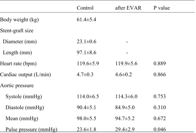
The change in hemodynamic parameters from control to after EVAR are shown in Table 1. There were no significant changes in heart rate, cardiac output, systolic, and diastolic aortic pressures after EVAR from control. On the other hand, there was a significant increase in pulse pressure (23.6 ± 1.8 to 29.4 ± 2.9 mmHg, p=0.046) after EVAR from control.
Change of aortic diameters and stiffness parameter β
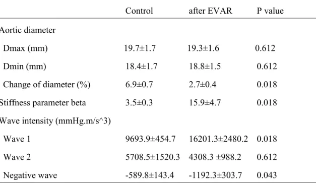
There was no significant difference between the Dmax of the control and after EVAR; 19.7±1.7 [12.6-23.6] to 19.3±1.6 [12.6-23.7] mm as well as Dmin of control and after EVAR; 18.4±1.7 [11.9-22.3] to 18.8±1.5 [12.2-22.8] mm at stent-graft site.
However, the change in aortic diameter significantly decreased after EVAR (control vs after EVAR; 6.9 ± 0.7 vs 2.7 ± 0.4%, p=0.018). The stiffness β also increased after EVAR (3.5 ± 0.3 vs 15.9 ± 4.7, p=0.018) (Table 2).
Wave intensity
There was significant increase in the W1 (first peak) after EVAR (control vs after EVAR; 9693.9 ± 454.7 vs 16201.3 ± 2480.2 mmHg-m/sec3, p=0.018), but not in the W2 (second peak). In contrast, the NW (negative reflected wave) was significantly enhanced after EVAR compared to control measurement (control vs after EVAR; - 589.8 ± 143.4 vs -1192.3 ± 303.7 mmHg-m/sec3, p=0.043) (Table 2).
Correlation with negative wave after EVAR
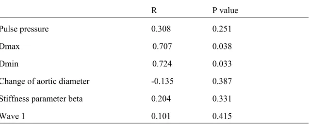
We correlated NW with other parameters. Before EVAR, there was no correlation between NW and any parameter. Among the parameters, which increased after EVAR (Table 3), the NW showed significant correlation with Dmax (R=0.707, p=0.038, Figure 4-1) and Dmin (R=0.724, p=0.033, Figure 4-2), however, it did not correlate with the length of stent-graft.
Discussions
In this study, we assessed the relationship between aortic diameter and the increase of reflected wave after EVAR. Our major findings were as follows: 1) Negative wave intensity, which is backward waves reflected toward the heart from the periphery, was significantly enhanced after EVAR compared to control; 2) Negative wave intensity correlated with the aortic diameters at stent-grafting site. To the best of our knowledge, this is the first study to investigate how the reflected wave after EVAR correlated with aortic diameter in vivo.
Wave intensity analysis considers waves as being composed of small ‘wavefronts’
that combine to produce a whole wave. The first peak, W1, occurs during early systole, the magnitude of which increases with increase in cardiac contractility 12. Jones et al clarified that administration of dobutamine increased W1 in dogs and a significant difference was observed between W1 and max dP/dt 15. In this study, W1 was increased while W2 did not changed after EVAR. However, we could not find the any correlation between W1 and the other hemodynamic parameters. Thus, the mechanism of enhanced W1 after EVAR remains unclear. Acute change in the arterial stiffness might be
influenced an increase W1.
Meanwhile, NW as reflected wave was enhanced after EVAR in this study. It has been reported that LV diastolic dysfunction related with reflected waves, i.e., cardio- arterial interaction, in patients after EVAR 4,5. In general, the reflected waves from peripheral vascular to the ascending aorta reach in early diastole. However, when the elastic properties of the aortic wall are diminished by EVAR, the reflected wave returns earlier to the ascending aorta, and is fused in the systolic phase 16. Alderson et al.
reported that the placement of a rigid stent within an elastic vessel produces wave reflection sites at the entrance to and exit from the stent. This phenomenon is produced by impedance mismatch, which is generated as a stepwise transition from elastic to rigid
boundary conditions as the flow enters the rigid segment and the reverse when it exits 17. Thus, the main reflection site moved from terminal aorta to the entrance and exit of the stent-graft after EVAR, and the reflected wave is enhanced.
PWV, defined as the speed with which the pulse travels in the aorta (distance/time), is directly related to the elastic properties of the aortic wall 16. Tzilalis et al. reported that PWV was greater in younger patients that reluctantly underwent EVAR due to thoracic aortic injury or aortic dissection compared to the healthy control group 16. Addition to that, Beaufort et al. described that the increase in PWV showed a positive linear correlation with the percentage of total aortic length covered by stent-graft, and assumed that the longer range of aorta become more rigid, thus, decrease in the aortic elasticity, leads to the increase in PWV 18. PWV is defined as Moens-Korteweg equation (PWV=√(Eh/2pr); E: elastic modulus, h: wall thickness, p: blood density, r:
vessel radius) 19. It means that the increased elastic modulus causes greater PWV and suggests that reflected wave is enhanced after EVAR. In this study, stiffness parameter β, as an index of arterial elasticity, was increased. This finding in healthy aorta without arteriosclerosis was accorded with that in the patients with arteriosclerosis after EVAR.
We clarified directly that the negative wave intensity as reflected wave is increased according to the change of elastic modulus by EVAR.
In addition, we showed the significant correlation between negative wave intensity and aortic diameter in healthy goat model. PWV is greater when the aortic diameter (r) is getting smaller, by Means-Korteweg equation 19. Hacham et al. also reported that reflected wave intensity showed a negative correlation with the diameter in an elastic tube of stenosis model 11. Thus, our findings suggest that the assessment of aortic diameter is important in patients who underwent EVAR. When EVAR is performed in patients with small aortic diameters, the reflected waves might increase. It is possible that these lead to poor prognosis according to LV diastolic dysfunction. Therefore,
further research will be needed to confirm the relationship between the aortic diameter and prognosis in patients with aneurysm after EVAR.
There was no correlation between the aortic diameter and the reflected wave before EVAR. Moreover, the reflected wave was related to the aortic diameter after EVAR.
This phenomenon agrees with clinical cases 9, reflected waves do not affect the cardio- arterial interaction even if healthy subjects have small aortic diameter. Meanwhile, arteriosclerotic patients might be influenced by the aortic diameter due to increase in aortic stiffness.
EVAR is established as a less-invasive procedure thoroughly exhibiting an effectiveness in especially frail elders, however, the change of cardiac property produced by the expansion of reflected wave should be considered.Particularly, our data suggests that EVAR give a significant influence in patients who have the smaller aorta such as elderly women. Thus, there is need to assess the change of cardiac function, i.e., LV diastolic function or exercise tolerability, in long-term follow-up period.Moreover, it will be necessary to develop tenderer stent-graft to preserve the vascular function to prevent the enhanced reflected wave.
Limitations
This study has several limitations. First, this was an experimental study using healthy goats, neither atherosclerotic nor aneurysmal models. Thus, the hemodynamic effect after EVAR may be underestimated because healthy aorta may compensate for the adverse effect by EVAR. However, in clinical case, it is difficult to evaluate the change in cardiovascular function produced solely by EVAR since various factors are mutually related. It is conceivable that the results of this study could lead to the future study, if EVAR is performed in the patients with arteriosclerosis. Secondly, this study was focused on the acute change of hemodynamics after EVAR. Thus, it remains unclear whether our findings are observed in chronic phase. Thirdly, we include the small
numbers of goats. Therefore, our findings will need to be confirmed in studies with a larger sample size.
Conclusion
Our study revealed that the reflected wave toward the heart from the periphery is significantly enhanced after EVAR, and the reflected wave associated with the aortic diameter of stent-grafting site. This highlights the importance of considering the possibility of developing LV dysfunction in patients with small aortic diameter who undergo EVAR, and the need to focus on diastolic dysfunction during follow-up period.
Acknowledgements
I appreciate Dr. Masumi Iwai-Takano, MD, PhD; Dr. Yusuke Tsuboko, PhD; Dr.
Yasuyuki Shiraishi, PhD; Dr. Tomoyuki Yambe, MD, PhD; Dr. Hitoshi Yokoyama, MD, PhD; for contribution of constructing this study and writing article. Additionally, I express my gratitude to Mr. Arakawa, MSc; and Ms. Watanabe contributed to data analysis.
Funding
This work was supported by Fukuda Foundation for Medical Technology, Japan (in part); and the Cooperative Research Project Program of Joint Usage/ Research Center at the Institute of Development, Aging and Cancer, Tohoku University, Japan (in part).
Conflicts of interest
Tomohiro Takano was supplied TX-2○R thoracic stent-grafts by COOK Medical LLC.
References
1. Farber MA, Lee WA, Szeto WY, Panneton JM, Kwolek CJ. Initial and midterm results of the Bolton Relay Thoracic Aortic Endovascular Pivotal Trial. J Vasc Surg.
2017;65(6):1556-1566.e1
2. O'Driscoll JM, Bahia SS, Gravina A, Di Fino S, Thompson MM, Karthikesalingam A, Holt PJ, Sharma R. Transthoracic Echocardiography Provides Important Long- Term Prognostic Information in Selected Patients Undergoing Endovascular Abdominal Aortic Repair. Circ Cardiovasc Imaging. 2016;9(2) e003557.
3. Healy GM, Redmond CE, Gray S, Iacob L, Sheehan S, Dowdall JF, Barry M, Cantwell CP, Brophy DP. Midterm Analysis of Survival and Cause of Death Following Endovascular Abdominal Aortic Aneurysm Repair. Vasc Endovascular Surg. 2017;51(5):274-281.
4. Takeda Y, Sakata Y, Ohtani T, Tamaki S, Omori Y, Tsukamoto Y, Aizawa Y, Shimamura K, Shirakawa Y, Kuratani T, Sawa Y, Yamamoto K, Mano T, Komuro I.
Endovascular aortic repair increases vascular stiffness and alters cardiac structure and function. Circ J. 2014;78(2):322-328.
5. Deery SE, Shean KE, Wang GJ, Black JH 3rd, Upchurch GR Jr, Giles KA, Patel VI, Schermerhorn ML; Society for Vascular Surgery Vascular Quality Initiative. Female sex independently predicts mortality after thoracic endovascular aortic repair for intact descending thoracic aortic aneurysms. J Vasc Surg. 2017 Jul;66(1):2-8.
6. van Bakel TMJ, Arthurs CJ, Nauta FJH, Eagle KA, van Herwaarden JA, Moll FL, Trimarchi S, Patel HJ, Figueroa CA. Cardiac remodelling following thoracic
endovascular aortic repair for descending aortic aneurysms. Eur J Cardiothorac Surg. 2019;55(6):1061-1070.
7. Lantelme P, Dzudie A, Milon H, Bricca G, Legedz L, Chevalier JM, Feugier P.
Effect of abdominal aortic grafts on aortic stiffness and central hemodynamics. J Hypertens. 2009;27(6):1268-1276.
8. Mokotedi L, Gunter S, Robinson C, Michel F, Solomon A, Norton GR, Woodiwiss AJ, Tsang L, Dessein PH, Millen AME. Early Wave Reflection and Pulse Wave Velocity Are Associated with Diastolic Dysfunction in Rheumatoid Arthritis. J Cardiovasc Transl Res. 2019;12(6):580-590.
9. Ayer JG, Harmer JA, Marks GB, Avolio A, Celermajer DS. Central arterial pulse wave augmentation is greater in girls than boys, independent of height. J Hypertens.
2010;28(2):306-313.
10. Davies JE, Alastruey J, Francis DP, Hadjiloizou N, Whinnett ZI, Manisty CH, Aguado-Sierra J, Willson K, Foale RA, Malik IS, Hughes AD, Parker KH, Mayet J.
Attenuation of wave reflection by wave entrapment creates a "horizon effect" in the human aorta. Hypertension. 2012;60(3):778-785.
11. Hacham WS, Abdulla NN, Salam Al-Ammri A, Khir AW. Wave speed and
reflections proximal to aneurysm and stenosis of elastic tubes. Conf Proc IEEE Eng Med Biol Soc. 2015;1009-1012.
12. Niki K, Sugawara M, Chang D, Harada A, Okada T, Sakai R, Uchida K, Tanaka R, Mumford CE. A new noninvasive measurement system for wave intensity:
evaluation of carotid arterial wave intensity and reproducibility. Heart Vessels.
2002;17(1):12-21.
13. Du GQ, Li HR, Xue JY, Chen S, Du P, Wu Y, Tian JW. Wave Intensity Analysis Can Identify Eccentric Cardiac Hypertrophy in Hypertensive Patients With Varied Left Ventricular Configurations. J Ultrasound Med. 2015;34(11):2019-2027.
14. Hirai T, Sasayama S, Kawasaki T, Yagi S. Stiffness of systemic arteries in patients with myocardial infarction. Circulation. 1989;80(1):78-86.
15. Jones CJ, Sugawara M, Kondoh Y, Uchida K, Parker KH. Compression and expansion wavefront travel in canine ascending aortic flow: wave intensity analysis.
Heart Vessels. 2002 Mar;16(3):91-8.
16. Tzilalis VD, Kamvysis D, Panagou P, Kaskarelis I, Lazarides MK, Perdikides T, Prassopoulos P, Boudoulas H. Increased pulse wave velocity and arterial
hypertension in young patients with thoracic aortic endografts. Ann Vasc Surg.
2012;26(4):462-467.
17. Alderson H1, Zamir M. Effects of stent stiffness on local haemodynamics with particular reference to wave reflections. J Biomech. 2004;37;(3):339-348.
18. de Beaufort HWL, Conti M, Kamman AV, Nauta FJH, Lanzarone E, Moll FL, van Herwaarden JA, Auricchio F, Trimarchi S. Stent-graft deployment increases aortic stiffness in an ex vivo porcine model. Ann Vasc Surg 2017;43:302-308.
19. Bramwell J.C and Hill A.V. The velocity of the pulse wave in man. Proceedings of the Royal Society, London, B93(1922):298-306.
Table 1 Hemodynamic parameters at control and after EVAR
Control after EVAR P value
Body weight (kg) 61.4±5.4
Stent-graft size
Diameter (mm) 23.1±0.6 -
Length (mm) 97.1±8.6 -
Heart rate (bpm) 119.6±5.9 119.9±5.6 0.889
Cardiac output (L/min) 4.7±0.3 4.6±0.2 0.866 Aortic pressure
Systole (mmHg) 114.0±6.5 114.3±6.0 0.753
Diastole (mmHg) 90.4±5.1 84.9±5.0 0.310
Mean (mmHg) 98.0±5.5 94.7±5.2 0.672
Pulse pressure (mmHg) 23.6±1.8 29.4±2.9 0.046 EVAR: endovascular aortic repair, LV: left ventricle, Value: mean ± SE
Table 2 Change of aortic diameters and wave intensity at control and after EVAR
Control after EVAR P value Aortic diameter
Dmax (mm) 19.7±1.7 19.3±1.6 0.612
Dmin (mm) 18.4±1.7 18.8±1.5 0.612
Change of diameter (%) 6.9±0.7 2.7±0.4 0.018 Stiffness parameter beta 3.5±0.3 15.9±4.7 0.018 Wave intensity (mmHg.m/s^3)
Wave 1 9693.9±454.7 16201.3±2480.2 0.018
Wave 2 5708.5±1520.3 4308.3 ±988.2 0.612
Negative wave -589.8±143.4 -1192.3±303.7 0.043 Value: mean ± SE
Table 3 Univariate analysis for relationship with negative wave intensity
R P value
Pulse pressure 0.308 0.251 Dmax 0.707 0.038
Dmin 0.724 0.033
Change of aortic diameter -0.135 0.387 Stiffness parameter beta 0.204 0.331
Wave 1 0.101 0.415
Figure 1.
Figure 1. A schematic overview of the animal preparation, stent-grafting and study protocol. A gout was placed in the right decubitus position. Flowmeter probe was attached to the aortic root (a), and common cervical arterial line (b) and terminal arterial line (c) were introduced. Aortic flow, aortic pressure, and ECG, which were recorded via a polygraph system, were fed into a hard disc of a personal computer (PC) through an analogue-to-digital converter at a sampling frequency of 1 kHz. Stent-graft was deployed between at distal of the cervical trunk (A) and the celiac trunk (B).
Figure 2-1.
Figure 2-2.
Figure 2. Representative case of wave intensity
1) ECG, ascending aortic pressure (AoP), aortic velocity (AoV) and calculated wave intensity (WI)obtained from eight successive beats in the ascending aorta.
2) Representative case of wave intensity at control (a) and after EVAR (b). There was the increasing of W1 and the decreasing of NW after EVAR.
W1: first peak during early systole, W2: second peak during end of ejection, NW:
backward wave in mid systole.
Figure 3.
Figure 3. Ultrasound imaging of a cross section of aorta at control (a) and after EVAR (b). The internal aortic diameters in stent-grafting site were measured as aortic diameter at control and after EVAR.
Figure 4.
Figure 4. Correlation with negative wave and aortic diameters after EVAR.
The NW had significant correlations with maximum aortic diameter (a) and minimum aortic diameter (b).