• 検索結果がありません。

Bone Regeneration by Low-dose Recombinant Human Bone Morphogenetic Protein-2 Carried on Octacalcium Phosphate Collagen Composite

N/A
N/A
Protected

Academic year: 2021

シェア "Bone Regeneration by Low-dose Recombinant Human Bone Morphogenetic Protein-2 Carried on Octacalcium Phosphate Collagen Composite"

Copied!
8
0
0

読み込み中.... (全文を見る)

全文

(1)

2020 The Hard Tissue Biology Network Association Printed in Japan, All rights reserved.

CODEN-JHTBFF, ISSN 1341-7649

Original

Bone Regeneration by Low-dose Recombinant Human Bone Morphogenetic Protein-2 Carried on Octacalcium Phosphate Collagen Composite

Nguyen Dien Bien

1)

, Kei-ichiro Miura

1)

, Yoshinori Sumita

2)

, Yuya Nakatani

1)

, Rena Shido

1)

, Fumihiko Kajii

3)

, Shinji Kamakura

4)

and Izumi Asahina

1)

1)

Department of Regenerative Oral Surgery, Nagasaki University Graduate School of Biomedical Sciences, Nagasaki, Japan

2)

Basic and Translational Research Center for Hard Tissue Disease, Nagasaki University Graduate School of Biomedical Sciences, Nagasaki, Japan

3)

TOYOBO Co., Ltd., Medical Equipment & Devices Production Center, Otsu, Japan

4)

Division of Bone Regenerative Engineering, Tohoku University Graduate School of Biomedical Engineering, Sendai, Japan (Accepted for publication, March 13, 2020)

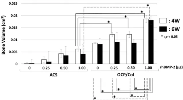
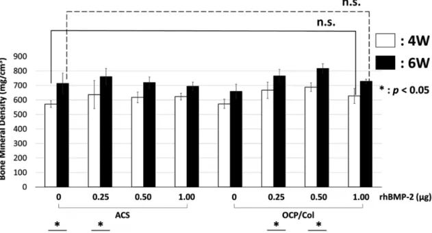
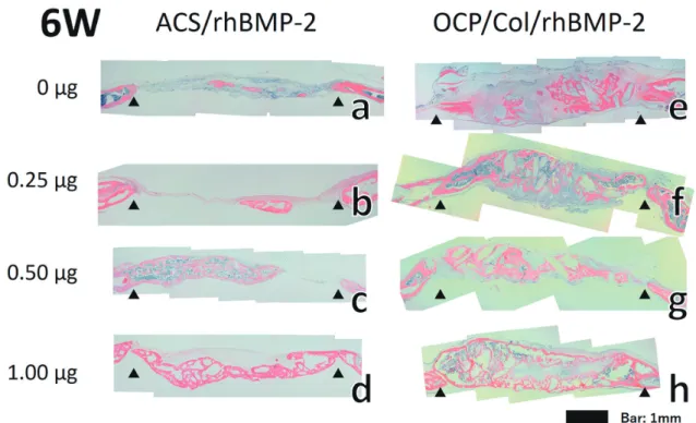
Abstract: Bone morphogenetic protein-2 (BMP-2) has diverse functions and is especially important in bone and cartilage development. Recombinant human BMP-2 (rhBMP-2) is an osteoinductive growth factor that has been clinically applied as a bone graft substitute. However, high-dose rhBMP-2 can cause complications such as induction of significant swelling that can endanger the patient’s life. Atelocollagen sponge (ACS) is the commercially provided standard carrier of rhBMP-2 in clinical applications. However, a large concentration of rhBMP-2 is required to be clinically effective with ACS as the carri- er. Octacalcium phosphate/collagen (OCP/Col) has been shown to be an excellent bone substitute compared with other bone substitute materials such as hydroxyapatite or β-tricalcium phosphate due to its biological properties. In this study, we eval- uated the use of OCP/Col as a carrier to minimize the effective dose of rhBMP-2. ACS or OCP/Col discs impregnated with different rhBMP-2 concentrations were implanted in mice calvarial bone defects. Morphological analysis with micro-CT both at 4 and 6 weeks post-implantation showed homogenous hard tissue formation in the defects of the OCP/Col group at all rhBMP-2 concentrations tested (0, 0.25, 0.50, or 1.00 μg). In contrast, ACS alone or with 0.25 μg of rhBMP-2 showed almost no bone formation. However, bone mineral density in all groups of ACS and OCP/Col was not dependent on rhB- MP-2 concentration. Histological evaluation indicated that bone formation progressed depending on rhBMP-2 concentra- tion in the defects of both the ACS and OCP/Col groups, although the newly formed bone area was significantly higher in the OCP/Col group than in the ACS group. These results indicate that OCP/Col could be an effective carrier of rhBMP-2, minimizing the application dose of rhBMP-2 in clinical settings and avoiding the complications caused by high-dose rhB- MP-2.

Key words: Bone regeneration, Carrier, Collagen, Octacalcium phosphate, Bone morphogenetic protein-2

Introduction

Bone regeneration is challenging in oral and maxillofacial surgery because there are many cases that require bone regeneration in this re- gion. In current clinical settings, autologous bone grafting remains the gold standard for bone regeneration of jawbone defects because of its superior osteoinductivity and osteoconductivity. However, this tech- nique has several disadvantages such as limited availability and donor site morbidity. Hydroxyapatite (HA) and β-tricalcium phosphate (β-TCP) have been widely applied in clinical settings

1-3)

; however, these materials have not replaced autogenous bone grafts in light of their su- perior biocompatibility and osteoconductivity but not osteoinductivity

4)

. The clinical application of recombinant human bone morphogenetic protein-2 (rhBMP-2), which is one of the most important bone inductive proteins, has been expected, and it has been approved for clinical use in extraction socket preservation and maxillary sinus floor augmentation

5)

. rhBMP-2 shows superior bone regeneration, however, several adverse

events have been reported such as local edema, seroma, and cancer in- duction at high-dose rhBMP-2

6-8)

. Atelocollagen sponge (ACS) is pres- ently used as a carrier of rhBMP-2; however, it is reported to release rh- BMP-2 instantaneously

9)

. Accordingly, it is necessary to identify a carrier that will spontaneously release rhBMP-2 and can act as a bone substitute

10)

.

Octacalcium phosphate (OCP) is a direct precursor of biological ap- atite, which sustainably and irreversibly converts into biological apatite under physiological conditions

11)

. Moreover, OCP has been demonstrat- ed to enhance osteoblastic cell differentiation

12)

and is effective for bone regeneration because of its high bone regenerative ability and rapid ab- sorbability compared with HA or β-TCP

13)

. OCP possesses many desira- ble properties as a bone substitute; however, it cannot be molded using sintering processes because of its crystal structure. In order to improve its handling property, a composite comprising OCP and collagen (OCP/

Col) was developed. OCP/Col was shown to yield significantly en- hanced bone regeneration compared with β-TCP or HA collagen com- posite

14)

. OCP/Col has also been shown to be both safe and efficacious in human tooth extraction sockets, cystic cavities, and maxillary sinus floor elevation

15-18)

. BMP-2 is known to absorb calcium phosphate and Correspondence to: Dr. Kei-ichiro Miura, Department of Regenerative Oral

Surgery, Nagasaki University Graduate School of Biomedical Sciences, Japan, 1-7-1, Sakamoto, Nagasaki 852-8588, Japan; Phone: +810958197704; E-mail:

[email protected]

(2)

also collagen

19, 20)

. Therefore, we propose that OCP/Col could be a supe- rior carrier for rhBMP-2 to minimize the required rhBMP-2 dose. The objective of the present study is to assess the osteogenic potential of low-dose rhBMP-2 carried on OCP/Col compared with that carried on ACS.

Materials and Methods

Preparation of OCP, OCP/Col, atelocollagen, and their combination with/without rhBMP-2

OCP was synthesized by direct precipitation as previously de- scribed

21)

, and sieved granules (particle size: 300–500 μm) were ob- tained. Collagen solution was prepared from NMP collagen PS (Nippon Meat Packers, Tsukuba, Ibaraki, Japan), a lyophilized powder of pep- sin-digested atelocollagen isolated from porcine dermis. Collagen was dissolved in distilled water and adjusted to 3% of the final concentration at pH 7.4. Then, sieved OCP granules were added to the collagen solu- tion and mixed. The proportion of OCP in OCP/Col was adjusted to 77 weight%. This mixture was then lyophilized and molded into discs (di- ameter: 9 mm, thickness: 1 mm). OCP discs then underwent dehydro- thermal treatment (150°C, 24 h) in a vacuum drying oven and steriliza- tion using gamma-ray irradiation (5 kGy). The resultant discs were punched to 5 mm in diameter using a HANDY PUNCH KIT (product number: 3H-PS10, H.H.H. Manufacturing Co., Osaka, Japan). rhBMP-2 (produced by INFUSE

®

Bone Graft, Medtronic, Memphis, TN, USA) was dissolved in distilled water at concentrations of 0, 0.01, 0.02, or 0.04 μg/μl. Then, 25 μl of rhBMP-2 solution was dripped onto the OCP/

Col or ACS disc (control group; diameter: 5 mm, thickness: 1 mm).

These materials were defined as OCP/Col, 0.25 OCP/Col, 0.50 OCP/

Col, 1.00 OCP/Col, ACS, 0.25 ACS, 0.50 ACS, and 1.00 ACS.

Animals and implantation procedures

All animal experiments were performed at the Nagasaki University Animal Experiment Facility and were in accordance with protocols ap- proved by the Local Institutional Animal Care and Use Committee of Nagasaki University (approval no. 1606211320). Eighty 10-week-old male C57BL/6J mice (CLEA Japan, Inc., Tokyo, Japan) were used.

Mice were housed in standard rodent cages in a light- and tempera- ture-controlled room in the Biomedical Research Center, Center for Frontier Life Sciences, Nagasaki University. Animals had free access to standard laboratory chow and tap water. Transplantation experiments were conducted under general anesthesia. General anesthetics were pre- pared by mixing 1.875 ml of medetomidine hydrochloride (1 mg/ml), 2 ml of midazolam (5 mg/ml), 2.5 ml of butorphanol (5 mg/ml), and 18.625 ml of physiological saline to yield a final liquid volume of 25 ml. The mixture was then subcutaneously or intraperitoneally injected at a concentration of 0.1 ml/10 g body weight. After disinfection of the op- erative field, an arc-shaped skin incision was made from the left to right preauricular region via the forehead region. The periosteum of the cal- varium was ablated and a full-thickness standardized trephine defect, 5 mm in diameter, was made in the calvarium under continuous saline buffer irrigation. Each material was then implanted into the trephine de- fect. Five mice were treated per group. After the defects were treated, the ablated periosteum and skin were repositioned and sutured. For the OCP/Col and ACS groups, the tissues were collected at four and six weeks postoperatively.

Morphological and quantitative analysis by micro-computed tomogra- phy The morphological and quantitative image analysis of newly formed

bone was performed using micro-computed tomography (CT) [micro X-ray CT machine for laboratory animals (R mCT), RIGAKU, Tokyo, Japan] under standardized conditions (90 kV, 150 mA, two minutes). In three-dimensional analysis using structural analysis software (TRI/3D- BON, RATOC System Engineering, Osaka, Japan), the newly formed bone area was analyzed after sampling using bone mineral density (BMD) standards, of established densities of between 300 mg/cm

3

and 1,500 mg/cm

3

, and the extracted range of bone was defined between 300 mg/cm

3

and 1,100 mg/cm

3

.

Tissue preparation and histological examination

After micro-CT examination, samples were immersed in PBS over- night and fixed in 4% paraformaldehyde for 24 h. Samples were then decalcified in EDTA for 10 days and washed with distilled water. There- after, samples were cut coronally into two pieces at the center of the de- fect and embedded in paraffin. Serial sections 5 μm thick were cut coro- nally and stained with hematoxylin and eosin. Images were taken using a photomicroscope (Axiocam ERc 5s, ZEISS, Oberkochen, Germany).

Quantitative measurement of cortical bone and bone marrow area The amount of cortical bone and bone marrow area in the implanted material was measured from the histological sections prepared near the center of the defect. The cortical bone and bone marrow data was ex- tracted using Adobe Photoshop

®

CS6 Extended. After converting to JPEG images, the data volume (Kilobytes) of cortical bone and bone marrow was measured with Windows 10 photo viewer.

Statistical analysis

Statistical analysis of all micro-CT data was performed using JMP

®

version 13 (SAS Institute, Cary, NC, USA). The temporal change of each OCP/Col group’s BMD value was analyzed by a nonparametric multiple comparison test. Values are expressed as mean ± standard devi- ation (SD). A p-value less than 0.05 was statistically significant.

Results Morphological analysis by micro-CT

Micro-CT analysis revealed that the ACS and 0.25 ACS groups showed almost no hard tissue formation, whereas the 0.50 ACS and 1.00 ACS groups showed hard tissue formation. The formation area was slightly wider in the 1.00 ACS group at both 4 and 6 weeks postopera- tively compared with the 0.50 ACS group. In contrast, hard tissue for- mation was recognized within the defect in the OCP/Col groups at all rhBMP-2 concentrations, and the radio-lucent area was decreased rhB- MP-2 dose-dependently (Fig. 1).

Morphometric analysis of newly formed bone volume by micro-CT analysis

The OCP/Col groups produced a greater volume of newly formed bone than the ACS groups. Namely, 0.25 OCP/Col, 0.50 OCP/Col, and 1.00 OCP/Col showed significant increases in newly formed bone vol- ume compared with 1.00 ACS, which showed the highest bone volume among the ACS groups, at four weeks, while 1.00 OCP/Col showed sig- nificant increases in bone volume compared with 1.00 ACS at 6 weeks (Fig. 2).

Bone mineral density (BMD)

BMD tended to increase from 4 to 6 weeks postimplantation in all

groups. There were also no significant differences in BMD between the

ACS and OCP/Col groups. For example, BMD of 1.00 OCP/Col, which

(3)

produced the highest bone volume, was not significant different com- pared with ACS alone both at four and six weeks (Fig. 3).

Histological evaluation

ACS and 0.25 ACS showed no bone formation in the defect, where- as 0.50 and 1.00 ACS showed a certain amount of new bone formation at four weeks. On the other hand, in the OCP/Col groups, the area of new bone gradually increased as the concentration of rhBMP-2 in- creased (Fig. 4). ACS and 0.25 ACS showed slight bone formation in the defect, and 1.00 ACS demonstrated bone bridging of the defect with new bone at six weeks. In contrast, the OCP/Col group showed remark- able osteogenesis compared with the ACS group (Fig. 5). Almost all im- planted ACS was absorbed in all ACS groups, while OCP/Col remained, especially at the lower concentrations of rhBMP-2. Additionally, newly formed bone in the OCP/Col groups was thicker than that in the ACS groups. Although OCP/Col without rhBMP-2 showed new bone forma- tion, OCP/Col with rhBMP-2 showed more mature bone formation with bone marrow.

Quantitative measurement of cortical bone and bone marrow area The area of newly formed cortical bone and bone marrow area meas- ured using histological specimens showed a similar tendency, that is, bone volume measured with micro-CT increased in a rhBMP-2 dose-de- pendent manner for both the ACS and OCP/Col groups. The area of the OCP/Col groups was higher than that of the ACS groups at the respec- tive concentrations of rhBMP-2. Statistically significant differences were observed between the lower (0, 0.25) and higher (0.50, 1.00) con- centrations of rhBMP-2 in both the ACS and OCP/Col groups (Fig. 6).

0.25 OCP/Col, which was the lowest concentration of rhBMP-2, showed similar bone area with 0.50 and 1.00 ACS at 6 weeks.

Figure 1. Tissue formation in the ACS and OCP/Col groups with and with- out rhBMP-2 at 4 and 6 weeks postoperatively. The ACS and 0.25 ACS groups showed almost no hard tissue formation, whereas the 0.50 ACS and 1.00 ACS groups showed hard tissue formation. The formation area was slightly wide in the 1.00 ACS group at both 4 and 6 weeks postoperatively.

In contrast, uniform hard tissue formation was recognized within the defect in the OCP/Col groups at all rhBMP-2 concentrations.

Figure 2. Bone volume in the ACS and OCP/Col groups with and without rhBMP-2 at 4 and 6 weeks postoperatively. At 4 weeks, 0.25 OCP/Col, 0.50 OCP/Col, and 1.00 OCP/Col showed significant increases in newly formed bone volume compared with 1.00 ACS. At 6 weeks, 1.00 OCP/

Col showed significant increases in bone volume compared with 1.00 ACS, whereas, OCP/Col, 0.25 OCP/Col, and 0.50 OCP/Col did not. * :

p<0.05

(4)

Figure 3. Bone mineral density in the ACS and OCP/Col groups with and without rhBMP-2. At both 4 and 6 weeks postoperatively, the bone mineral density of 1.00 OCP/Col with rhBMP-2 was not significantly different compared with ACS without rhBMP-2. * : p<0.05

Figure 4. Effect of ACS and OCP/Col with and without rhBMP-2 on bone formation in defects at 4 weeks postoperatively. At 4 weeks ACS and

0.25 ACS showed no bone formation in the defect, whereas 0.50 and 1.00 ACS showed slight new bone formation. On the other hand, in the

OCP/Col groups, the area of new bone gradually increased as the concentration of rhBMP-2 increased.

(5)

Discussion

In the current study, we examined the potential application of OCP/

Col as an rhBMP-2 carrier to minimize the effective dose of rhBMP-2 on bone regeneration. Our findings demonstrate that low-concentration rhBMP-2 impregnated on OCP/Col discs could induce sufficient bone formation. According to the qualitative analysis using micro-CT and

histological findings, the concentration of rhBMP-2 could be decreased to less than one-fourth using OCP/Col as the carrier compared with ACS (Figs. 2 and 6). In terms of clinical application of BMP, selection of an appropriate carrier is extremely important for the following reasons.

Since rhBMP-2 diffuses within a short time when topically administered without a carrier

22)

, an appropriate carrier for proper delivery, retention, Figure 5. Effect of ACS and OCP/Col with and without rhBMP-2 on bone formation in defects at 6 weeks postoperatively. At 6 weeks the ACS and 0.25 ACS groups showed slight formation in the defect, and the 1.00 ACS group showed bone bridging of the defect with new bone. In con- trast, the OCP/Col group showed remarkable osteogenesis compared with the ACS group. Although OCP/Col without rhBMP-2 showed remark- able new bone formation, OCP/Col with rhBMP-2 showed more mature bone formation with bone marrow. Additionally, newly formed bone in the OCP/Col groups was thicker than that in the ACS groups.

Figure 6. Effect of ACS and OCP/Col with and without rhBMP-2 on areas of cortical bone and bone marrow at 4 and 6 weeks postoperatively.

The areas of cortical bone and bone marrow were increased in an rhBMP-2 dose-dependent manner in both the ACS and OCP/Col groups, and

the OCP/Col groups showed higher bone areas than the ACS groups. Statistically significant differences were observed between the lower (0,

0.25) and higher (0.50, 1.00) concentrations of rhBMP-2 in both the ACS and OCP/Col groups. * : p<0.05

(6)

and sustained released of BMP-2 is needed

19)

. The carrier must be non- toxic, noncarcinogenic, nonantigenic, proinflammatory, and preferably biodegradable and moldable

23)

. There are four major categories of rhB- MP-2 carriers: natural-origin polymers, inorganic materials, synthetic biodegradable polymers, and composites

24)

. In current clinical settings, ACS is the commercially provided standard carrier for rhBMP-2 among the presently available carriers that fulfill the requirements described above

25)

. However, a high rhBMP-2 concentration is required when ACS is used as the carrier

17

. As previously demonstrated, high doses of BMP- 2 cause severe clinical side effects such as ectopic bone formation, oste- oclast-mediated bone resorption, inappropriate adipogenesis, and can-

cer

19, 26)

. Additionally, high-dose rhBMP-2 can induce acute edema and

swelling, leading to fatal airway obstruction in the oral and maxillofa- cial region

6, 27)

. Therefore, low-dose rhBMP-2 should be applied to avoid these adverse effects. In the present clinical settings, the concentration of rhBMP-2 with ACS as a carrier is 37.5 μg/μl. Even taking into an ac- count the differences between humans and mice in the response to BMP, it is expected that the concentration of rhBMP-2 can be reduced when OCP/Col is used as a carrier.

There are a number of reasons why OCP/Col is a potential superior carrier for rhBMP-2 compared with ACS. First, in accordance with a previous report, OCP/Col itself has bone inductive activity

28)

. As shown in Figs. 1, 4 and 5, new bone formation was observed apart from the rim of the bone defect in the OCP/Col only group (without rhBMP-2), indi- cating that OCP/Col shows osteoconductivity as well as osteoinductivi- ty. It is apparent that the bone inductive activity of rhBMP-2 augments that of OCP/Col, since OCP/Col with rhBMP-2 induced more mature bone formation with cortical bone and bone marrow compared with OCP/Col without rhBMP-2 (Figs. 1, 4 and 5). BMD tended to increase time-dependently, but significant differences were not observed. There were also no significant differences among the ACS and OCP/Col groups with different concentrations of rhBMP-2 (Fig. 3). This suggests that while the quality of bone is similar, the quantity is different between ACS and OCP/Col as carriers. Second, the composition of OCP/Col is favorable for use as an rhBMP-2 carrier because rhBMP-2 is known to bind tightly to calcium phosphate and collagen, which are major compo- nents of OCP/Col

19, 20)

. It is speculated that rhBMP-2 is released as OCP and collagen are degraded, allowing the slow release of rhBMP-2

26, 27)

. Remnants of OCP/Col were observed at 6 weeks postimplantation, while ACS was completely absorbed. This might assist in the sustaina- ble release of rhBMP-2 with OCP/Col as the carrier. Furthermore, rhB- MP-2 influences chemotaxis and mesenchymal stem cells proliferation and differentiation into osteoblasts, thus, the porous structure of OCP/

Col, composed of collagen, is a suitable environment for cell migra- tion

29)

. Lastly, OCP/Col has superior mechanical strength to ACS

30)

. We observed that newly formed bone in the OCP/Col implant group was thicker than that in the ACS implant groups (Figs. 4 and 5). This finding suggests that OCP/Col is sufficiently strong to resist pressure from the skin flap. rhBMP-2/ACS has been approved for clinical application for socket preservation and sinus floor augmentation in the oral and maxil- lofacial region

31)

. In these areas, the implant material does not suffer from pressure because it is surrounded by the bone wall. However, the implant material needs to have certain mechanical strength in the case of alveolar bone ridge augmentation. In this respect, rhBMP-2 with OCP/Col could be a good implant material for guided bone regeneration (GBR) because it possesses both strength and flexibility.

In the present study, we demonstrated superior bone regeneration of rat calvarial bone defects with rhBMP-2 carried on OCP/Col. Although OCP/Col itself has bone inductive activity, low-dose rhBMP-2 enhances

this activity. The minimization of the effective dose of rhBMP-2 reduces its side effects. Recently, there has been increasing demand for alveolar bone regeneration in response to the popularization of implant dentistry.

We suggest that rhBMP-2 with OCP/Col could be an ideal implant ma- terial for GBR because of its superior osteoinductivity and mechanical properties such as strength and flexibility. In conclusion, though OCP/

Col itself is a good bone substitute, it could also be an effective carrier for rhBMP-2, thereby reducing the effective dose of rhBMP-2 and re- sulting in its safe clinical application for more reliable bone augmenta- tion.

Acknowledgements

This study was partially supported by Grants-in-Aid for Scientific Research from the Japan Society for the Promotion of Science (No.

17H01604).

Conflict of Interest The authors have declared that no COI exists.

References

1. LeGeros RZ. Properties of osteoconductive biomaterials: calcium phosphates. Clin Orthop Relat Res 395: 81-98, 2002

2. Ogose A, Hotta T, Kawashima H, Kondo N, Gu W, Kamura T and Endo N. Comparison of hydroxyapatite and beta tricalcium phos- phate as bone substitutes after excision of bone tumors. J Biomed Mater Res B Appl Biomater 72: 94-101, 2005

3. Ogose A, Kondo N, Umezu H, Hotta T, Kawashima H, Tokunaga K, Ito T, Kudo N, Hoshino M and Gu W. Histological assessment in grafts of highly purified beta-tricalcium phosphate (OSferion®) in human bones. Biomaterials 27: 1542-1549, 2006

4. Lichte P, Pape HC, Pufe T, Kobbe P and Fischer H. Scaffolds for bone healing: concepts, materials and evidence. Injury 42: 569-573, 2011

5. Asahina I. Bone morphogenetic proteins: their history and charac- teristics. J Hard Tissue Biol 23: 283-286, 2014

6. Woo EJ. Adverse events reported after the use of recombinant hu- man bone morphogenetic protein 2. J Oral Maxillofac Surg 70: 765- 767, 2012

7. Carreira A, Lojudice F, Halcsik E, Navarro R, Sogayar M and Gran- jeiro J. Bone morphogenetic proteins: facts, challenges, and future perspectives. J Dent Res 93: 335-345, 2014

8. Sugamori Y, Mise-Omata S, Maeda C, Aoki S, Tabata Y, Murali R, Yasuda H, Udagawa N, Suzuki H, Honma M and Aoki K. Peptide drugs accelerate BMP-2-induced calvarial bone regeneration and stimulate osteoblast differentiation through mTORC1 signaling. Bi- oessays 38: 717-725, 2016

9. Brown KV, Li B, Guda T, Perrien DS, Guelcher SA and Wenke JC.

Improving bone formation in a rat femur segmental defect by con- trolling bone morphogenetic protein-2 release. Tissue Eng Part A 17: 1735-1746, 2011

10. Kawai T, Kamakura S, Matsui K, Fukuda M, Takano H, Iino M, Ishikawa S, Kawana H, Soma T, Imamura E, Kizu H, Michibata A, Asahina I, Miura K, Nakamura N, Kibe T, Suzuki O and Takahashi T. Clinical study of octacalcium phosphate and collagen composite in oral and maxillofacial surgery. J Tissue Eng 11:

2041731419896449, 2020

11. Brown WE, Smith JP, Lehr JR and Frazier AW. Octacalcium phos-

phate and hydroxyapatite: crystallographic and chemical relations

between octacalcium phosphate and hydroxyapatite. Nature 196:

(7)

1050-1055, 1962

12. Suzuki O, Kamakura S, Katagiri T, Nakamura M, Zhao B, Honda Y and Kamijo R. Bone formation enhanced by implanted octacalcium phosphate involving conversion into Ca-deficient hydroxyapatite.

Biomaterials 27: 2671-2681, 2006

13. Kamakura S, Sasano Y, Shimizu T, Hatori K, Suzuki O, Kagayama M and Motegi K. Implanted octacalcium phosphate is more resorb- able than β‐tricalcium phosphate and hydroxyapatite. J Biomed Ma- ter Res Part A 59: 29-34, 2002

14. Kamakura S, Sasaki K, Homma T, Honda Y, Anada T, Echigo S and Suzuki O. The primacy of octacalcium phosphate collagen compos- ites in bone regeneration. J Biomed Mater Res A 83: 725-733, 2007 15. Kawai T, Echigo S, Matsui K, Tanuma Y, Takahashi T, Suzuki O

and Kamakura S. First clinical application of octacalcium phosphate collagen composite in human bone defect. Tissue Eng Part A 20:

1336-1341, 2014

16. Kawai T, Suzuki O, Matsui K, Tanuma Y, Takahashi T and Kamaku- ra S. Octacalcium phosphate collagen composite facilitates bone re- generation of large mandibular bone defect in humans. J Tissue Eng Regen Meddoi: 10.1002/term.2110, 2015

17. Kawai T, Tanuma Y, Matsui K, Suzuki O, Takahashi T and Kamaku- ra S. Clinical safety and efficacy of implantation of octacalcium phosphate collagen composites in tooth extraction sockets and cyst holes. J Tissue Eng 7: 2041731416670770, 2016

18. Miura K-i, Sumita Y, Kajii F, Tanaka H, Kamakura S and Asahina I.

First clinical application of octacalcium phosphate collagen com- posite on bone regeneration in maxillary sinus floor augmentation:

A prospective, single-arm, open-label clinical trial. J Biomed Mater Res B Appl Biomater 108: 243-252, 2020

19. Lee YH, Lee BW, Jung YC, Yoon BI, Woo HM and Kang BJ. Ap- plication of alginate microbeads as a carrier of bone morphogenetic protein-2 for bone regeneration. J Biomed Mater Res B Appl Bio- mater 107: 286-294, 2019

20. Kamakura S, Nakajo S, Suzuki O and Sasano Y. New scaffold for recombinant human bone morphogenetic protein-2. J Biomed Mater Res A 71: 299-307, 2004

21. Suzuki O, Nakamura M, Miyasaka Y, Kagayama M and Sakurai M.

Bone Formation on Synthetic Precursors of Hydroxyapatite. Tohoku J Exp Med 164: 37-50, 1991

22. Takaoka K, Nakahara H, Yoshikawa H, Masuhara K, Tsuda T and Ono K. Ectopic bone induction on and in porous hydroxyapatite combined with collagen and bone morphogenetic protein. Clinical orthopaedics and related research 234: 250-254, 1988

23. Takahashi K. Bone morphogenetic protein (BMP): from basic stud- ies to clinical approaches. Folia Pharmacologica Japonica 116: 232- 240, 2000

24. Haidar ZS, Hamdy RC and Tabrizian M. Delivery of recombinant bone morphogenetic proteins for bone regeneration and repair. Part A: Current challenges in BMP delivery. Biotechnol Lett 31: 1817- 1824, 2009

25. Bessa PC, Casal M and Reis R. Bone morphogenetic proteins in tis- sue engineering: the road from laboratory to clinic, part II (BMP de- livery). J Tissue Eng Regen Med 2: 81-96, 2008

26. Boerckel JD, Kolambkar YM, Dupont KM, Uhrig BA, Phelps EA, Stevens HY, García AJ and Guldberg RE. Effects of protein dose and delivery system on BMP-mediated bone regeneration. Biomate- rials 32: 5241-5251, 2011

27. Yaremchuk K, Toma M and Somers M. Acute airway obstruction associated with the use of bone-morphogenetic protein in cervical spinal fusion. Laryngoscope 120: S140-S140, 2010

28. Kouketsu A, Matsui K, Kawai T, Ezoe Y, Yanagisawa T, Yasuda A, Takahashi T and Kamakura S. Octacalcium phosphate collagen composite stimulates the expression and activity of osteogenic fac- tors to promote bone regeneration. J Tissue Eng Regen Med 14: 99- 107, 2020

29. Neovius E, Lemberger M, Skogh AD, Hilborn J and Engstrand T.

Alveolar bone healing accompanied by severe swelling in cleft chil- dren treated with bone morphogenetic protein-2 delivered by hydro- gel. J Plast Reconstr Aesthet Surg 66: 37-42, 2013

30. Kamakura S, Sasaki K, Honda Y, Anada T and Suzuki O. Octacalci- um phosphate combined with collagen orthotopically enhances bone regeneration. J Biomed Mater Res B Appl Biomater 79: 210-217, 2006

31. Triplett RG, Nevins M, Marx RE, Spagnoli DB, Oates TW, Moy PK

and Boyne PJ. Pivotal, randomized, parallel evaluation of recombi-

nant human bone morphogenetic protein-2/absorbable collagen

sponge and autogenous bone graft for maxillary sinus floor augmen-

tation. J Oral Maxillofac Surg 67: 1947-1960, 2009

(8)

Figure 2. Bone volume in the ACS and OCP/Col groups with and without rhBMP-2 at 4 and 6 weeks postoperatively
Figure 3. Bone mineral density in the ACS and OCP/Col groups with and without rhBMP-2
Figure 6. Effect of ACS and OCP/Col with and without rhBMP-2 on areas of cortical bone and bone marrow at 4 and 6 weeks postoperatively

参照

関連したドキュメント