Οriginal P aper
Key Words:
Acute cholangitis, choledocholi- thiasis, endoscopic nasobiliary drain- age, endoscopic biliary stent, meal intake rate.
Background/Aims: To clarify whether or not use of an endoscopic biliary stenting (EBS) is superior to endoscopic nasobiliary drainage (ENBD) in cases of acute cholangitis due to choledocholithiasis. Method- ology: Of 447 patients with choledocholithiasis who were treated in the Department of Gastroenterology, Fukuoka University Chikushi Hospital between Janu- ary 1994 and September 2006, the subjects were 99 moderate acute cholangitis patients who underwent endoscopic drainage as initial treatment. Clinical ef- ficacy, complications and patient satisfaction (meal intake rete) were investigated in the EBS group (67
patients) and the ENBD group (32 patients). Results:
There were no significant differences in the improve- ment in inflammation, total bilirubin, or biliary en- zymes between the EBS and ENBD groups. Catheter occlusion was seen in three patients (4%) in the EBS group, and the catheter was self-extracted by three patients (10%) in the ENBD group. Conclusion: In moderate acute cholangitis due to choledocholithisis, the treatment efficacy and safety of EBS are equal to those of ENBD, and EBS appears to be a better choice in elderly patients in particular.
ABSTRACT
Hepato-Gastroenterology 2015; 62:4-9 doi 10.5754/hge13464
© H.G.E. Update Medical Publishing S.A., Athens-Stuttgart
Comparison Between Endoscopic Biliary Stenting and Nasobiliary
Drainage in Patients with Acute
Cholangitis due to Choledocholithiasis:
Is Endoscopic Biliary Stenting Useful?
Keisuke Otani, Toshiharu Ueki, Keiichiro Matsumura, Toru Maruo, Ryohei Minoda, Yuichiro Otsuka, Kenichiro Kawamoto, Eijiro Noma, Tomoko Mitsuyasu and Toshiyuki Matsui
Department of Gastroenterology, Chikushi Hospital, Fukuoka University, Fukuoka, Japan
Corresponding author: Dr. Toshiharu Ueki, Department of Gastroenterology, Chikushi Hospital, Fukuoka University, 1-1-1 Zokumyoin, Chikushino, Fukuoka 818-8502, Japan;
Tel.: +81-92-9211011; Fax: +81-92-9292630; E-mail: [email protected] The aim of this study is to clarify
whether or not use of an endoscopic biliary stenting (EBS) is superior to en- doscopic nasobiliary drainage (ENBD) in cases of acute cholangitis due to cho- ledocholithiasis.
INTRODUCTION
Biliary drainage is very important in acute suppura- tive cholangitis, and a patient’s general condition can de- teriorate rapidly, especially in cases of severe cholangi- tis, often leading to an unfortunate outcome unless ade- quate biliary drainage is done promptly (1–3). The types of biliary drainage are endoscopic drainage, percutane- ous transhepatic drainage, and open drainage. There are no randomized, controlled studies (RCTs) compar- ing endoscopic drainage and percutaneous transhepatic drainage, and no definitive conclusion has been reached as to which should be the first choice. However, many reports have stated that endoscopic treatment should be preferred in cases when an endoscopic approach is possible since it is associated with fewer serious com- plications, such as intraperitoneal hemorrhage or bi- leperitonitis, and significantly shorter hospital stays (4–6). Endoscopic drainage procedures include EBS and ENBD. In the Tokyo Guidelines for acute cholangitis it is said that both choices are good (7), but there are few RCTs comparing EBS and ENBD or investigations of the effects of endoscopic sphincterotomy (EST) performed prior to EBS or ENBD (8-11). According to published re- ports, EBS and ENBD have equal biliary decompression effects, but the catheter sizes in EBS and ENBD were not uniform, and acute cholangitis from malignant tumors was also included in the published studies. In this study, we retrospectively assessed whether or not EBS is safe
and beneficial for patients with moderate acute cholan- gitis due to choledocholithiasis.
METHODOLOGY
Of a cohort of 447 consecutive patients with cho- ledocholithiasis treated in our hospital department be- tween January 1994 and September 2006, 343 patients were diagnosed with acute cholangitis, of whom 117 had moderate acute cholangitis according to the Tokyo Guidelines for acute cholangitis (12). Of these 117 pa- tients, two had concomitant biliary cancer, eight had un- dergone endoscopic papillary balloon dilation (EPBD) (13), three had undergone percutaneous transhepatic biliary drainage (PTBD), and seven had undergone EST only. These patients were excluded, leaving 97 patients who had undergone endoscopic biliary drainage (EBD;
ENBD group, 30 patients; EBS group, 67 patients) as subjects. The patients were treated with ENBD from January 1994 to 2002 and with EBS from 2003 to 2006.
The For ENBD, a 7-F pigtail type nasal biliary drainage tube (PBD-21Z; Olympus Medical Systems Corp., To- kyo, Japan) was used. For EBS, a 7-F straight type bili- ary stent tube (RX Biliary Stent; Boston Scientific Japan Corp., Tokyo Japan) was used. All patients received sys- temic antibiotics for three days as a rule, and sedation with diazepam was given as needed (14, 15). In patients with a tendency to bleed and patients with restlessness, EBD was done without EST during the initial treatment.
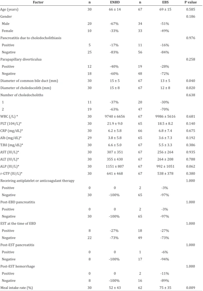
Factor n ENBD n EBS P value
Age (years) 30 66 ± 14 67 69 ± 15 0.585
Gender 0.186
Male 20 -67% 34 -51%
Female 10 -33% 33 -49%
Pancreatitis due to choledocholithiasis 0.976
Positive 5 -17% 11 -16%
Negative 25 -83% 56 -84%
Parapapillary diverticulua 0.258
Positive 12 -40% 19 -28%
Negative 18 -60% 48 -72%
Diameter of common bile duct (mm) 30 15 ± 5 67 13 ± 5 0.040
Diameter of choledocolith (mm) 30 15 ± 8 67 12 ± 8 0.020
Number of choledocholiths 0.638
1 11 -37% 20 -30%
2 19 -63% 47 -70%
WBC (/L) * 30 9740 ± 6656 67 9986 ± 5616 0.681
PLT (104/L)* 30 21.9 ± 9.0 65 18.5 ± 8.2 0.140
CRP (mg/dL)* 30 6.2 ± 5.8 66 6.8 ± 7.4 0.675
Alb (mg/dL)* 29 3.8 ± 5.8 65 3.6 ± 7.3 0.192
T.Bil (mg/dL)* 30 6.6 ± 5.0 67 5.5 ± 3.3 0.386
AST (IU/L)* 30 307 ± 351 67 256 ± 264 0.935
ALT (IU/L)* 30 355 ± 430 67 264 ± 208 0.788
ALP (IU/L)* 30 1151 ± 807 67 992 ± 1051 0.062
r-GTP (IU/L)* 30 641 ± 468 67 538 ± 378 0.380
Receiving antiplatelet or anticoagulant therapy 1.000
Positive 0 0 2 -3%
Negative 30 -100% 65 -97%
Post-EBD pancreatitis 1.000
Positive 0 0 2 -3%
Negative 30 -100% 65 -97%
EST at the time of EBD 1.000
Positive 8 -27% 18 -27%
Negative 22 -73% 49 -73%
Post-EST pancreatitis 1.000
Positive 0 0 1 -6%
Negative 8 -100% 17 -94%
Post-EST hemorrhage 1.000
Positive 0 0 2 -11%
Negative 8 -100% 16 -89%
Meal intake rate (%) 30 52 ± 43 62 75 ± 35 0.009
TABLE 1. Comparison of clinical background characteristics between the endoscopic nasobiliary group and the endoscopic biliary stent group (means ± SD).
*: At the time of EBD.
Factor n ENBD n EBS P value WBC (/µL)* 30 4120 ± 6521 62 5030 ± 5300 0.236 PLT (´104/µL)* 30 -4.2 ± 8.4 62 -4.6 ± 7.1 0.927 CRP (mg/dL)* 30 5.0 ± 5.8 62 4.7 ± 6.8 0.963 Alb (mg/dL)* 22 0.33 ± 0.38 50 0.26 ± 0.52 0.620 T.Bil (mg/dL)* 29 3.7 ± 3.9 62 3.9 ± 3.1 0.409 AST (IU/L)* 29 211 ± 335 63 209 ± 277 0.759 ALT (IU/L)* 29 177 ± 244 63 186 ± 189 0.430 ALP (IU/L)* 28 435 ± 478 60 493 ± 1039 0.417
r-GTP (IU/L)* 27 312 ± 279 62 293 ± 253 0.929 Number of days
to alleviate fever 15 2.3 ± 2.1 40 2.3 ± 2.8 0.338 TABLE 2. Comparison of clinical outcomes between the endoscopic nasobiliary group and the endoscopic biliary stent group (means ± SD).
Laboratory parameter (before EBD – 7 days after EBD).
The basic incision for EST was a medium incision 916).
The ENBD tubes were not washed out as a rule, although when occlusion was suspected, they were washed out with a small amount of physiological saline.
Factors analyzed
1. In both the ENBD and EBS groups, the following factors were analyzed: clinical background [age, sex, the presence/absence of cholecystolithiasis, the presence/
absence of pancreatitis due to choledocholithiasis, the presence/absence of parapapillary diverticula, diam- eter of the common bile duct (mm), diameter of the choledocholith (mm), number of choledocholiths, WBC (/μL), platelet count (PLT; ×104/μL), CRP (mg/dL), to- tal bilirubin (T. Bil; mg/dL), AST (IU/L), ALT (IU/L), ALP (IU/L), r-GTP (IU/L) at the time of EBD], the presence/
absence of receiving antiplatelet or anticoagulant thera- py, the presence/absence of post-EBD pancreatitis, the presence/absence of EST at the time of EBD, the pres- ence/absence of post-EST pancreatitis, the presence/
absence of post-EST hemorrhage, and the meal intake rate (%).
2. Factors that were considered to be related to the effects of EBD included improved laboratory parame- ters [WBC (/μL), PLT (×104/μL), CRP (mg/dL), Alb (mg/
dL), T. Bil (mg/dL), AST (IU/L), ALT (IU/L), ALP (IU/L), r-GTP (IU/L): before EBD – 7 days after EBD ], and time from EBD until resolution of fever (<37°C).
3. Factors that were considered to be related to the mean rate of meal intake during 7 days after EBD in- cluded age, gender, the presence/absence of pancreati- tis due to choledocholithiasis, diameter of the common bile duct (mm), diameter of the choledocholith (mm), number of choledocholiths, WBC (/μL), PLT (×104/
μL),CRP (mg/dL), T. Bil (mg/dL), AST (IU/L), ALT (IU/L),
ALP (IU/L), r-GTP (IU/L), the presence/absence of re- ceiving antiplatelet or anticoagulant therapy, the pres- ence/absence of EST at the time of EBD, the presence/
absence of procedure-related pancreatitis, and EBD (ENBD vs. EBS).
Procedure-related pancreatitis was defined as ab- dominal pain persisting for at least 24 h after the ERCP and associated with an elevation of the serum amylase level to at least three times the upper limit of normal at 18 h after ERCP15, (17). Post-EST hemorrhage was de- fined as any of the following conditions: 1) appearance of tarry stool; 2) decrease in hemoglobin by 2 mg/dL or more; 3) necessity for blood transfusion; and 4) hemo- static treatments including heat probe (18). The meal intake rate was used as an indicator of patient satisfac- tion. The mean meal intake rate was calculated from the meal intake rate for three meals (breakfast, lunch, and dinner) from Day 1 to Day 7 after EBD, according to in- patient records.
Statistical Analysis
In both the ENBD and EBS groups, the χ2 and Fish- er’s exact tests were used for comparisons of categori- cal data. All continuous data values were expressed as means ± SD. Differences in the mean values were exam- ined by the Mann-Whitney U test.
The relationships between the rate of meal intake and age, sex, the presence/absence of cholecystolithia- sis, pancreatitis due to choledocholithiasis, parapap- illary diverticula, diameter of the common bile duct, diameter of the choledocholith, number of choledocho- liths, WBC, PLT, CRP, T. Bil, AST, ALT, ALP, r-GTP, receiving antiplatelet or anticoagulant therapy, EST at the time of EBD, procedure-related pancreatitis, post-EST hemor- rhage, and EBD (ENBD vs. EBS) were investigated by simple regression analysis. Multivariate regression analysis was conducted on the factors that may have af- fected the rate of meal intake according to the findings of univariate analysis using simple regression analysis.
A forward selection method was used for variable se- lection, and analyses were conducted using stentry (P
= 0.20) and slstay (P = 0.25). Statistical significance was defined as a P-value <0.05. The software used for the statistical analysis was PASW Statistics 17 for Windows.
RESULTS
1. Of the clinical background characteristics, there were no significant differences in age, gender, the pres- ence/absence of pancreatitis due to choledocholithiasis, the presence/absence of parapapillary diverticula, num- ber of choledocholiths, administration of antithrom- botic agents, presence/absence of post-EBD pancre- atitis, presence/absence of EST at the time of EBD, the presence/absence of post-EST pancreatitis, presence/
absence of post-EST hemorrhage, or laboratory param- eters before EBD, but the diameter of the common bile duct, the diameter of choledocholiths and meal intake rate were greater in the ENBD group than in the EBS group (P = 0.040, P = 0.020, and P = 0.009: Table 1). All cases of procedure-related pancreatitis were mild (17).
2. There were no significant differences in the levels of improvement in WBC, PLT, CRP, T. Bil, AST, ALT, ALP, and γ-GTP or decrease in Alb during the first week after drainage. The number of patients with fever at the time of admission was 14 in the ENBD group and 40 in the EBS group. The mean time until resolution of the fever was 2.3 days in both groups, with no significant differ-
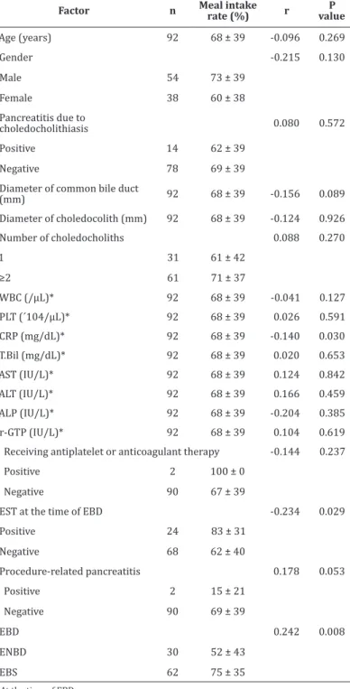
Factor n Meal intake
rate (%) r P
value
Age (years) 92 68 ± 39 -0.096 0.269
Gender -0.215 0.130
Male 54 73 ± 39
Female 38 60 ± 38
Pancreatitis due to
choledocholithiasis 0.080 0.572
Positive 14 62 ± 39
Negative 78 69 ± 39
Diameter of common bile duct
(mm) 92 68 ± 39 -0.156 0.089
Diameter of choledocolith (mm) 92 68 ± 39 -0.124 0.926
Number of choledocholiths 0.088 0.270
1 31 61 ± 42
≥2 61 71 ± 37
WBC (/µL)* 92 68 ± 39 -0.041 0.127
PLT (´104/µL)* 92 68 ± 39 0.026 0.591
CRP (mg/dL)* 92 68 ± 39 -0.140 0.030
T.Bil (mg/dL)* 92 68 ± 39 0.020 0.653
AST (IU/L)* 92 68 ± 39 0.124 0.842
ALT (IU/L)* 92 68 ± 39 0.166 0.459
ALP (IU/L)* 92 68 ± 39 -0.204 0.385
r-GTP (IU/L)* 92 68 ± 39 0.104 0.619
Receiving antiplatelet or anticoagulant therapy -0.144 0.237
Positive 2 100 ± 0
Negative 90 67 ± 39
EST at the time of EBD -0.234 0.029
Positive 24 83 ± 31
Negative 68 62 ± 40
Procedure-related pancreatitis 0.178 0.053
Positive 2 15 ± 21
Negative 90 69 ± 39
EBD 0.242 0.008
ENBD 30 52 ± 43
EBS 62 75 ± 35
TABLE 3. Univariate analysis for predictive factors associated with the meal intake rate during 7 days after EBD (means ± SD).
*: At the time of EBD.
ence (Table 2).
3. The meal intake rate could be investigated in 30 patients (100%) in the ENBD group and 62 patients (93%) in the EBS group. On simple regression analysis, the factors significantly related to the mean meal intake rate from Day 1 to Day 7 after EBD were EBS (P = 0.008), EST (P = 0.029), and CRP level (P = 0.030). That is, the meal intake rate was higher in the EBS group, patients who underwent EST, and patients with low CRP levels.
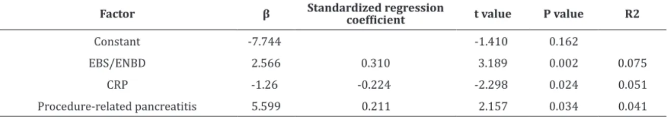
On multiple regression analysis, EBS (P = 0.002), CRP (P
= 0.024), and procedure-related pancreatitis (P = 0.034) were significant independent factors (Tables 3, 4). That is, the meal intake rate was higher in the EBS group and lower in patients with procedure-related pancreatitis and patients with high CRP levels.
4. In the ENBD group, 3 of the 30 patients (10%) pulled out their ENBD catheter, 2 of whom were very old (≥85 years old). There was no catheter occlusion or migration. In the EBS group, occlusion occurred in 3 of the 67 patients (4%). One of these patients had purulent bile in the biliary tract, and the another one had a giant parapapillary diverticulum. There was no catheter mi- gration. The incidence of complications was not signifi- cantly different between the groups (Table 5).
DISCUSSION
ENBD and ERBD (EBS) are widely used for biliary decompression in acute cholangitis. ENBD was estab- lished by Wurbs and Classen (19) in 1977, and its utility has also been reported in the world (1-3, 11). EBS was developed by Sooehendra et al. (20) in 1979 and is in widespread use today. Reports to date have shown no significant differences between the two in comparisons of success rate, incidence of complications, and drain- age effect, and both are considered to be good choices for acute cholangitis (7-11). In previous reports, how- ever, the catheter size in ENBD and EBS was not uniform, and cases of acute cholangitis due to malignant tumor were also included. The subjects in the present study were patients with moderate acute cholangitis due to choledocholithiasis, and patients with concomitant biliary tract malignancies were excluded. The catheter diameter was the same (7-F) for ENBD and EBS. The di- ameter of the common bile duct and the diameter of the choledocholith were larger in the ENBD group than in the EBS group, but there were no significant differences in the proportion of combination EST or other clinical background factors. A comparison of improvement in blood biochemistry tests and time until resolution of fe- ver from before EBD to seven days after EBD showed no significant differences between the two groups.
The discomfort of patients with transnasally placed ENBD is significant, and problems such as self-extrac- tion of the ENBD catheter by the patient are often seen (9-11). Lee et al. conducted a questionnaire survey of ENBD and ERBD (EBS) patients in which discomfort was rated numerically, and they reported that discom- fort was greater in the ENBD group (9). In the present study, a questionnaire survey of patients was not con- ducted, but the meal intake rate was used as an indi- cator of patient satisfaction. Daily caloric intake was not calculated; instead, given that the amount of food provided to individual patients differs, the mean meal intake rate from Day 1 to Day 7 after EBD was investi- gated. On multivariate analysis, significant independent factors related to the meal intake rate were EBS, CRP level, and procedure-related pancreatitis. As mentioned
above, there were no significant differences between the groups in the biliary decompression effect or im- provement in inflammation. Therefore, the meal intake rate is thought to have been higher in the EBS group than in the ENBD group because the EBS group had less discomfort. With respect to nutritional status, the decrease in the post-EBD serum albumin level was not significantly different between the two groups, but this was probably because EBD was done for a short period.
It is thought that patients with high CRP levels did not have sufficient meal intake because there was insuffi- cient improvement in acute cholangitis even after EBD.
Factor ENBD EBS P value Pulled out
catheter 3/30 (10%) 0 0.69
Catheter
obstruction 0 3/67 (4%)
Catheter
migration 0 0
Factor β Standardized regression
coefficient t value P value R2
Constant -7.744 -1.410 0.162
EBS/ENBD 2.566 0.310 3.189 0.002 0.075
CRP -1.26 -0.224 -2.298 0.024 0.051
Procedure-related pancreatitis 5.599 0.211 2.157 0.034 0.041
TABLE 4. Multiple regression analysis for factors associated with the meal intake rate.
TABLE 5. Complications in the endoscopic nasobiliary group and the endoscopic biliary stent group.
Patients who developed procedure-related pancreatitis abstained from food even after EBD because of abdomi- nal and back pain due to pancreatitis.
ENBD is an external biliary drainage procedure. With ENBD, the catheter can be washed out, and bile cultures can be done, but bodily fluids are lost and bile does not flow into the intestine. Thus, there is the possibility that lipid digestion and absorption disorders and motil- ity disorders of the intestine will appear during fasting (12). In contrast, EBS is an internal drainage procedure that may lead to the prevention of endotoxemia, since increases in intestinal bacteria and damage to the in- testinal mucosa are less likely to occur (21). With long- term placement in ENBD or EBS, spontaneous migra- tion of the tube is often seen, but in the present study, spontaneous migration did not occur in any cases be- cause of the short placement time of one week. In the ENBD group, there were no cases of catheter occlusion and no cases in which the catheter was washed out, but in the EBS group, catheter occlusion was seen in 4% of patients. In contrast, the catheter was self-extracted by the patient in 10% of cases in the ENBD group, of whom
about 70% was very old patients, similar to previous re- ports (8-11). The incidence of complications was lower in the EBS group than in the ENBD group, although the difference was not statistically significant. The reason that patients removed their catheters was discomfort in the nasal region, and as mentioned above, this discom- fort is thought to have affected the meal intake rate.
In the present study, no patients died from moderate acute cholangitis. There was no significant difference in the incidence of procedure-related complications between the ENBD and EBS groups. Overall, post-EST bleeding was 8% and post-EST pancreatitis was 4%, similar to previous reports (17). Park et al. (11) found a significant difference in the incidence of hyperamy- lasemia between two groups with differences in the incidence of EST and the size of catheters. Hyperamy- lasemia was not investigated in the present study, but similar to three previous RCTs, (9-11) there was no sig- nificant difference in the incidence of procedure-related pancreatitis between the two groups, and its pancreati- tis was mild in both cases in which it occurred. In ad- dition, the rate of pancreatitis was not affected by the conduction of EST together with EBD. The reasons for this are thought to be that the catheter diameter was the same in both groups, the bile duct and pancreatic duct openings were separated because EST was gener- ally done with a medium incision, and EST was done by endoscopists with more than 10 years of experience.
In conclusion, judging from the biliary decompres- sion effect, the incidence of procedure-related compli- cations, and the meal intake rate (patient satisfaction) in the ENBD and EBS groups, EBS would seem to be the better choice in moderate acute cholangitis due to cho- ledocholithiasis, especially in older patients.
REFERENCES
1. Lygidakis NJ. Acute Suppurative Cholangitis: Comparison of Internal and External Biliary Drainage. Am J Surgery. 1982;
143:304-306.
2. Lai ECS, Paterson IA, Tam PC: Severe acute cholangitis.
The role of emergency nasobiliary drainage. Surgery 1989;
107(3):268-272.
3. Lai ECS, Mok FPT, Tan ESY: Endoscopic Biliary Drainage for Sever Acute Cholangitis. N Engl J Med 1992; 326:1582-1586.
4. Lau JYW, Ip SM, Chung SCS: Endoscopic drainage aborts endotoxaemia in acute cholangitis. British J Surgery 1996;
83:181-184.
5. Sugiyama M, Atomi Y: Treatment of Acute Cholangitis Due to Choledocholithiasis in Elderly and Younger Patients. Arch Surg 1997; 132:1129-1133.
6. Hanau LH, Steigbigel NH: Acute cholangitis. Infect Dis Clin North Am 2000; 14:521-546.
7. Nagino M, Takada T, Kawarada Y: Methods and timing of bil- iary drainage for acute cholangitis. Tokyo Guidelines. J Hepa- tobiliary Pancreas Surg 2007; 14:68-77.
8. Misra SP, Dwivedi M: Biliary Endoprosthesis as an Alter- native to Endoscopic Nasobiliary Drainage in Patients with Acute Cholangitis. Endoscopy 1996; 28:746-749.
9. Lee DWH, Chan ACW, Lam Y-H: Biliary decompression by nasobiliary catheter or biliary stent in acute suppurative chol-
angitis: a prospective randomized trial. Gastrointest Endosc.
2002 56:361-365.
10. Sharma BC, Kumar R, Agarwal N: Endoscopic Biliary Drain- age by Nasobiliary Drain or by stent Placement in patients with Acute Cholangitis. Endoscopy 2005; 37(5):439-443.
11. Park SY, Park C-H, Cho S-B: The safety and effectiveness of endoscopic biliary decompression by plastic stent placement in acute suppurative cholangitis compared with nasobiliary drainage. Gastrointest Endsc 2008; 68(6):1076-1080.
12. Wada K, Takada T, Kawarada Y: Diagnostic criteria and se- verity assessment of acute cholangitis: Tokyo Guidelines. J Hepatobiliary Pancreas Surg 2007; 14:52-58.
13. Komatsu Y, Kawabe T, Toda N: Endoscopic Papillary Balloon Dilation for the Management of Common Bile Duct Stones: Ex- perience of 2226 Cases. Endoscopy 1998; 30:12-17.
14. Van Lent AUG, Bartelsman JFWM, Tytgat GNJ: Duration of antibiotic therapy for cholangitis after successful endo- scopic drainage of the biliary tract. Gastrointest Endosc 2002;
55(4):518-522.
15. Ueki T, Otani K, Fujimura N: Comparison between emer- gency and elective endoscopic sphincterotomy in patients with acute cholangitis due to choledocholithiasis: is emer- gency endoscopic shincterotomy safe?. J Gastroenterol 2009;
44:1080-1088.
16. Ikeda S, Tanaka M, Itoh H: A Newly Devised Cutting Probe for Endoscopic Sphincterotomy of the Ampulla of Vater. En- doscopy 1977; 98:238-241.
17. Cotton PB, Lehman G, Vennes J: Endoscopic sphincterotomy complications and their management: an attempt at consen- sus. Gastrointest Endosc 1991; 37(3):383-393.
18. Sugiyama M, Atomi Y: The Benefits of Endoscopic Nasobil- iary Drainage Without Sphincterotomy for Acute Cholangitis.
Am J Gastroenterology 1998; 93(11):2065-2068.
19. Wurbs, D., Glassen M: Transpapillary long standing tube for hepatobiliary drainage. Endoscopy 1977; 12:192-193.
20. Soehendra N, Reynders-Frederix V: Palliative Gallengangdrainage,Eine neue Methode zur endoskopis- chen Einfuhrung eines inneren Drains.Dtsch. med. Wschr.
Deutsche Medizinische Wochenschrift 1979; 104:206-207.
(in German)
21. Gouma DJ, Coelho JCU, Fisher JD: Endotoxemia After Relief of Biliary Obstruction by Internal and External Drainage in Rats. Am J Surgery1986; 151:476-479.