Acta med. Nagsaki. 12: 83-98
An Electron Microscopic Study of Pathological Changes of the Great Occipital Nerve Following Local Cooling
Masashichi KAWANO and Hidekatsu MATSUMURA
The Second Department of Surgery, Nagasaki University School of Medicine, Nagasaki, Japan
Received for publication, March 4, 1968
On electron microscopy the great occipital nerve of the rabbit was studied in its relation to local cooling of the head on the occipital region of the scalp to learn the ill effect of unduly prolonged bed rest using ice pillow following head injury as a part of investigation of chronic post-traumatic headache.
After three hours of occipital cooling in the temperature ranging between 2°C. and 5°C. the mylin sheath is swollen, the lamella is torn and vacuoles are formed. The axon becomes atrophic, axon-Schwann interface is enlarged, and spiral invagination of the lamella is common. Endoplasmic reticulums are increased in the axon and mitochondria are swollen and cristae are destroyed. These changes are found in all fibers with no discrimination of their size.
After 6 hours of cooling, these changes are brought to the extreme, and most of these changes remain long after cooling is stopped, or they are irreversible.
Recovery after three hour cooling is much better. Swelling of the axon and vacuoles in the lamella disappear within two weeks, and axon-Schwann interface returns to their normal relation within two months, although spi- ral invagination of the lamella may remain
I. INTRODUCTION
As the result of striking increase of traffic accident in civilized nations a large number of head injury patients are created every year.
Aside from its menace to the lives of victims head injury leaves various symptoms even after the acute sick stage passed. Among these post-traumatic complications chronic headache is the most annoy-
ing problem both for patients and physicians, because it implies inse- curity or danger of forthcoming mental difficiency in addition to the pain persistence, thus complicating its clinical picture with emotional in- volvement. The situation is worsened when the physician is undecided because of difficult understanding and interpretation of the problem and fails to win the trust of the patients, or when the patient is irritative or upset by prolonged and ineffective treatment, or when a legal pr- oblem of compensation is involved. Most of physicians try to locate
*川 野 正 七、松 村 豪 一
the source of headache in the intracranial structure, in vain except in chronic subdural hematolna which can be discovered easily by cerebral angiography and treated immediately by burr hole and irrigation, and after a long trial and confusion hand the patients to the psychiatrist labelling them as post-traumatic neurosis. There is no doubt that post- traumatic headache has a unique psychosomatic aspect, but it can not be justified to ignore the presence of headache and attribute to psycho- genic or neurotic factor alone.
In his monumental monograph WoIFF discussed this problem in details." He estimates between one-third and one half of all persons who injure their heads sufficiently to warrant hospitalization develop chronic post-traumatic headaches. He classified them into three types:
(1) muscle contraction headache, (2) painful site of injury, and (3) vascular headache. There is, however, another type of headache often encountered in Japan, headache associated with tenderness of the great occipital nerve and the first branch of the trigeminal nerve, the former being predominant. This type of headache called attention of a few neurosurgeons including ARAKI2' and ASANO who named it great occipi- tal and trigeminal syndrome, or GOTS. The incidence of this neuralgic post-traumatic headache was 28 percent according to TAKETOMO and SAKATA3), and 34.7 percent after HIRATA4'. In our study of 104 chronic post-traumatic headache patients 45.1 per cent or 47 cases proved tender- ness of either the great occipital nerve or the trigeminal nerve or both,". This may include vascular headache patients, but it is justifia- ble to assume that there are a considerable number of post-traumatic headache patients whose pain is attributable to neuralgia or irritable condition of the nerve as a result of aseptic neuritis or degeneration.
In another survey of post-traumatic headache we discovered a striking fact that 85 per cen of all non-operative post-traumatic patients had cooled their head for various length of time, from a few hours to 83 days, mostly for a week or two regardless the type of injury or the
state of consciousness, using icepacked pillow or bag6'. And the inci- dence of post-traumatic neuralgic headache is significantly high among these local cooling cases.
With a hypothesis that localized cooling of a region of the skin with ice may give untoward effect to the underlying sensory nerves, as well as the motor nerves as illustrated in certain cases of BELL'S palsy, the following experiments were conducted to see under electron micro- scope how the nerve reacts to local cooling in the course of time and it recovers from the damage.
II. MATERIALS AND METHOD
Fifty seven adult rabbits weighing 2.8 to 3.2 kg were subjected to
the experiments. Under anesthesia with intraperitoneal injection of Nembutal, 50 ml per kg. body weight, each rabbit was immobilized on a stereotaxic apparatus in prone position. Then, a polyethylene bag containing ice fragments mixed with salt was placed on the occipital re- gion of the scalp and an alcohol thermometer was inserted under the skin to record the subcutaneous temperature, and another to mea- sure rectal temperature. Ice bag was, then, manipulated to maintain the temperature between 2°C. and 5°C. often requiring addition or exchange of ice fragments. Usually subcutaneous temperature dropped to 5°C. or below within 30 minutes.
When proposed length of cooling hours was reached, the cooling was stopped, and immediately the great occipital nerve was exposed by midline incision of the occipital and suboccipital area and careful atraumatic dissection of the nerve. For fixation of the nerve 3% glut- araldehyde was applied for 20 minutes before the nerve was ligated., divided and stored in 3% glutaraldehyde for 16 hours. The nerve was
then sliced to fine blocks of Imm3 and rinsed in phosphate buffer for 20 minutes, before they were fixed again in 2% Os04 solution for two hours and rinsed in phosphate buffer. Then followed dehydration with aceton for two hours. The specimens went through three different ratios of aceton resin mixture; 7:3, 5:5, and 3:7 in 6 hours, and aceton was evaporated in a desiccator for 15 to 17 hours. When they were dried, 100% resin was added and mixed in an electric shaker for 3 hours. These specimens were dropped one by one into gelatin capsule, containing resin, and undesirable air bubble was removed by suction for one hour, before they were warmed for 24 hours in 60°C. The final speimen was imbedded in epon epoxy resin, and the block was sliced into rather thick sections of 2 to 3,u before staining with truisin blue to select the area to be examined. The selected area of the specimen was then sliced with LKB-ultratome, stained with uranyl acetate and Pb (OH)2 and then examined under electron microscope, JEM-6C type made of Japan Electronic Company. As buffer solution, Harvard Uni- versity phosphate buffer (PH 7.4) was used exclusively because of its superiority over the Millonig's buffer solution. In interpretation of the electron microscopic findings of the occipital nerves the authors owe to the ultra-microscopic description of the nerve fibers in the muscle by ROBERTSON and of the sciatic nerves by HONJIN3 .
III. RESULTS
Before describing and discussing the abnormal change following local cooling of the occipital area of the rabbits, normal electron micro- scopic findings of the control must be described. The ultra-struc- ture of the minute anatomical unit of the nerve fibers, such as the lamella of the myelin sheath, fine structure of the axon, and the mi-
tochondria, can not be satisfactorily demonstrated under the ordinary microscope. Only electron microscopic study can reveal these micro structure of the nerve fibers.
1. Normal electron microscopic findings of the great occipital nerve of the rabbit.
1) Schwann cells: The Schwann cell neurilemma sheath is covered with two layers of the cell membranes, two nucleus membranes, and it contains a collection of small granules called the nucleus corpusculum and other granules distributed evenly in the protoplasm. The cell substance is found around the nucleus in large quantity and contains mitochondria of oval or round shape and many endoplasmic reticulums (Fig. 1).
2) Henle's sheath: Outside of the neurilemma sheath there are found collagen fibrils, a kind of connective tissues (Fig. 1).
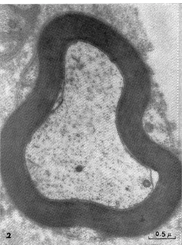
3) Myelin sheath: In higher magnification the myelin sheath is composed of a lamellar structure with a roll of many fine membranes (Fig. 2). On the outer surface of the myelin sheath the lamella runs across the Schwann cell protoplasm and is connected with the Schwann cell membrane. This outer mesaxon is distinctly recognized in our oc- cipital nerve specimen (Fig. 2). The vacuoles between the lamellae and distorted or torn lamella were occasionally found in the normal larger nerve fibers, but rare in medium-size fibers and none in small- size fibers .
4) Axon membrane: There lies a 150 to 250A thin membrane be- tween the axon and the myelin sheath. There is also found the inner mesaxon, a continuation of extraverted two axon membranes to the myelin sheath lamella, in the great occipital nerve (Fig. 2).
5) Axon: Contained in the axoplasm are axon fibrils, endoplasmic reticulums and mitochondria with cristae (Fig. 2).
2. Changes after 3 hours of local cooling.
There are found several abnormal findings when the great occipital nerve is inspected under electron microscope after three hours of occipi- tal cooling. They are swelling of the myelin sheath, seperation and
vacuole formation in the lamella, atrophy of the axon, enlarged axon- Schwann interface with spiral invagination of the lamella, increased endoplasmic reticulum in the axon, and swelling of the mitochondria.
These findings are found in all myelinated nerve fibers with no regard to their size (Fig. 3).
3. Changes after 6 hours of local cooling.
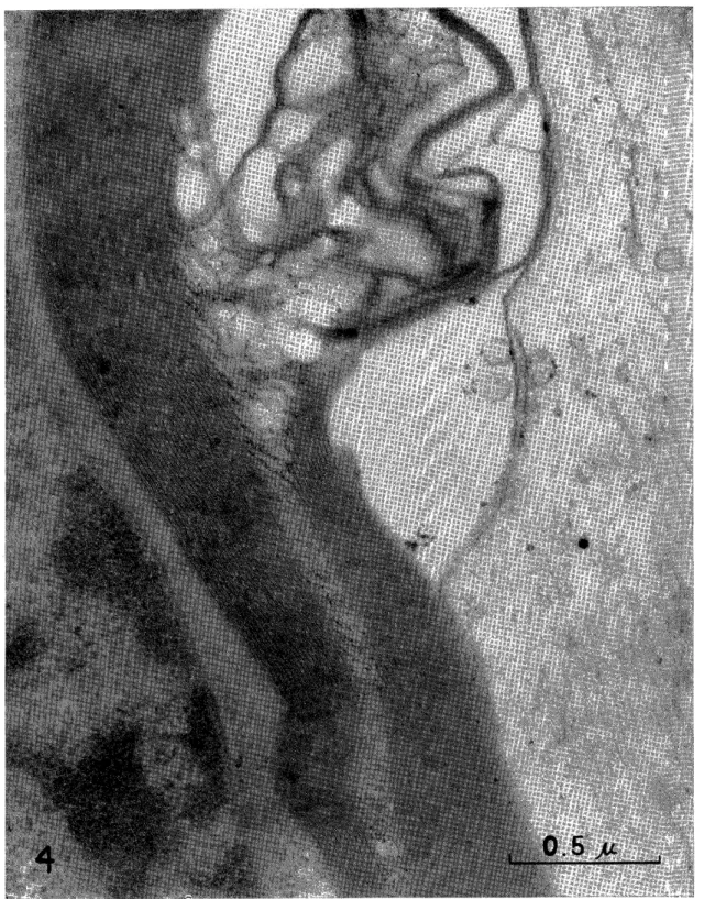
The electron microscopic specimen of the great occipital nerve of the rabbit taken out after 6 hours of occipital cooling reveals advanced stage of the above-mentioned changes including maximum increase of axon-Schwann interface. Occasionally, there is found invagination of Mauthner sheath from Schmidt-Lantermann cleft (Fig. 4).
4. Recovery of pathological changes after cessation of cooling.
A number of animals were 'kept alive for different periods after
cessation of cooling with the aim to see how the nerve recover from the damage due to local cooling. Of 6-hour-cooled rabbits none revealed sign of recovery on electron microscopic examination in 3 to 7 days after cooling was stopped, indicating that the damage was irreversible.
There were, however, signs of recovery in the 3-hour-cooling group as follows:
1L) After two days, most of the changes are still remained, but mild changes are already disappeared.
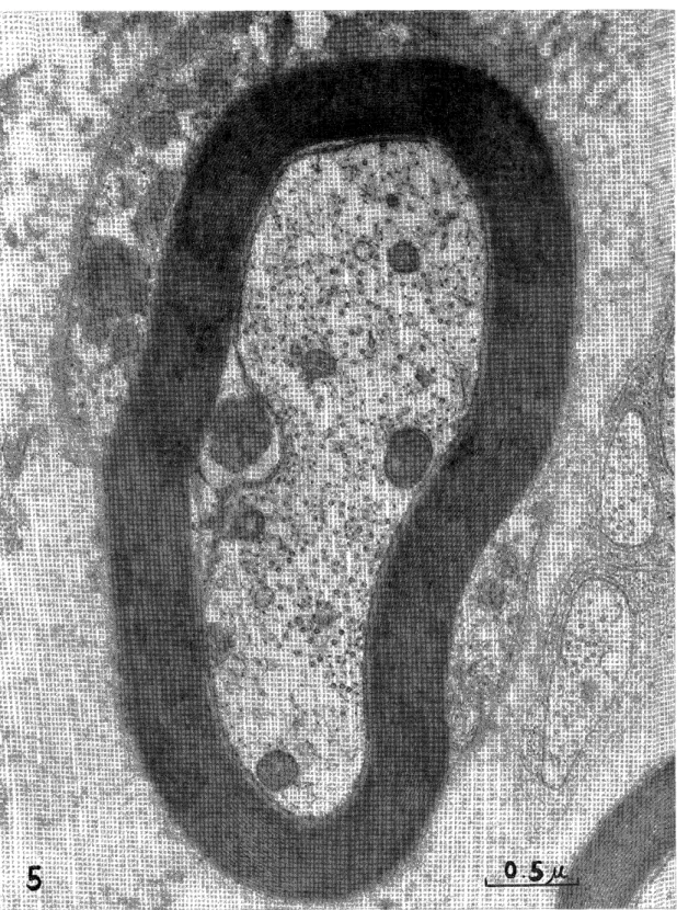
2) After two weeks, swelling of the axon and vacuoles in the lamella of small-size nerve fibers can not be seen, although spiral in- vagination of the lamella remains near the outer and inner mesaxon of the small-size and medium-size fibers (Fig. 5).
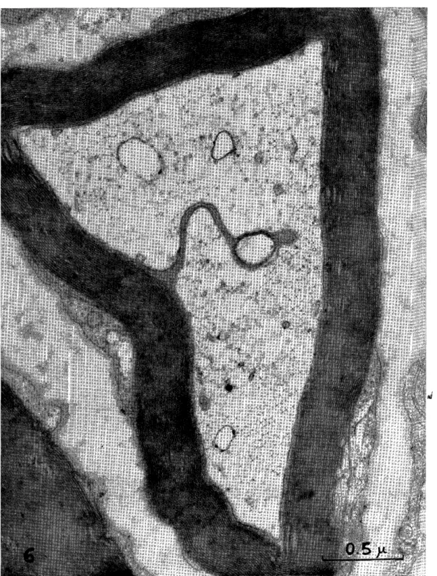
3) After two months, axon-Schwann interface is no longer en- larged, although folds are occasionally seen in the myelin sheath (Fig. 6 and 7).
IV. DISCUSSION
Cold temperature affects the body in many ways even in our daily life, and the effect of cold had been a subject of study since old days.
Studies on this subject, however, flourished during and after the first and second World War when frost-bite and chilblain tortured thousands of soldiers on land and sea. So-called "immersion foot", a vaso-neuro- pathy due to immersion of feet in the battle-field trench, was a com- mon malady in winter fronts, and many studies were made on this particular subjectsalone.9 15) Among others. the most study on the pathology of cold injury to the peripheral nerve would be that of DENNY- BROWN and others 16) , who cooled a segment of the sciatic nerve of the cat to non-freezing degree and studied the mode of degeneration and dysfunction of the various nerve fibers. One of his conclusions that the largest-size medullated nerve fibers are most sensitive to the exposure to cold was challenged later by SCHAUMBURG et all" who insisted that damage to the medullated fibers was diffuse and non-selective with no regard to the size of nerve fibers.
With the advance of electron microscopy, ultra-structure of the myelinated nerve fibers were discussed in details7- , enabling us to reevaluate the pathological changes in the minute nerve fibers, and this is true with damages due to the exposure to cold.
In our experimental study of the effect of cooling to the peri- pheral nerves, we selected the great occipital nerve from the following two reasons: (I) It is the major sensory nerve to convey the pain sen- sation in the posterior half of the head, (2) its main trunk is in close contact with the scalp so that the nerve can be cooled from the surface
of the skin without difficulty, and (3) the setting can simulate the clinical situation where the head injury patients lie on ice pillow as
mentioned in the introduction.
Although there has been no literature concerning the effect of cold to the great occipital nerve, electron microscopic findings of our speci- men after occipital cooling with ice bag revealed a great morphological resemblance to the report by SgAUMBURG et al 171 on the sciatic nerve.
But their study was only observed under light microscope and they did not observe under electron microscope. Damages to the nerve was dif- fuse and non-discriminative to the size of nerve fibers, even consider- ing the fact that this nerve contains not only large-size fibers but also medium-and small-size nerva fibers.
There is a difference, however, between their findings and ours.
They reported that nerves cooled below 20°C. showed vacuolization and gaps in myelin with swelling and fragmentation of axons, but we were unable to find swelling but atrophy of axons. ROBERTSON") cooled the sciatic nerve fibers of the frog to 0 to 4°C. and found swelling of myelin and atrophy of axons, similar to our findings but more signifi- cant. There is no question that myelin breaks up into thin layers of 150A by cooling as demonstrated by FINEAN19 in his x-ray diffraction technique. The same phenomenon was confirmed on electron microscopy by ROBERTSON20). According to his explanation, the surface membrane of Schwann cell combines with the next unit membrane to form the internal compound membrane, and gaps in the mesaxon widen and reach axon, when myelin was immersed in cold hypotonic solution.
As to unmyelinated fibers, a gap opens in hypotonic solution and closes in hypertonic solution, reversible processes. These dynamic action of the membrane should be kept in mind when changes due to cooling is studied and interpreted.
There is a question, however, whether the change of the nerve after cooling is the direct effect of cold or the secondary effect caused by ischemia due to cold. DENNY-BROWN et al'-6' feel that the effect of cold is more abrupt than ischemia in its transition from the normal state to necrosis. LAKE"' found that tissue cultures were damaged by temperatures of 5 to 10°C. though they could be preserved without damage at temperatures from -5 to 50C., suggesting interference of ac- cumulated metabolites. DENNY-BROWN et all" condisers the action of cold on myelin is probably direct with liberation of diffusible fat, citing the observations of Harvard Medical School that the blood lipid splits from certain lipo-proteins at low temperature.
Nevertheless, in the nerve by cooling are much similar to the da- mages due to ischemia, and DENNY-BROWN et al 161 do not deny that "is- chemia is indeed a likely factor in the production of tissue damage"
found in the nerve following cooling. We shall report in another paper how microcirculation of the great occipital nerve is affected by cooling22'2s' The process is exactly that of skin under the effect of cold: vasoconstric- tion, vasodilatation, sludge of venous blood, hemostasis, extravasation of corpuscles and other substances, and finally occlusion ensue s22,23
In clinical situation, cooling of the occipital region and the neck with ice pillow does not cool the occipital nerves to near-zero tempera- ture. In our clinical experiment the difference between the arm-pit temperature and occipital subcutaneous temperature was 11 degrees. In other word, the great occipital nerve, the major trunk at least, seldom reaches temperature below 20°C. This differs greatly from the condi- tion in our animal experiments in which the temperature around the nerve was maintained between 2° and 5°C. to see the effect of cold in shorter time than in clinical situation. As well known, however, the grade of damage by cold depends on duration as well as intensity of cold. Even the cooling in mild degree, if continued long, may pro- duce as severe damage as would be produced by near-zero cooling of short duration. We found the damages produced by one hour cooling is negligible, three hour cooling is reversible, but 6 hour cooling leaves several irreversible damages in our below 5°C animal experiments. It would be reasonable to assume that there is a possibility of leaving ir- reversible damage to the great occipital nerve and lower the threshold to pain, if the back of the head and neck be cooled with ice pillow for a long period, as long as two months in some, in the post-traumatic care of the patient.
We do not yet make our experiment on physiological change of the great occipital nerve in its relation to local cooling, although we postulate that prolonged cooling of. the occipital area may increase irritability of the nerve by lowering its threshold to pain. WOLFF advo- cates the presence of a pain-threshold-lowering substance, a polypeptide, in the surface tissues of the scalp near and about the dilated cranial arteries in migraine patients. If he is right, occipital cooling of the head will indirectly as well as directly lower the pain threshold by dilating the cranial artery and by producing a local edema.
Pressure is another factor to be considered. in the present series of experiment the effect of pressure was excluded in control experiment, but pressure of ice pillow or hard Japanese pillow upon the overlying heavy head (nearly 4kg.) will be able to influence both occipital circu- lation and nuchal muscles in the patient of prolonged bed rest.
We should not underestimate, if not overestimate, the important role of psychosomatic aspect of head injury, particularly when the conscious healthy individuals were isolated from his normal activity
and environment and forced to stay in bed to chagrin over the acci- dent which might have been avoided and brood over the insecure future and fear of the damage to the highest integrate organ of their own, with remorse, resentment and frustration. The relation of these psychology and neurotic personality to chronic post-traumatic headache, particularly to vascular and muscular headache has been studied by many authors and well reviewed by Woz.FF. In this paper we should
he satisfied to limit our interest to the relation of local cooling of head to the possible damage of the great occipital nerve which may lead to neuralgic headache, aside from effects to enforce other types of head- ache.
V. CONCLUSION
1. The great occipital nerve, the major sensory nerve to convey pain sensation of the posterior regions of the head, is affected by local cooling of the occipital area of the head in the temperature between 2° and 5°C.
2. The longer the duration of cooling, the greater damage of the nerve is produced.
3. The mild changes produced by cooling of less than 3 hours are reversible and disappear after various periods following cessation of cooling.
4. Some of the severe damages are irreversible even leaving long after cooling. These include extreme atrophy of axon, enlarged axon- Schwann interface, spiral invagination of the lamella and intrusion of Mauthner sheath.
5. Cooling longer than 3 hours increases these irreversible changes.
6. In the great occipital nerve, the damage is not selective but affects fibers of all sizes.
7. These changes are compatible with the damage due to ischemia.
8. Prolonged bell rest on ice pillow may produce similar or milder changes in head injury patient and may contribute to chronic post-trau- matic headache .
9. Studies of microcirculation and physiological change in conduc- tion of pain impulse must be made to evaluate these electron micro- scopic findings.
REFERENCES
1. WOLFF, H.E.: Headache and other head pain. New York, Oxford University Press, 2. ARAKI, C.: Nippon Geka 7ensho. 10: 1, Tokyo. Kanehara, 1954. (Japanese) 3. TAKETOMO, T. and SAKATA, K.: Nippon Rinsho, 23: 1981, 1965. (Japanese) 4. HIRATA, M.: Kumamoto Igakukai Zasshi, 35: 439, 1961. (Japanese)
5. KAwANO, M. et al: Neurologia medico-chirurgica 7: 156, 1965.
6. KAWANO, M.: Chiryo, 48: 1989, 1966. (Japanese)
7. ROBERTSON, J.D.: J. Biophysic. Biochem. Cytol,. 1: 271, 1955.
8. HONJIN, R.: Seitai no Kagaku, 8: 119, 1957.
9. BLACKWOOD, W.: Proc. Roy. Soc. Med. Lond., 36: 521, 1943.
10. BLACKWOOD, W. and RUSSELL, H.: Edinburgh M.J., 50: 385, ].943.
11. UNGLEY, C. C. and BLACKWOOD, W.: Lancet, 2: 447, 1942.
12. UNGLEY, C.C.: Proc. Roy. Soc. Med., Lond., 36: 518, 1943.
13. WEBSTER, D.R., WOOLHOUSE, F.M., and JOHNSTON, J. L.: J. Bone and Joint Surg., 24 785, 1942.
I.4. WRITE, J.C.: New Eng. J. Med., 228: 213, 1943,
15. WHITE, J.C., and WARREN, S.: War Med., 5: 6, 1944.
16. DENNY-BROWN, D. et al: J. Neuropath. Exp. Neurol., 4 305, 1945.
17. SCHAUMBURG, H. and BYCK, R. et al: Arch. Neurol. 16: 103, 1967.
18. ROBERTSON, J.D.: J. Biophysic. Biochem. Cytol., 4 349, 1957.
19, FINE N, J.B. and MILLINGTON, P.F.: J. Bioqhysic. and Biochem. Cytol. 3. 89, 1957.
20. ROBERTSON, J.D.: Biochemeial Society Symposia, 16: 1, 1959.
21. LAKE, N.C.: Lancet, 2: 557, 1917.
23. MATSUMURA, H. and KAWANO, M.: Neurologia medico••chirurgica 9: 1967 ingress.
22. MATSUMURA, H. and RAWANO, M.: J. Electron Microscopy 17 (1): 87, 1968.
Fig. 1. The normal structure of the great occipital nerve of a rabbit fixed witn 3%
glutaraldehyde and 2% 0sO4x20,800.
Fig 2 The normal structure of the great occipital nerve of a rabbit, demonstrating distinct inner mesaxon and outer mesaxon x41,100.
Fig. 3. Changes of the great occipital nerve of a rabbit after 3 hours of local coo- ling. x47,000.
Fig. 4. Changes after 6 hours of local cooling. x63,700,
Fig 5 2 weeks after 3 how's of local cooling, x38,400,
Fig 6 2 months after 3 hours of local cooling x 54, 900
Fig 7. 2 months after 3 hours of local cooling x48,000,