Acta Med. Nagasaki 47 : 67-69
Case Report
Adult T-Cell Leukemia/Lymphoma with Gastric Lesion
Takayuki ASAKAWA 1), Masazumi TERADA1), Shigeki MINAMI1), Hidenobu OSHIBUCHI1), Takeshi MATSUO2)
1) Department of Surgery, Oshibuchi Hospital
2) Department of Pathology, Kyushu Microbiological Laboratory
A case of adult T-cell leukemia/lymphoma with a gastric tumor is presented. The patient was a 72-year-old woman who had been admitted to our hospital because of epigastric pain. Upper gastrointestinal endoscopy disclosed polypoid re- gion in the antrum of the stomach. Specimens taken from this region revealed malignant lymphoma. Although the pa- tient was serologically positive for human T-cell leukemia virus type I antibody. A chest X-ray, abdominal ultrasonography, and whole body computed tomography re- vealed no lymphadenopathy. We diagnosed her to have pri- mary gastric malignant lymphoma and thus performed a total gastrectomy with regional lymph node excision and a splenectomy. Both histological and immunohistochemical studies of the resected specimen showed the lymphoma to be a diffuse pleomorphic type with a T cell phenotype. A radical surgical resection could be performed. At nine months postoperatively, she was again admitted to our hos- pital because of abdominal pain. Both ultrasonography and computed tomography revealed lymph node swelling around the surrounding the para aorta. She was treated with sys- tematic chemotherapy. Unfortunately, she died at one year after the operation.
ACTA MEDICA NAGASAKIENSIA 47: 67-69, 2002
Key Words: adult T-cell Leukemia/lymphoma, gastric lesion
Introduction
Primary lymphoma of the stomach is a rare disease among gastric malignancies' ' z'.
However, gastrointestinal involvement in patients with leukemia and lymphoma of B-cell type frequently occurs, however T-cell type remains rare3,-5.
Gastrointestinal involvement in adult T-cell leuke- mia/lymphoma (ATL) which is caused by human T- Adress Correspondence: Takayuki Asakawa, M.D.
Department of Surgery, Oshibuchi Hospital, 37-1 Satomen Mikuriya-machi, Matuura 859-4752, Japan
TEL: +81-956-75-0311 FAX: +81-956-75-0100
cell lymphotropic virus type I (HTLV-I) is not rare'"'.
We herein present a case of adult T-cell leukemia/
lymphoma (ATL) with malignant cell infiltration into stomach.
Case report
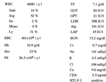
A 72-years-old woman visited our hospital with epigastric discomfort in September 13, 1995. On physi cal examination, no abdominal mass, hepatosplenomegaly or systemic lymphadenopathy was observed. The labo- ratory data are shown in Table 1. The white blood cell count was 6000/mm' with normal differentiation.
The human T-cell lymphotrophic virus type I (HTLV- I) antibody was positive in the serum. The serum LDH and calcium levels were normal. Upper gastrointesti- nal endoscopy revealed a gastric polypoid lesion of the antrum (Fig 1). Several specimen obtained by endscopic biopsies showed a diffuse infiltration of malignant lymphoma. Preoperatively, ultrasonography (US) and computer tomography (CT) examinations showed no evidence of either swollen lymph nodes or organ me- tastasis. An operation was performed on November 17,
Table 1. Laboratory data on admission
WBC 6000 / [t 1 TP 7.1 g/dl
Stab 10% GOT 20 IU/1
Seg 52% GPT 21 IU/1
Eo 2 % LDH 298 IU/1
Mono 4 % Alp 191 IU/1
Ly 31% LAP 451U/1
RBC 401x104 / tt l BUN 15.2 mg/dl
Hb 10.9 g/dl Cr 0.7 mg/dl
Hct 35% Na 141 mEq/1
Pit 26.5 x10⁴ I K 4.3 mEq/1
Cl 106 mEq/1
Ca 9.0 mg/dl
CEA 2.5 ng/ml
HTLV-I positive
Figure 1. Endosopic findings of the stomach reveals a polypoid lesion on an antrum.
1995.
At operation, the liver, omentum, small and large bowels were normal. A total gastrectomy with a re- gional lymph node excision and splenectomy were performed.
A histological examination of the resected specimen showed that the lymphoma cell diffusely infiltrated into the submucosal layer of the stomach on polypoid lesion (Fig2a).
lymphoma cells (Fig2b).
Immunohistochemical examinations of the specimens were positive for UCHL-1 and negative for L-26. In addition, tumor showed no involvement of the re- gional lymph nodes.
During follow-up, a swollen lymph node surround- ing the para-aorta was detected by abdominal US and CT at 9 months after the initial operation. The patient was treated with systematic combination chemother- apy (750 mg/mm 2 of cyclophosphamide, 50 mg/mm 2 of doxorubicin hydrochloride, 1.4 mg/mm 2 of vincristine sulfate, and 100 mg/mm2 of prednisolone acetate).
Though two cycles of the chemotherapy was done, the size of the lymph node showed no change. Laboratory examinations revealed an elevated LDH and calcium level. Thereafter, the patient developed multiple organ failure and died 1 year after the operation.
Figure 2a. Histological examination of the resected specimen showed diffuse infiltration of abnormal lymphocytes into the submucosal layer.
Also the infiltration into specimens from other areas was noted.
The tumor was composed of medium to large sized
Discussion
Adult T-cell lymphoma is a new disease entity, re- ported by Uchiyama et al in 19779'. The clinical symp- toms of ATL are highly characteristic"'. For example, hepatosplenomegaly, systemic lymphadenopathy, rashes, and bone marrow involvement are usually observed and the gastrointestinal tract is frequently involved.
Sakata et al" describes endoscopic features of gastric involvement of ATL were classified into three types the types of ATL: diffuse type (including multiple ul- cers and diffuse erosions), tumor forming type (includ- ing submucosal tumor-like lesion, multiple polypoid le- sions, and tumors with ulceration), and giant fold type. Also 30.3% of patients with ATL had gastric in- volvement.
In another study, Dawson et al."' indicated five major
symptoms as the criteria for primary malignant
lymphoma of the intestinal tract: no palpable superfi- cial lymphadenopathy, no enlarged mediastinal lymph nodes evident on chest radiographs, normal total and differential WBC counts, predominant bowel lesions at laparotomy, and no tumor in the liver or spleen.
Our patient had a polypoid lesion in the stomach.
However, no other organ infiltration was found either at admission or during the operation. Consequently the present case met all these criteria. This case is consid- ered to be a case of primary gastric lymphoma with ATL. In addition, the staging was stage I based on the Ann-Arbor system"'. Otherwise pathological findings show the diffuse infiltration of lymphoma cell into submocosal layer on the polypoid lesions and the other lesions. Namely this case was not primary gastric lym- phoma and was gastric involvement of ATL.
Several reports have showed a combination of sur- gery, post operative irradiation or/and chemotherapy to be effective for treating the gastric ATL with re- gional node involvementl°'.14'.15'. Gastric involvement of ATL was one of the prognostic factors in acute type ATL, whereas it had no influence on the prognosis of lymphoma type ATL $'.
In case of gastric lymphoma positive for human T- cell leukemia virus type I, smear of periperal blood and endoscopic examination was important for the di- agnosis of leukemia type or lymphoma type. It is dif- ficult to judge the indication of operation for gastric lymphoma with ATL. Combination chemotherapy would be the best treatment for Gastric lymphoma with ATL.
References
1) Connors J, Wise L. Management of gastric lymphomas. Am J Surg 127:102-108, 1974
2) Contreary K, Nance FC, Becker WF. Primary lymphoma of gastro- intestinal tract. Ann Surg 191: 593-598, 1980
3) Seo IS, Binkley WB, Warner TFC, Warfel KA. A combined mor phologic and immunologic approach to the diagnosis of gstrointestinal
lymphomas. I . Malignant lymphoma of the stomach (a clinicopathologic
study of 22 cases). Cancer 49: 493-501, 1982
4) Dragosies B, Bauer P, Radaszkiewiez T. Primary gastrointestinal
non-Hodgkins Lymphomas: a retrospective clinicopathologic study of 150 cases. Cancer 55: 1960-1973, 1985
5) Kanavaros P, Lavergne A, Galian A, Houdart R, Bernard JF.
Primary gastric peripheral T-cell malignant lymphoma with
helper/inducer phenotype: first case report with a complete
histological ultrastructural and immunochemical study. Cancer 61:
1602-1610, 1988
6) Shimoyama M, Minato K, Saito H, et al. Comparison of adult T- cell leukemia-lymphoma and cutaneous T-cell lymphoma. Jpn J
Clin Oncol 9: 357-372, 1979
7) Jaffe ES, Blattner WA, Blayney DW, et al. The pathologic spec- trum of adult T-cell leukemia/lymphoma in the United States. Am
J Surg Pathol 8: 263-275, 1984
8) Sakata H, Fujimoto K, Iwakiri R, et al. Gastric lesions in 76 pa- tients with adult T-cell leukemia/lymphoma. Cancer 78: 396-402 9) Uchiyama T, Yodoi J, Sagawa K, Takatsuki K, Uchino H. Adult T-
cell leukemia: Clinical and hematologic features of 16 cases. Blood
50: 481-491, 1977
10) Kubonishi I, Daibata M, Yano S, et al. Gastric lymphoma associ- ated with human T-cell leukemia VIrus Type I. Arch Intern
Med 147: 603-605, 1987
11) Shimoyama M. Diagnostic criteria and classification of clinical subtypes of adult T- cell leukemia-lymphoma: a report from the
Lymphoma Study Group. Br J Haematol 79: 428-437,1991 12) Dawson IM, Cornes JS, Morson BC. Primary malignant lymphoid
tumors of the intestinal tract: report of 37 cases with a study of
factors influencing prognosis. Br J Surg 49: 80-89, 1961
13) Carbone PP, Kaplan HS, Musshoff K, Smithers DW, Tubiana M.
Report of the committee on Hodgkin staging classification. Cancer
Res 31: 1860-1861, 1971
14) Katabami S, Hinoda Y, Ohe Y, et al. Adult T-cell leukemia/lym-
phoma (lymphoma type) with remarkable gastric lesions: A case report. Gastroenterologia Japonica 27: 95-101, 1992
15) Kojima H, Ikeda H, Kubo T, et al. HTLV-I associated gastric lym- phoma. Zentralbl Pathol 138: 351-354, 1992