• 検索結果がありません。

Adrenomedullin is not Related to Acute Hypoxic Pulmonary Vascular Response in Patients with Chronic Respiratory Disease

N/A
N/A
Protected

Academic year: 2021

シェア "Adrenomedullin is not Related to Acute Hypoxic Pulmonary Vascular Response in Patients with Chronic Respiratory Disease"

Copied!
6
0
0

読み込み中.... (全文を見る)

全文

(1)

Acta Med. Nagasaki 44 : 55-60

Adrenomedullin is not Related to Acute Hypoxic Pulmonary Vascular Response in Patients with Chronic Respiratory Disease

Katsuyuki FURUKAWA, Satoshi IKEDA, Tadasu IWASAKI, Tetsuro MATSUSHITA, Kazuaki YAKABE, Kenji YAMAGUCHI, Yoshiyuki MIYAHARA, Shigeru KOHNO

Second Department of Internal Medicine, Nagasaki University School of Medicine

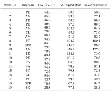
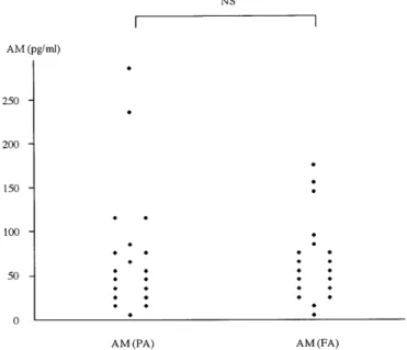
In the present study, acute hypoxia was induced in 19 pa- tients with chronic respiratory disease to evaluate the corre lation between pulmonary circulation kinetics and adrenomedullin (AM) levels. Using radioimmunoassay (RIA), pulmonary cir- culation kinetics were evaluated before and after hypoxic loading (13% oxygen for 15 minutes) by determining AM levels in plasma obtained from the pulmonary artery (PA) and the right femoral artery (FA). There were no signifi- cant differences in pre-hypoxia plasma AM levels between samples obtained from the PA and FA, and plasma AM lev- els did not change after hypoxic loading. Subjects were classi- fied into two groups [responders (R) and non-responders (NR) ] to evaluate changes in the mean pulmonary arterial

pressure(△MPAP).  There  were  no  changes  in  AM  levels  be‑

tween these two groups in either the PA or FA after hypoxic loading. These results suggest that AM do not appear to be related to hypoxic pulmonary vascular response to acute hypoxic loading in patients with chronic respiratory disease.

Key words: human adrenomedullin, hypoxic pulmonary vaso constriction, acute hypoxia

to be clarified.

Adrenomedullin (AM) is a newly-identified vasodilative peptide isolated from human pheochromocytoma tissues."

Markedly potent human AM mRNA expression has been observed in pheochromocytoma tissue and the adrenal medulla as well as in the lungs'. Specific recep- tors for AM have been reported in cultured rat vascu- lar endothelial cells (EC) as well as in vascular smooth muscle cells (VSMC)." The vasodilative action of AM is considered to result from nitric oxide (NO) produc- tion in EC and increased cAMP levels in VSMC.8' The level of AM receptors is reportedly high in the rat lung." Moreover, plasma AM levels are significantly low in the right heart circulatory system compare to that in the left."' Thus, the pulmonary circulation may intrinsically involve in plasma AM clearance.

To evaluate the pathophysiological significance of AM, hypoxia was induced in patients with chronic respira- tory disease, and pulmonary circulation kinetics were determined together with plasma AM levels before and after hypoxic loading.

Subjects and Methods Introduction

In 1946, Von Euler and Liljestrand first reported that pulmonary vascular constriction was induced by a rapid decrease in alveolar oxygen levels, resulting in in- creased pulmonary arterial pressure." This phenome- non was referred to thereafter as hypoxic pulmonary vasoconstriction (HPV), and several studies have been conducted.'-" However, the mechanism of HPV remains

Address Correspondence: Yoshiyuki Miyahara, M.D.

Second Department of Internal Medicine, Nagasaki University School of Medicine, 1-7-1 Sakamoto, Nagasaki 852-8501, Japan TEL: +81-95-849-7280 FAX: +81-95-849-7285

E-mail: [email protected]

Subjects

Subjects consisted of 19 patients with chronic respi- ratory disease (8 men and 11 women). Cases included pulmonary emphysema (n=6), diffuse panbronchiolitis

(n=2), pulmonary tuberculosis sequela (n=2), atypi- cal mycobacteriosis (n=4), pulmonary fibrosis (n=1), pulmonary aspergilloma (n=2) and collagen disease of the lung (n=2). Diagnoses were based on physical ex- aminations, hematological examination, chest roentgen- ography, computed tomography, pulmonary function test, transbronchial lung biopsy and open lung biopsy.

Examinations were performed in each patient during

clinically stable phases of the disease course. Table 1

(2)

shows patient ages and findings on arterial blood gas analysis, while Table 2 shows findings on pulmonary function test. Informed consent was obtained from all patients.

Table 1. Patient characteristics (n=19).

Age (years) 65 ± 7

PaCO2 (Torr) 41.1 ± 4.7

Pa02 (Torr) 86.5 ± 13.9

Pv02 (Torr) 42.3 ± 8.4

pH 7.417 ± 0.03

Values are means±SD. PaCO2: arterial carbon dioxide tension, Pa02: ar- terial oxygen tension, Pv02: pulmonary arterial oxygen tension.

Table 2. Pulmonary function tests of all subjects.

atient No. Diagnosis FEV,/FVC(%) VC(%predicted) DLCO(%predicted)

1 PE 54.8 50.6 58.8

2 AM 85.2 93.6 72.1

3 PE 85.2 58.6 96.8

4 PE 59.0 97.0 88.5

5 AM 69.5 102.0 84.8

6 CL 70.0 45.8 72.6

7 AM 86.1 51.5 52.4

8 PT 94.2 104.3 109.1

9 DPB 64.5 115.8 88.5

10 AM 79'3 45.7 122.0

11 PE 79.9 60 124.6 73.0

.4

12 PE

571 101.7 70.8

13 PA 84 .6 84.6 83.5

14 PA 84 .8 78.9 67.1

15 PT 72 .8 79.8 72.8

16 CL 94 .6 57.4 27.0

17 PF 82.7 79.4 49.7

18 DPB 78.8 67.2 64.7

19 PE 25.8 77.1 28.2

PE: pulmonary emphysema, AM: atypical mycobacterosis, CL: collagen disease of the lung, PT: pulmonary tuberculosis sequela, DPB: diffuse panbronchiolitis, PA: pulmonary aspergilloma, PF: pulmonary fibrosis, FEV,/FVC: forced vital capacity expired in is, VC: vital capacity, DLCO: carbon monoxide diffusing capacity.

Methods

Administration of drugs that influence circulation ki- netics or the central nervous system was discontinued on the day before examination. All examinations were performed on fasting patients without premedication.

Right heart catheterization was performed using a Swan- Ganz catheter (model TF002H-7F, Baxter Healthcare) via

the right femoral vein with patients in the supine rest- ing position to determine pulmonary capillary wedge pressure (PCWP), mean pulmonary arterial pressure

(MPAP ), and mean right atrial pressure (MRAP). Cardiac output (CO) was determined by the thermodilution method, using REF-1 ejection fraction/CO computer

(Edwards Critical Care Division). Body surface area (BSA) was calculated according to the by DuBois formula,"' and cardiac index (CI) was obtained by the

following equation based on the CO and BSA values:

CI = CO/BSA (1/min/m2)

Total pulmonary vascular resistance (TPR) and pul- monary vascular resistance (PVR) were calculated ac- cording to the following equations: TPR = MPAP/CO X80 (dyne'sec'cm-5). PVR = (MPAP - PCWP) /COX 80 (dyne'sec'cm-5).

A 20G catheter (Arterial Line Kit, USA) was in- serted via the right femoral artery (FA) to determine the mean arterial pressure (MAP) as well as to collect blood samples. Blood samples were also collected from the pulmonary artery (PA) for AM level determina- tion and blood gas analysis. Blood gases were analyzed using a Ciba-Corning pH/blood gas analyzer fitted with a cooximeter. Subjects were instructed to inhale mixed gas consisting of 13% oxygen and nitrogen, which was filled in a Douglas bag, via a face mask connected to the Douglas bag with a one-way valve. Pulmonary func- tion test was performed using an Autospirometer System 9 (Minato Medical Corporation) within one week after the right heart catheterization.

Protocol

When subjects were instructed to inhale the room air, PCWP, MPAP, MRAP, MAP and CO were deter- mined. Then, a Swan-Ganz catheter was left in the main pulmonary artery (MPA) to collect blood sam- ples from the PA for AM level determination and for blood gas analysis. Blood samples were also collected from the FA. Subsequently, patients were instructed to inhale a mixed gas containing 13% oxygen and nitro- gen using a face mask, and MPAP and MAP were de-

termined continuously. Fifteen minutes later, PCWP, MPAP, MRAP, MAP and CO were redetermined. Simul- taneously, blood samples were again collected from the

PA and FA for AM level determination and for blood gas analysis. The face mask was removed from the subjects immediately after the final collection. There were no early or late side effects observed after right heart catheterization or 13% oxygen inhalation in any patient.

Measurement of AM

Plasma AM concentrations were analyzed before and

after hypoxia. Blood samples for analysis of AM were

drawn by ice-chilled syringe, transferred into polypro-

pylene tubes containing EDTA and Aprotinin and

stored at 0°C. Plasma was separated by centrifugation

for 15 minutes at 01C and immediately frozen and

stored at -70'C until radioimmunoassay (RIA). Plasma

AM concentrations were determined by RIA, as previ-

(3)

ously described and compared with mean peripheral blood concentrations reported for normotensive sub- jects (18 ± 2 pg/ml).121

Statistical analysis

All data are expressed as mean ± SD. Within-group comparisons were made using paired t-tests, and between- group comparisons were made using unpaired t-tests.

P < 0.05 was considered significant.

hypoxic loading.

Based on changes in MPAP before and after hypoxic loading ( AMPAP), subjects were classified into two groups [responders (R group, AMPAP z 5mmHg) and non-responders (NR group, AMPAP < 5mmHg)]. When subjects inhaled room air, TPR and PVR were higher in the R group than in the NR group. The magnitude of change in TPR (ATPR) was significantly greater in the R group compared with the NR group (95.5 ± 58.2 dyne'sec'cm-5, and 27.5 ± 44.0 dyne'sec'cm-5, respectively;

P < 0.05). In contrast, the change in CI (ACI) did not

Results

During room air inhalation, the circulation kinetics of all subjects remained within the normal range (heart rate, HR: 77.3 ± 11.3 beats/min, PCWP: 5.2 ± 2.8 mmHg, MPAP: 14.3 ± 4.2 mmHg, MRAP: 2.4 ± 1.9 mmHg, MAP: 103.8 ± 13.9 mmHg, CI: 3.3 ± 0.8 1/min /m2). Plasma AM levels did not differ significantly be- tween samples taken from the PA and FA (71.9 ± 69.8 pg/ml, and 61.1 ± 46.5 pg/ml, respectively) (Fig. 1).

Table 3 shows circulation kinetics, blood gases and AM levels determined 15 minutes after hypoxic load- ing. Arterial oxygen tension (Pa02) and pulmonary ar- terial oxygen tension (Pv02) were significantly de- creased under hypoxic loading. The increased HR re- flected the decreases in Pa02 and Pv02, PCWP, MRAP, MAP and CI did not change under hypoxic loading;

however, MPAP, TPR and PVR were increased signif- icantly. AM levels in PA and FA did not change after

Table 3. Pre-hypoxic and hypoxic hemodynamic values and

blood gas and plasma AM levels.

Room air 13%02 P value

HR (beats/min) 77.3 ± 11.3 91.5 ± 12.0 < 0.01

PCWP (mmHg) 5.2 ± 2.8 5.4 ± 2.8 NS

MPAP (mmHg) 14.3 ± 4.2 19.7 ± 6.0 < 0.01

MRAP (mmHg) 2.4 ± 1.9 2.2 ± 2.1 NS

MAP (mmHg) 103.8 ± 13.9 107.7 ± 18.1 NS

CI (1/min/m2) 3.3 ± 0.8 3.5 ± 0.5 NS

TPR (dyne'sec'cm-5) 249.0 ± 84.2 315.9 ± 115.3 < 0.01 PVR (dyne'sec'cm-5) 156.5 ± 65.2 228.9 ± 97.1 < 0.01 PaCO2 (Torr) 41.1 ± 4.7 39.2 ± 3.0 < 0.01 Pa02 (Torr) 86.5 ± 13.9 44.1 ± 6.2 < 0.01 Pv02 (Torr) 42.3 ± 8.4 31.3 ± 2.9 < 0.01 AM (PA) (pg/ml) 71.9 ± 69.8 65.3 ± 57.5 NS AM (FA) (pg/ml) 61.1 ± 46.5 56.2 ± 34.8 NS Values are means±SD. HR: heart rate, PCWP: pulmonary capillary wedge pressure, MPAP: mean pulmonary arterial pressure, MRAP: mean right atrial pressure, MAP: mean arterial pressure, CI: cardiac index, TPR: total pulmonary resistance, PVR: pulmonary vascular resistance, PaCO2: arterial carbon dioxide tension, PaO2: arterial oxygen tension, Pv02: pulmonary arterial oxygen tension, AM: adrenomedullin, PA: the pulmonary artery, FA: the right femoral artery, NS: not-significant.

Table 4. Main functional and hemodynamic values in re-

sponders and non-responders.

Responders Non-responders P value

(n=11) (n=8)

Age (years) 66.2 ± 7.7 62.8 ± 7.1 NS

FEV,/FVC (%) 74.3 ± 14.3 70.6 ± 22.0 NS

VC (%predicted) 77.5 ± 27.3 83.3 ± 21.9 NS DLCO (%predicted) 72.7 ± 25.1 72.8 ± 29.8 NS

PaCO2 (Torr) 41.6 ± 5.0 40.4 ± 4.5 NS

Pa02 (Torr) 86.7 ± 13.5 86.3 ± 15.3 NS

Pv02 (Torr) 40.8 ± 2.5 44.3 ± 12.7 NS

MPAP (mmHg) 15.4 ± 4.8 12.9 ± 2.6 NS

CI (1/min/m2) 3.1 ± 0.6 3.5 ± 1.1 NS

TPR (dyne'sec'cm5) 281.4 ± 86.9 204.6 ± 59.7 < 0.05 PVR (dyne'sec'cm-5) 189.9 ± 62.1 110.6 ± 35.3 < 0.01

Fig. 1. Comparison of baseline plasma PA and FA AM con- centrations in 19 patients with chronic respiratory disease.

AM: adrenomedullin, PA: the pulmonary artery, FA: the right femoral artery, NS: not-significant.

Values are means±SD. FEV,/FVC: forced vital capacity expired in ls, VC: vital capacity, DLCO: carbon monoxide diffusing capacity, PaCO2:

arterial carbon dioxide tension, Pa02: arterial oxygen tension, Pv02: pul-

monary arterial oxygen tension, MPAP: mean pulmonary arterial pres-

sure, CI: cardiac index, TPR: total pulmonary resistance, PVR: pulmo-

nary vascular resistance, NS: not-significant.

(4)

differ significantly between these two groups (0.28 ± 0.38 1/min/m2, and 0.15 ± 0.87 1/min/m2, respectively).

There were no significant differences in age, pulmo- nary function or blood gases (Table 4). There were no significant differences between these two groups in plasma AM levels from the PA and FA samples [R (PA): 61.9 ± 73.7 pg/ml, and NR (PA): 85.8 ± 66.4 pg/ml, and R (FA) : 50.6 ± 42.4 pg/ml, and NR (FA) : 75.5 ± 50.7 pg/ml, respectively) ]. PA and FA plasma AM levels did not change significantly after hypoxic loading (Fig. 2).

Fig. 2. Changes in AM after acute hypoxia. AM: adrenomedullin, PA: the pulmonary artery, FA: the right femoral artery, Responders: changes in mean pulmonary arterial pressure be- fore and after hypoxic loading ( OMPAP) z 5 mmHg, Non- responders: LMPAP < 5 mmHg, NS: not-significant.

Discussion

Adrenomedullin (AM), consisting of 52 amino acids, has one intramolecular disulfide bond and shows slight homology with calcitonin gene-related peptide (CGRP).5' The vasodilative action of AM is equivalent to that of CGRP, the most potent previously-known vasodilative

peptide. Plasma AM levels have been reported to in- crease in heart failure and hypertension, 13,14) and have

been positively correlated with pulmonary arterial pres- sure (PAP).") Moreover, when pulmonary hypertensive rats treated with monocrotaline were compared with healthy control rats, AM levels in the right ventricle and peripheral blood were observed to be significantly higher in the monocrotaline-treated rats than in the control rats, and high mRNA expression levels were observed in the right ventricle of the treated rats.") When human AM was injected into rats via the ca- rotid artery, PAP did not change."' However, AM de- creased PAP dose-dependently after pulmonary vascu- lar contractility had been increased by U-46619, an analog of thromboxane A2.17"8' The action of AM in the pulmonary circulation was contrary to the increase in PAP.

It is generally accepted that the rise in PAP during acute hypoxia in healthy subjects is due to the com- bined effect of increased PVR and increased CL19-2" In patients with chronic respiratory disease with pulmonary hypertension, Saadjian et al.") showed that hypoxic breathing further increased MPAP but did not affect CI. A significant elevation in MPAP, TPR and PVR but not significant elevation in CI was observed during hypoxic breathing in ours subjects. ATPR was signifi- cantly higher in the R group compared with the NR group, while ACI was not different between these two groups. This finding suggests that the magnitude of the rise in PAP during hypoxia depends on pulmonary vascular reactivity.

It has been reported that pulmonary vascular re- sponse to acute alveolar hypoxia differs among indi- viduals and species. The number of VSMC of the PA as well as different reactivities of substances that were released in response to acute hypoxia can be attributed to these differences in pulmonary vascular response .25,26) In the present study, pulmonary vascular response to acute hypoxia varied, resulting in the classification of 42% of patients as non-responders. Beard et al. re- ported 28% of healthy subjects that did not respond to hypoxia [fractional concentration of oxygen in in- spired gas (FiO2) = 12 % ],21) Fishman et al. reported 50% (Fi02 = 12 - 14%). Furthermore, Weitzenblum et al. reported 50% of patients with chronic bronchitis to

be non-responders to hypoxia (Fi02 = 13% ) .28) In the present study, plasma AM levels were higher in patients with chronic respiratory disease than in normotensive subjects and there were no significant differences between PA and FA AM levels in patients with chronic respiratory disease. This differed from re- sults reported in previous studies. Moreover, AM levels in PA and FA did not change after hypoxic loading.

No significant differences in plasma AM levels were

observed between the R and NR groups (Fig. 2). These

(5)

results suggest that pulmonary vascular response to acute hypoxia is not related to AM release. Moreover, AM did not appear to decrease the acute hypoxia- induced pulmonary vascular response in patients with chronic respiratory disease. When AMPAP was evalu- ated after inhalation of air containing 2% oxygen for 8 minutes in rats, AMPAP was significantly decreased in the rats treated with AM in comparison to that in rats without the treatment. The decrease in AMPAP tended to be AM dose-dependent. In rats exposed to hypoxia for 7 days, AMPAP was further markedly de- creased. In a binding experiment using 125I-AM, al- though the number of AM receptors in the lung showed a greater increase in chronic hypoxic rats than in control rats, levels of AM mRNA expression did not change .21' These findings indicate that AM might be related to acute hypoxia-induced pulmonary vascular response in some way. In the present study, we deter- mined AM levels in PA and FA blood. During acute hypoxia, if AM is released into the pulmonary tissue or the number of AM receptors increase in the lung, such changes may not be reflected by plasma AM lev- els. Moreover, since hypoxia was induced for only a short time (15 minutes), plasma AM levels might not have been apparent.

When healthy persons without history of cardiopul- monary disease are subjected rapidly to high-altitudes, high-altitude pulmonary edema (HAPE) sometimes oc- curs. HPV is reported as a major factor in HAPE. Thus, it is reported that the pulmonary vascular response to acute hypoxia is increased in HAPE patients in com- parison to that in healthy subjects. Therefore, HAPE patients may have a constitutional abnormality in ad- aptation to high-altitudes."' When acute hypoxia (10%

oxygen for 15 minutes) was induced in both HAPE patients and healthy subjects to determine endothelin-

1 (ET-1) levels in the peripheral blood, no significant differences were observed between the two groups ei- ther before or after hypoxic loading."' In the cultured rat VSMC, rat or human AM was shown to inhibit ET-

1 production induced by thrombin and platelet-derived growth factors. Moreover, rat or human AM increased intracellular cAMP levels, which correlated with the inhibition of ET-1 production. However, neither rat nor human AM decreased baseline ET-1 production.32' Therefore, it is speculated that AM acts in part on ET-

1 production in VSMC in a paracrine manner, thus controlling vascular response. Since AM and ET-1 act in both paracrine and autocrine manners, such changes in AM may not be reflected in plasma AM levels.

In summary, AM did not appear to be related to acute pulmonary vascular response in patients with chronic respiratory disease. However, various issues

remain to be clarified. Therefore, further evaluations are necessary.

Acknowledgment

We would like to thank the doctors who contributed to this study.

References

1) Von Euler VS, Liljestrand G: Observations on the pulmonary ar- tery pressure in the cat. Acta Physiol Scand 23:301-320, 1946.

2) Fishman AP: Hypoxia on the pulmonary circulation. How and where it acts. Circ Res 38:221-231, 1976.

3) Voelkel NF: Mechanisms of hypoxic pulmonary vasoconstriction.

Am Rev Respir Dis 133:1186-1195, 1986.

4) Naeije R: Pulmonary circulation in hypoxia. Int J Sports Med 13:S27-S30, 1992.

5) Kitamura K, Kangawa K, Kawamoto M, et al: Adrenomedullin; a novel hypotensive peptide isolated from human pheochromocytoma.

Biochem Biophys Res Commun 192:553-560, 1993.

6) Kitamura K, Sakata J, Kangawa K, Kojima M, Matsuo H, Eto T:

Cloning and characterization of cDNA encording a precursor for human adrenomedullin. Biochem Biophys Res Commun 194:720-

725, 1993.

7) Eguchi S, Hirata Y, Kano H, et al: Specific receptors for adre- nomedullin in cultured rat vascular smooth muscle cells. FEBS

Lett 340:226-230, 1994.

8) Ishizaka Y, Tanaka M, Kitamura K, et al: Adrenomedullin stimlates cyclic AMP formation in rat vascular smooth cells. Biochem

Biophys Res Commun 200:642-646, 1994.

9) Owji AA, Smith DM, Coppock HA: An abundant and specific bind- ing site for the novel vasodilator adrenomedullin in the rat.

Endocrinology 136:2127-2134, 1995.

10) Nishikimi T, Kitamura K, Saito Y, et al: Clinical studies on the sites of production and clearance of circulating adrenomedullin in

human subjects. Hypertension 24:600-604, 1994.

11) DuBois D, DuBois EF: A formula to estimate the approximate sur- face area if height and weight be known. Arch Intern Med 17:863-

871, 1916.

12) Kohno M, Hanehira T, Kano H, et al: Plasma adrenomedullin con- centrations in essential hypertension. Hypertension 27:102-107,

1996.

13) Nishikimi T, Saito Y, Kitamura K, et al: Increased plasma levels of adrenomedullin in patients with heart failure. J Am Coll Cardiol

26:1424-1431, 1995.

14) Ishimitu T, Nishikimi T, Saito Y, et al: Plasma levels of adre- nomedullin, a newly id entified hypotensive peptide, in patients

with hypertension and renal failure. J Clin Invest 94:2158-2161,

1994.

15) Yoshibayashi M, Kamiya T, Kitamura K, et al: Plasma levels of adrenomedullin in primary and secondary pulmonary hypertension

in patients < 20 years of age. Am J Cardiol 79:1556-1558, 1997.

16) Shimokubo T, Sakata J, Kitamura K, Kangawa K, Matsuo H, Eto T:

Augmented adrenomedullin concentrations in right ventricle and plasma of experimental pulmonary hypertension. Life Sci 57:1771-

1779, 1995.

17) Heaton J, Lin B, Chang J.K, Steinberg S, Hyman A, Lippton H:

Pulmonary vasodilation to adrenomedullin: a novel peptide in hu- mans. Am J Physiol 268:H2211-H2215, 1995.

18) Nossaman BD, Feng CJ, Cheng DY, et al: Comparative effects of adrenomedullin analog, and CGRP in the pulmonary vascular bed of the cat and rat. Life Sci 56:63-66, 1995.

19) Doyle JT, Wilson JS and Warren TV: The pulmonary vascular re- sponses to short-term hypoxia in human subjects. Circulation

5:263-270, 1952.

20) Fishman AP, Fritts HW Jr, Cournand A: Effects of acute hypoxia

(6)

and exercise on the pulmonary circulation. Circulation 12:204-215, 1960.

21) Westcott RN, Fowler NO, Scott RC, Hauenstein VD and Mcquire J:

Anoxia and human pulmonary vascular resistance. J Clin Invest

30:957-970, 1951.

22) Saadjian A, Philip-Joet F, Levy S and Arnaud A: Vascular and car- diac reactivity in pulmonary hypertension due to chronic obstruc-

tive lung disease: assessment with various oxygen concentrations.

Eur Respir J 5:525-530, 1992.

23) Grover RF, Wagner WW, McMurty IF, Reeves JT: Pulmonary cir- culation: The cardiovascular system: Handbook of physiology, Am

Physiol Soc 3:103-136, 1983.

24) Peake MD, Harabin AL, Brennan NJ, Sylvester JT: Steady state vascular responses to graded hypoxia in isolated lungs of five spe-

cies. J Appl Physiol 51:1214-1219, 1981.

25) Tucker A, McMurty IF, Reeves JT, Alexander AF, Will DH, Grover RF: Lung vascular smooth muscle as a determinant of pulmonary

hypertension at high altitude. Am J Physiol 228:762-767, 1975.

26) Ahmed T, Oliver W Jr, Wanner A: Variability of hypoxic pulmo- nary vasoconstriction in sheep. Am Rev Respir Dis 127:59-62,

1983.

27) Beard JT, Newman JH, Loyd JE, Byrd BF: Doppler estimation of changes in pulmonary artery pressure during hypoxic breathing. J

Am Soc Echocardiogr 4:121-130, 1991.

28) Weitzenblum E, Schrijen F, Mohan-Kumar T, Colas VF, Lockhart A: Variability of the pulmonary vascular response to acute

hypoxia in chronic bronchitis. Chest 94:772-778, 1988.

29) Zhao L, Brown LA, Owji AA, et al: Adrenomedullin activity in chronically hypoxic rat lungs. Am J Physiol 271:H622-H629, 1996.

30) Kawashima A, Kubo K, Kobayashi T, Sekiguchi M: Hemodynamic

responses to acute hypoxia, hypobaria, and exercise in subjects susceptible to high-altitude pulmonary edema. J AppI Physiol 67:

1982-1989, 1989.

31) Droma Y, Ri-li G, Tanaka M, et al: Acute hypoxic pulmonary vas- cular response does not accompany plasma endothelin-1 elevation

in subjects susceptible to high altitude pulmonary edema. Intern

Med 35:257-260, 1996.

32) Kohno M, Kano H, Horio T, Yokokawa K, Yasunari K, Takeda T:

Inhibition of endothelin production by adrenomedullin in vascular

smooth muscle cells. Hypertension 25:1185-1190, 1995.

Table  2.  Pulmonary  function  tests  of  all  subjects.
Table  3.  Pre-hypoxic  and  hypoxic  hemodynamic  values  and
Fig.  2.  Changes  in  AM  after  acute  hypoxia.  AM:  adrenomedullin,  PA:  the  pulmonary  artery,  FA:  the  right  femoral  artery,  Responders:  changes  in  mean  pulmonary  arterial  pressure   be-fore  and  after  hypoxic  loading  (  OMPAP)  z  5

参照

関連したドキュメント

The other is to find an associated lesion in patients with typical IgG4-related disease, such as autoimmune pancreatitis or IgG4-related chronic sclerosing dacryoadenitis and

10) Takaya Y, et al : Impact of cardiac rehabilitation on renal function in patients with and without chronic kidney disease after acute myocardial infarction. Circ J 78 :

Medicine (Baltimore).. A model to predict survival in patients with end-stage liver disease. Urinary neutrophil gelatinase-associated lipocalin as a marker of acute

Abstract: In this paper, we prove several inequalities in the acute triangle by means of so- called Difference Substitution.. As generalization of the method, we also consider

Infinitesimal actions of quadratic forms is computed in Weyl ordering and normal ordering, and these define involutive distributions on the space of exponential functions.. We

Zaltus SX, applied as part of a burndown program, may be used for residual weed control, as well as to assist in postemergence burndown of many weeds where field corn will be

 Failing to provide return transportation or pay for the cost of return transportation upon the end of employment, for an employee who was not a national of the country in which

Chronic obstructive pulmonary disease is associated with severe coronavirus disease 2019 (COVID-19). Characteristics of hospitalized adults With COVID-19 in an Integrated Health