© The Author(s) 2019. Published by Oxford University Press on behalf of the European Society of Human Reproduction and Embryology. All rights reserved.
For permissions, please e-mail: [email protected].
Advance Access Publication on December 7, 2019 doi:10.1093/humrep/dez229
ORIGINAL ARTICLE Early pregnancy
Preimplantation genetic testing for aneuploidy: a comparison of live birth rates in patients with recurrent
pregnancy loss due to embryonic
aneuploidy or recurrent implantation failure
Takeshi Sato
1, Mayumi Sugiura-Ogasawara
1,*, Fumiko Ozawa
1, Toshiyuki Yamamoto
2, Takema Kato
3, Hiroki Kurahashi
3,
Tomoko Kuroda
4, Naoki Aoyama
4, Keiichi Kato
4, Ryota Kobayashi
5, Aisaku Fukuda
5, Takafumi Utsunomiya
6, Akira Kuwahara
7,
Hidekazu Saito
8, Toshiyuki Takeshita
9, and Minoru Irahara
71Department of Obstetrics and Gynecology, Graduate School of Medical Sciences, Nagoya City University, Nagoya, Japan2Institute of Medical Genetics, Tokyo Women’s Medical University, Tokyo, Japan 3Division of Molecular Genetics, Institute for Comprehensive Medical Science, Fujita Health University, Toyoake, Japan4Kato Ladies Clinic, Tokyo, Japan5IVF Osaka Clinic, Osaka, Japan6St. Luke Clinic, Oita, Japan7Department of Obstetrics and Gynecology, Tokushima University, Tokushima, Japan8Center for Maternal-Fetal, Neonatal and Reproductive Medicine, National Center for Child Health and Development, Tokyo, Japan9Department of Obstetrics and Gynecology, Nippon Medical School, Tokyo, Japan
*Correspondence address. E-mail: [email protected]
Submitted on July 3, 2019; resubmitted on September 17, 2019; editorial decision on September 25, 2019
STUDY QUESTION:Can preimplantation genetic testing for aneuploidy (PGT-A) improve the live birth rate and reduce the miscarriage rate in patients with recurrent pregnancy loss (RPL) caused by an abnormal embryonic karyotype and recurrent implantation failure (RIF)?
SUMMARY ANSWER:PGT-A could not improve the live births per patient nor reduce the rate of miscarriage, in both groups.
WHAT IS KNOWN ALREADY:PGT-A use has steadily increased worldwide. However, only a few limited studies have shown that it improves the live birth rate in selected populations in that the prognosis has been good. Such studies have excluded patients with RPL and RIF. In addition, several studies have failed to demonstrate any benefit at all. PGT-A was reported to be without advantage in patients with unexplained RPL whose embryonic karyotype had not been analysed. The efficacy of PGT-A should be examined by focusing on patients whose previous products of conception (POC) have been aneuploid, because the frequencies of abnormal and normal embryonic karyotypes have been reported as 40–50% and 5–25% in patients with RPL, respectively.
STUDY DESIGN, SIZE, DURATION:A multi-centre, prospective pilot study was conducted from January 2017 to June 2018. A total of 171 patients were recruited for the study: an RPL group, including 41 and 38 patients treated respectively with and without PGT-A, and an RIF group, including 42 and 50 patients treated respectively with and without PGT-A. At least 10 women in each age group (35–36, 37–38, 39–40 or 41–42 years) were selected for PGT-A groups.
PARTICIPANTS/MATERIALS, SETTING, METHODS:All patients and controls had received IVF-ET for infertility. Patients in the RPL group had had two or more miscarriages, and at least one case of aneuploidy had been ascertained through prior POC testing. No pregnancies had occurred in the RIF group, even after at least three embryo transfers. Trophectoderm biopsy and array comparative genomic hybridisation (aCGH) were used for PGT-A. The live birth rate of PGT-A and non-PGT-A patients was compared after the development of blastocysts from up to two oocyte retrievals and a single blastocyst transfer. The miscarriage rate and the frequency of euploidy, trisomy and monosomy in the blastocysts were noted.
.. .. .. .. .
Downloaded from https://academic.oup.com/humrep/article-abstract/34/12/2340/5663604 by Fujita Health University user on 28 May 2020
MAIN RESULT AND THE ROLE OF CHANCE:There were no significant differences in the live birth rates per patient given or not given PGT-A: 26.8 versus 21.1% in the RPL group and 35.7 versus 26.0% in the RIF group, respectively. There were also no differences in the miscarriage rates per clinical pregnancies given or not given PGT-A: 14.3 versus 20.0% in the RPL group and 11.8 versus 0% in the RIF group, respectively. However, PGT-A improved the live birth rate per embryo transfer procedure in both the RPL (52.4 vs 21.6%, adjusted OR 3.89; 95% CI 1.16–13.1) and RIF groups (62.5 vs 31.7%, adjusted OR 3.75; 95% CI 1.28–10.95). Additionally, PGT-A was shown to reduce biochemical pregnancy loss per biochemical pregnancy: 12.5 and 45.0%, adjusted OR 0.14; 95% CI 0.02–0.85 in the RPL group and 10.5 and 40.9%, adjusted OR 0.17; 95% CI 0.03–0.92 in the RIF group. There was no difference in the distribution of genetic abnormalities between RPL and RIF patients, although double trisomy tended to be more frequent in RPL patients.
LIMITATIONS, REASONS FOR CAUTION:The sample size was too small to find any significant advantage for improving the live birth rate and reducing the clinical miscarriage rate per patient. Further study is necessary.
WIDER IMPLICATION OF THE FINDINGS:A large portion of pregnancy losses in the RPL group might be due to aneuploidy, since PGT-A reduced the overall incidence of pregnancy loss in these patients. Although PGT-A did not improve the live birth rate per patient, it did have the advantage of reducing the number of embryo transfers required to achieve a similar number live births compared with those not undergoing PGT-A.
STUDY FUNDING/COMPETING INTEREST(S):This study was supported by the Japan Society of Obstetrics and Gynecology and grants from the Japanese Ministry of Education, Science, and Technology. There are no conflicts of interest to declare.
TRIAL REGISTRATION NUMBER:N/A
Key words: preimplantation genetic testing for aneuploidy/recurrent miscarriage/recurrent pregnancy loss/recurrent implantation failure/live birth rate
Introduction
Preimplantation genetic screening (PGS) by blastomere biopsy and fluorescence in situ hybridisation (FISH) analysis was initiated as a means of preventing miscarriages in patients with unexplained recur- rent pregnancy loss (RPL, Munné et al., 2005). PGS has been per- formed worldwide, although there has been controversy regarding whether it can improve the live birth rate and prevent miscarriage in patients with RPL or infertile patients (Mastenbroeket al., 2011).
Recently, several molecular techniques, such as array comparative genomic hybridisation (aCGH), the digital polymerase chain reaction (dPCR), single-nucleotide polymorphism (SNP) array, real-time quan- titative PCR (qPCR) and next generation sequencing (NGS), have been utilised for preimplantation genetic testing for aneuploidy (PGT- A) (American Society of Reproductive Medicine, 2018). Furthermore, trophectoderm (TE) biopsy of blastocysts has been found to be superior to cleavage-stage embryo biopsy (Scott Jret al., 2013a).
After the improvement of these techniques, several randomised control trials (RCT) revealed that PGT-A improved the live birth rate in limited infertile populations with a favourable prognosis (Yanget al., 2012;Scott Jret al., 2013b; Formanet al., 2013;Rubioet al., 2017).
However, several RCTs were unable to demonstrate any benefit to the live birth rate from PGT-A (Kanget al., 2016;Kushniret al., 2016;
Verpoestet al., 2018;Murphyet al., 2019). The largest RCT showed that the chance of having a baby with and without PGT-A was similar in infertile women of an advanced maternal age (Verpoestet al., 2018). In addition, the use of PGT-A is not recommended for all infertile women (ESHRE, 2017; ASRM, 2018).
Identifiable causes of RPL include antiphospholipid syndrome (APS), uterine anomalies, parental chromosomal abnormalities and abnormal embryonic karyotypes (Sugiura-Ogasawara et al., 2004; Sugiura- Ogasawaraet al., 2010;Sugiura-Ogasawaraet al., 2012; ESHRE Early Pregnancy Guideline Development Group, 2017; Popescu et al., 2018). However, the actual cause in over half of RPL cases has been
.. .. .. .. .. .. .. .. .. .. .. .. .. .. .. .. .. .. .. .. .. .. .. .. .. .. .. .. .. .. .. .. .. .. .. .. .
considered unknown in patients when their products of conception (POC) have not been karyotyped. However, when the POC have been analysed, 40–50% have been found to be caused by an abnormal embryonic karyotype (Sugiura-Ogasawaraet al., 2012;Popescuet al., 2018). When using PGT-A for unexplained RPL, only one retrospective cohort study has indicated a similar live birth rate (63 versus 68%) and similar miscarriage rate (18 versus 25%) between patients with IVF and PGT-A and those solely under expectant management (Murugappan et al., 2016). The limitation of this study was that it included patients with embryonic euploidy because the embryonic karyotype is seldom analysed clinically.
Regarding recurrent implantation failure (RIF), exclusion crite- ria in studies of patients with a favourable outcome included patients with RIF and patients who were poor responders. RIF can involve complex pathological symptoms affected by numerous, frequently unknown factors. Aneuploidy might be one of the causes because it increases according to women’s age and the rate of aneuploidy in blastocysts reaches 58% at 40 years of age (Franasiaket al., 2014).
The present pilot study was therefore conducted to compare the live birth rates with and without the use of PGT-A in patients with RPL caused by embryonic aneuploidy and patients with RIF. To the best of our knowledge, this is the first study focusing on the live birth rates of these two groups of patients.
Materials and Methods
Design
Patients were recruited to participate in this multicentre, prospective study between January 2017 and June 2018. All patients were seen at Nagoya City University Hospital, Kato Ladies Clinic, IVF Osaka Clinic or St. Luke Clinic for investigation of the cause of the RPL or infertility.
Downloaded from https://academic.oup.com/humrep/article-abstract/34/12/2340/5663604 by Fujita Health University user on 28 May 2020
At least 10 patients of each group, aged 35–36, 37–38, 39–40 and 41–
42 years, were selected for inclusion in the group receiving PGT-A.
Matched patients with the same inclusion and exclusion criteria were enrolled as controls who were not to undergo PGT-A (non-PGT-A group).
Whole genome amplification (WGA) and aCGH were performed in Nagoya City University, Tokyo Women’s Medical University and Fujita Medical University.
Oocyte retrievals were performed for up to two cycles for each patient according to the number of obtained blastocysts and the couple’s wishes. Cases in which blastocysts were not provided within 6 months after temporary registration were regarded as dropouts, and the subsequent full registration was not permitted.
A single ET of a thawed blastocyst was planned for each patient; in the PGT-A group, only euploid blastocysts were transferred.
Recurrent pregnancy loss caused by embryonic aneuploidy Women included in the RPL protocol had no previous live birth but two or more previous clinical miscarriages, where at least one miscarriage was caused by embryonic aneuploidy, and where the pregnancies were the result of in vitro fertilisation and embryo transfer (IVF-ET), were included in the RPL protocol. All patients underwent a systematic examination, including 4D-ultrasound sonog- raphy and/or hysterosalpingography, chromosome analysis of both partners, diagnostic tests for APS including screening for lupus anticoagulant by activated partial thromboplastin time and dilute Russell’s viper venom time and (β2 glycoprotein I–dependent) anticardiolipin antibody and blood tests for hypothyroidism and diabetes mellitus, before a subsequent pregnancy was attempted.
Exclusion criteria were an abnormal chromosome in either or both partners, a congenital uterine anomaly, APS and other severe complications.
Recurrent implantation failure
Patients with a history of three or more implantation failures after IVF-ET treatment were enrolled in the RIF protocol. The inclusion criterion was that no pregnancy had occurred after three or more good quality blastocyst transfers. Exclusion criteria were an abnormal chromosome in either or both partners, a congenital uterine anomaly and azoospermia.
Ethics statement
The protocol was approved by the Research Ethics Committee of the Japan Society of Obstetrics and Gynecology ( JSOG) and Nagoya City University, Graduate School of Medical Sciences and all partic- ipating institutes. This study was registered at ClinicalTrials.gov. as UMIN000026104. Couples provided their written informed consent to participate in this study.
Ovarian stimulation, oocyte retrieval, embryo culture and trophectoderm biopsy
Patients underwent ovarian stimulation, oocyte retrieval and ET per standard protocol. Protocols used were based on the physician’s preference. Ovarian stimulation was performed with a long protocol of gonadotropin-releasing hormone (GnRH) agonist, a short protocol
.. .. .. .. .. .. .. .. .. .. .. .. .. .. .. .. .. .. .. .. .. .. .. .. .. .. .. .. .. .. .. .. .. .. .. .. .. .. .. .. .. .. .. .. .. .. .. .. .. .. .. .. .. .. .. .. .. .. .. .. .. ..
of GnRH agonist, a GnRH antagonist protocol or a clomiphene citrate (CC) protocol (Sawada et al., 2018). Each protocol was selected according to the patient age and the ovarian reserve predicted by the serum anti-Mullerian hormone and/or basal follicular stimulating hor- mone (FSH) level on Day 3 of the menstrual cycle. Oocyte maturation using 5000 IU human chorionic gonadotropin (hCG) depended on the protocol employed when the leading follicle reached a diameter of more than 20 mm as measured by transvaginal ultrasonography.
At 36 h after the injection of hCG, transvaginal ultrasonography–
assisted oocyte retrieval was performed, and following the removal of cumulus cells, intracytoplasmic sperm injection (ICSI) was performed for oocytes at the MII stage. Normal fertilisation was assessed 16–
18 h after ICSI by the presence of two pronuclei, and all zygotes were cultured to the blastocyst stage.
On Day 5 or 6 after oocyte retrieval, a TE biopsy was performed on a good quality blastocyst from that around five TE cells located apart from the inner cell mass (ICM) were aspirated gently and separated from the blastocyst by applying multiple pulses of a noncontact 1.48- μm diode laser (Saturn 5 ActiveTM, Cooper Surgical, Inc., CT, USA) through a zona pellucida opening created by the laser. The biopsied TE cells were washed three times in 1×phosphate buffered saline (PBS) (Life Technologies, NY, USA), transferred to a PCR tube containing 2.5 μl 1×PBS and cryopreserved at−80◦C until analysis. After the TE biopsy, blastocysts were vitrified using the Cryotop method as described previously (Kuwayamaet al., 2005).
Whole genome amplification and comprehensive chromosome screening using an array comparative genomic hybridisation technique
WGA of the biopsied TE samples and male and female Human Refer- ence DNA (Agilent Technologies, Inc., CA, USA) was performed with the use of a PicoPLEX WGA Kit (Takara Bio USA Inc., CA, USA) in accordance with the manufacturer’s guidelines (Luet al., 2012). The WGA products of the TE samples and male and female reference DNA were labelled with Cyanine3 (Cy3) or Cyanine5 (Cy5) fluorophores for 2 h at 37◦C. Labelled DNA was purified using SureTag Purification Columns and then hybridised using a GenetiSure Pre-Screen Array Kit (Agilent Technologies, Inc., CA, USA) under cover slides for 16 h at 67◦C. After hybridisation, microarray slides were washed, dried and scanned using a SureScan Microarray Scanner (Agilent Technologies, Inc., CA, USA). The scanning data were analysed by CytoGenomics Single Cell Analysis software (Agilent Technologies, Inc., CA, USA) for obtaining the copy number of each chromosome.
Blastocyst classification and single embryo transfer
According to the results of the analysis, blastocysts were classified into four groups: A, euploids; B, euploids with suspicious mosaicism; C, aneuploids; or D, undiagnosable. Blastocysts that had results showing small variations, but that couldn’t be confirmed as aneuploids—for example, when mosaicism was suspected, were classified as belonging to group B. It was determined that the blastocysts in group A or B could be transferred. The blastocyst classification was determined by means of a web conference in that all researchers participated.
Downloaded from https://academic.oup.com/humrep/article-abstract/34/12/2340/5663604 by Fujita Health University user on 28 May 2020
For patients with one or more blastocysts classified as group A or B, a single ET of a thawed euploid blastocyst of the best morphological quality was performed for each patient. The priority of transfer was higher for group A than group B. Cryopreserved blastocysts were thawed in accordance with the manufacturer’s guidelines. In cases in that all the blastocysts were classified as group C or D, the ET was cancelled.
For patients in the non-PGT-A groups, a single ET of a thawed blastocyst with good quality was performed the same way.
Comparison and statistical analysis
The primary outcome was a live birth for each enrolled patient with one or two oocyte retrievals and one opportunity for ET in either of the two protocols. Secondary outcomes were live birth per ET, clinical pregnancy, biochemical pregnancy loss and clinical miscarriage. A case with a serum hCG level>4 mIU/ml on the 10thday after ET but tissue that never progressed to a gestational sac as viewed by transvaginal ultrasonography was diagnosed with a biochemical pregnancy loss. A clinical pregnancy was diagnosed as such when a gestational sac was ascertained by a transvaginal ultrasonography. A clinical miscarriage was diagnosed as a miscarriage after a gestational sac was ascertained.
Student’s t-test was used to analyse the difference between means.
Multiple logistic regression analyses were conducted to compare the outcomes of PGT-A and non-PGT-A groups after controlling for covariables withP<0.10.
The distribution of euploidy, trisomy, double (triple) trisomy and (at least one) monosomy was compared between patients with RPL and RIF. Euploidy with suspicious mosaicism was included as euploidy.
Adjusted residuals as a post hoc test was determined after chi-squared tests were calculated.
All analyses were carried out using the statistical software SPSS, Version 21. A P value <0.05 was considered to denote statistical significance.
Results
A total of 79 patients with a history of RPL were enrolled in the study.
Of these, 41 were selected for PGT-A and 38 were included as controls (Table I). The mean (SD) age and number of prior miscarriages were 39.2 (2.05) vs 39.3 (2.07) and 2.56 (0.78) vs 2.47 (0.92) for the PGT- A and non-PGT-A group, respectively. There were no differences in the baseline characteristics of the two groups. A total of 64 OR cycles were performed for the patients in the PGT-A group, and 174 good quality blastocysts were obtained from 33 patients. Among the 174 blastocysts, 161 (92.5%) were diagnosable by aCGH analysis. Of these, 47 (29.2%) were diagnosed as euploid, and 21 ETs were performed (Table II).
There was no difference in the live birth rate per patient between the PGT-A and non-PGT-A groups (26.8 vs 21.1%). There was also no difference in the miscarriage rate per clinical pregnancy (14.3 vs 20.0%). The live birth rate and clinical pregnancy rate per ET were significantly higher in the PGT-A group than in the non-PGT-A group (52.4 vs 21.6%, adjusted OR 3.89; 95% CI 1.16–13.1 and 66.7 vs 29.7%, 5.14; 1.52–17.3). The live birth rate per clinical pregnancy was similar in both groups. PGT-A reduced the biochemical pregnancy loss rate per biochemical pregnancy significantly (12.5 vs 45.0%, 0.14; 0.02–
.. .. .. .. .. .. .. .. .. .. .. .. .. .. .. .. .. .. .. .. .. .. .. .. .. .. .. .. .. .. .. .. .. .. .. .. .. .. .. .. .. .. .. .. .. .. .. .. .. .. .. .. .. .. .. .. .. .. .. .. .. ..
0.85). The rate of total pregnancy loss per patient, which included both clinical miscarriage and biochemical pregnancy loss, was significantly lower in the PGT-A group than in the non-PGT-A group (4/41 = 9.8%
vs 11/38 = 28.9%, 0.22; 0.06–0.82).
A total of 92 patients with a history of RIF were enrolled, of which 42 were chosen for PGT-A and 50 were included as controls (Table III). The mean (SD) age and number of prior ETs were 38.6 (2.06) vs 38.7 (2.15) and 5.00 (2.30) vs 4.34 (1.72) in the PGT-A and non-PGT-A groups, respectively. There were no differences in the baseline characteristics of the two groups. A total of 81 OR cycles were performed for patients in the PGT-A group, and 208 good quality blastocysts were obtained from 39 patients. Among the 208 blastocysts, 199 (95.7%) were diagnosable by aCGH analysis of that 42 (21.1%) were euploid, and 24 ETs were performed (Table IV).
There was no difference in the live birth rates per patient of the PGT-A and non-PGT-A groups (35.7 vs 26.0%). There was also no difference in the rate of miscarriage per clinical pregnancy (11.8 vs 0%).
The live birth rate and clinical pregnancy rate per ET were significantly higher in the PGT-A group compared to the non-PGT-A group (62.5 vs 31.7%, 3.75; 1.28–10.95 and 70.8 vs 31.7%, 5.62; 1.82–17.3). The live birth rate per clinical pregnancy was similar in both groups. The rate of biochemical pregnancy loss per biochemical pregnancy was significantly lower in the PGT-A group compared to the non-PGT-A group (10.5 vs 40.9%, 0.17; 0.03–0.92).
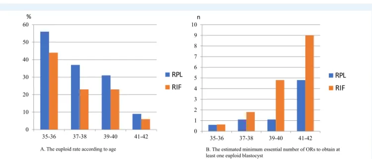
There was no significant difference of the distribution of blastocysts with euploidy, at least a single monosomy, trisomy or double trisomy between the RPL and RIF groups (Fig. 1). The frequency of double trisomy tended to be higher in the RPL group (adjusted residuals; 1.6).
The euploidy rate decreased from 56 to 37, 31 and 9% in patients with RPL and from 44 to 23, 23 and 6% in patients with RIF according to age (Fig. 2a). The estimated minimum essential number of ORs to obtain at least one euploid blastocyst was calculated to be 0.6, 1.1, 1.1 and 4.8 in patients with RPL, while it was 0.63, 1.8, 4.8 and 9.0 in patients with RIF, in groups aged 35–36, 37–38, 39–40 and 41–42 years, respectively (Fig. 2b).
Only trisomy was obtained in previous POC (Supplementary Figure SIb); however, a 1:1 ratio of trisomy to monosomy was ascertained in blastocysts subjected to PGT-A of both groups (Supplementary Figure SIa). The frequency of aneuploidy in POC and blastocysts increased according to the chromosome number.
Six patients had no embryos of category A, five of whom requested ET using embryos of category B embryos. Three cases resulted in live births, but in two cases, there was no pregnancy.
Discussion
We failed to show that PGT-A improves the live birth rate per patient or reduces the rate of clinical miscarriage significantly in both groups. The efficacy of PGT-A, at least in RPL patients, was expected because the present study focused only on patients whose POC were ascertained to be aneuploid. Mosaicism might be speculated to be one of the reasons why PGT-A showed limited efficacy. Recently, concordance between TE and the ICM was established in 62.1% of embryos analysed by PGT-A (Popovicet al., 2019). The reliability of the TE biopsy compared to the ICM biopsy in blastocysts is extremely high, but that of the cleavage-stage biopsy compared with the ICM biopsy is less so. The rate of false positive results between TE and ICM has
Downloaded from https://academic.oup.com/humrep/article-abstract/34/12/2340/5663604 by Fujita Health University user on 28 May 2020
Table I Baseline demographics of PGT-A and non-PGT-A patients with recurrent pregnancy loss.
PGT-A Non-PGT-A p-value
...
Number of enrolled patients 41 38
Mean age (SD, range) 39.2 (2.05, 35–42) 39.3 (2.07, 35–42) 0.71
Mean BMI (SD) 21.1 (2.86) 21.7 (2.45) 0.36
Mean (SD) number of previous miscarriages 2.56 (0.78) 2.47 (0.92) 0.65
Mean (SD) number of previous pregnancies with the use of IVF-ET
2.00 (0.87) 1.53 (0.92) 0.021
Mean (SD) number of previous live births 0 0
Smokers (n) 1 2
Bold indicates statistical significance.
Table II Comparison of clinical outcomes between PGT-A and non-PGT-A patients with recurrent pregnancy loss.
PGT-A (n= 41)a Non-PGT-A
(n= 38)b
Adjusted ORs (95% CI)∗, p-value
...
Number of patients with at least one good quality blastocyst
21 38
Diagnosed blastocysts/total number of blastocysts
161/174 (92.5%) -
Euploid blastocysts/diagnosed blastocysts 47/161 (29.2%) -
Embryo transfers/patients 21/41 (51.2%) 37/38 (97.3%) 0.03 (0.003–0.23), 0.001
Biochemical pregnancies/embryo transfers 16/21 (76.2%) 20/37 (54.1%) 2.45 (0.71–8.44), 0.16
Biochemical pregnancy losses/biochemical pregnancies
2/16 (12.5%) 9/20 (45.0%) 0.14 (0.02–0.85), 0.03
Clinical pregnancies/embryo transfers 14/21 (66.7%) 11/37 (29.7%) 5.14 (1.52–17.3), 0.008
Miscarriages/clinical pregnancies 2/14 (14.3%) 2/10 (20.0%) 0.68 (0.06–6.51), 0.68
47,XX,+12[13]/46,XX[7]
46,XX (21wIUFD)
47,XX,+20 47,X?,+18
Ectopic pregnancies/clinical pregnancies 1/14 (7.1%) 1/11 (9.1%) 5.67 (0.03–1014.5), 0.51
Live births/embryo transfers 11/21 (52.4%) 8/37 (21.6%) 3.89 (1.16–13.1), 0.028
Live births/patients 11/41 (26.8%) 8/38 (21.1%) 1.33 (0.45–3.91), 0.60
a,bBoth groups were followed up until the second oocyte retrieval and the first embryo transfer.
∗Adjusted for the number of previous pregnancies with the use of IVF-ET Bold indicates statistical significance.
been reported as 7.5% (Lawrenzet al., 2019). Damage by biopsy might influence the outcome although only cleavage-stage biopsy and not TE biopsy has been reported to reduce the live birth rate (Franasiaket al., 2014).
Over 70% of embryos are reported to be at least partially aneuploid by Day 3 because of prevalent errors of both meiotic and mitotic origins. On the other hand, aneuploidy observed in miscarried POC has been thought to be due to division errors of meiotic origin (Nagaoka et al., 2013). Variation of thePLK4gene was found to be associated with mitotic errors in human embryos, and infertile women with the high-risk genotype contribute fewer blastocysts for testing at Day 5, suggesting that their embryos were less likely to survive to blastocyst formation (McCoyet al., 2015). Blastocysts with monosomy can not survive after implantation (Franasiaket al., 2014). The present study
.. .. .. .. .. .. .. .. .. .. .. .. .. .. ..
indicated that natural selection or chromosome correcting pathways might be superior to PGT-A in the current situation.
Indeed, PGT-A has several ethical problems related to its use; false positives, due to mosaicism, and technical aspects of the process can lead to the abandonment of large numbers embryos that have the potential for live births (Rosenwakset al., 2018). In the present study, we had 3 healthy babies from 5 transferred mosaic embryos and, in the first clinical trials, 100 such babies were reported (Grecoet al., 2015;
Rosenwakset al., 2018).
PGT-A improved the live birth rate per ET both in the RPL and RIF groups. PGT-A has an advantage reducing the number of ETs (Forman et al., 2014). PGT-A also lowered the rate of biochemical pregnancy loss. This suggests that the cause of biochemical pregnancy loss might be speculated to be due to chromosome abnormality. Indeed, the
Downloaded from https://academic.oup.com/humrep/article-abstract/34/12/2340/5663604 by Fujita Health University user on 28 May 2020
Table III Baseline demographics of PGT-A and non-PGT-A patients with recurrent implantation failure.
PGT-A Non-PGT-A P-value
...
Number of enrolled patients 42 50
Mean age (SD, range) 38.6 (2.06, 35–42) 38.7 (2.15, 35–42) 0.78
Mean BMI (SD) 21.6 (2.68) 21.7 (3.07) 0.88
Mean (SD) number of previous embryo transfers 5.00 (2.30) 4.34 (1.72) 0.119
Mean (SD) number of previous pregnancies with the use of IVF-ET
0 0
Mean (SD) number of previous live births 0 0
Smokers (n) 2 0
Mean (SD) months of infertility 62.0 (39.1) 62.7 (47.5) 0.94
Cause of infertility % (n)
Male 28.6% (12) 30.0% (15) 0.713
Female 35.7% (15) 28.0% (14)
Unexplained 35.7% (15) 42.0% (21)
Table IV Comparison of clinical outcomes between PGT-A and non-PGT-A patients with recurrent implantation failure.
PGT-A (n = 42)a Non-PGT-A (n = 50)b Adjusted ORs (95% CI)∗, p-value
...
Number of patients with at least one good quality blastocyst
24 42
Diagnosed blastocysts/total number of blastocysts
199/208 (95.7%) -
Euploid blastocysts/diagnosed blastocysts 42/199 (21.1%) -
Embryo transfers/patients 24/42 (57.1%) 41/50 (82.0%) 0.29 (0.11–0.75), 0.01
Biochemical pregnancies/embryo transfers 19/24 (79.2%) 22/41 (53.7%) 3.28 (1.03–10.5), 0.05
Biochemical pregnancy losses/biochemical pregnancies
2/19 (10.5%) 9/22 (40.9%) 0.17 (0.03–0.92), 0.04
Clinical pregnancies/embryo transfers 17/24 (70.8%) 13/41 (31.7%) 5.62 (1.82–17.3) 0.003
Miscarriages/clinical pregnancies 2/17 (11.8%) 0/13 (0%) -, 0.999
46,XY not tested
Ectopic pregnancies/clinical pregnancies 0/17 (0%) 0/13 (0%) -
Live births/embryo transfers 15/24 (62.5%) 13/41 (31.7%) 3.75 (1.28–10.95) 0.016
Live births/patients 15/42 (35.7%) 13/50 (26.0%) 1.69 (0.68–4.20) 0.26
a,bBoth groups were followed up until the second oocyte retrieval and the first embryo transfer.
∗Adjusted for the number of previous pregnancies with the use of IVF-ET Bold indicates statistical significance.
rate of chromosome abnormality decreases according to the develop- mental stage: 70–80% of clinical miscarriages (Ogasawaraet al., 2000;
Azmanovet al., 2007), 4% of stillbirths and 0.3% of newborn babies (Nagaokaet al., 2013). Furthermore, trisomies and monosomies are equally prevalent in blastocysts; however, monosomies disappear after implantation (Franasiaket al., 2014). This evidence suggests that chro- mosome abnormality might be more frequent and of greater severity in biochemical pregnancy losses in this earlier stage of development compared with clinical miscarriages.
.. .. .. .. .. .. .. .. .. .
There was no difference in the distribution of abnormalities, although the frequency of double trisomy tended to be higher in patients with RPL than in those with RIF (Fig. 1). The euploidy rate was much lower in both RPL (29.2%) and RIF (21.1%) patients than in 15 169 blastocysts of a previous study (38-year-old women, 52.1%, 39-year-old women, 47.1%) (Franasiaket al., 2014). Thus, RPL caused by aneuploidy and RIF may be associated with a meiosis-specific genes. On the other hand, endometrial receptivity for euploid embryo implantation might be sufficiently high in both RPL and RIF patients since the clinical pregnancy
Downloaded from https://academic.oup.com/humrep/article-abstract/34/12/2340/5663604 by Fujita Health University user on 28 May 2020
Figure 1 The distribution of blastocysts with euploidy, at least one monosomy, trisomy or double trisomy after PGT-A in patients in the recurrent pregnancy loss (RPL) and recurrent implantation failure (RIF) groups.There was no significant difference in the distribution of blastocyst with euploidy, at least a single monosomy, trisomy or double trisomy between the RPL and RIF groups. The frequency of double trisomy tended to be higher in RPL (adjusted residuals: 1.6). If a blastocyst contained both monosomy and trisomy, it was classified as ‘at least one monosomy’.
Figure 2 The euploidy rate and estimated minimum essential number of oocyte retrievals required to obtain at least one euploid blastocyst according to the woman’s age.(A) The euploid rate decreased according to age from 56 to 37, 31 and 9% in patients with RPL and from 44 to 23, 23 and 6% in patients with RIF. (B) The estimated minimum essential number of oocyte retrievals required to obtain at least one euploid blastocyst in groups aged 35–36, 37–38, 39–40 and 41–42 years, respectively, was calculated to be 0.6, 1.1, 1.1 and 4.8 in patients with RPL and 0.6, 1.8, 4.8 and 9.0 in patients with RIF.
rate per embryo transfer after PGT-A was excellent (66.7 and 70.8%) compared with a previous study of women of advanced maternal age (38–41 years, 54.4%) (Rubioet al., 2017). The cause of RPL with aneu- ploidy might involve ‘superfertility’ due to high endometrial receptivity for aneuploidy embryo implantation (Teklenburget al., 2010).
.. .. .. .. ..
Monosomies disappeared in POC although both trisomies and monosomies were ascertained in blastocysts (Supplementary Figure SI).
The frequency of aneuploidy increased according to the chromosome number. Embryos with both trisomy and monosomy in larger chromosomes might have difficulty in developing.
Downloaded from https://academic.oup.com/humrep/article-abstract/34/12/2340/5663604 by Fujita Health University user on 28 May 2020
Regarding ethical considerations, there are no laws in Japan related to reproductive technology. The JSOG made a ruling on PGT in 1998 such that each case must be submitted to the JSOG and that the facilities can only initiate PGT after obtaining JSOG permission. PGT-A has been prohibited because the JSOG considered that the feelings of handicapped people might be resistant to PGT. In a total of 622 cases, PGT for monogenic/single gene defects was used for several extremely severe genetic disorders, and PGT for chromosomal structural rear- rangements for RPL caused by a translocation was permitted from 2006 to December 2018. The maternal age has increased year by year and older women with RPL or infertility desire PGT-A in spite of a lack of evidence. Thus, the JSOG decided to conduct the present pilot study and ordered the present facilities to carry it out before conducting a RCT to examine the effect of PGT-A on the live birth rate while also considering ethical issues in a committee open to patients and media.
The usefulness of PGT-A has been made public by its proponents not only in the USA but also in Japan (Rosenwakset al., 2018). Patients and physicians might have the misperception that PGT-A has advantages for all patients in spite of the lack of evidence. It also should be noted that physicians who administer the clinics or laboratories have a potential conflict of interest.
There were only two miscarriages (20.0%, 2/10) in the control group of the present RPL protocol. This was relatively low when compared with the 41.9% reported previously (in patients with a previous average of 2.9 miscarriages and who were 40 and older) (Sugiura-Ogasawara et al., 2009). The reason might be speculated to be that the miscarriage rate in patients with RPL due to embryonic aneuploidy was lower than in those with a normal embryonic karyotype (38.3 vs 62.0%, mean age 32) (Ogasawaraet al., 2000). There has been no data on the miscarriage rate in patients with RPL whose POC were aneuploid and who have received IVF-ET. There was no bias between the PGT- A group and non-PGT-A group because of the same inclusion and exclusion criteria.
One limitation of this study is that the sample size was too small to find any significant advantage in improving the live birth rate and reducing the clinical miscarriage rate because this work was conducted as a pilot study to calculate the sample size for subsequent RCT. The JSOG decided not to continue this pilot study and not to conduct an RCT because of the difficulty involved. It did not change the ruling that PGT-A is prohibited. Thus, it was impossible for us to increase the sample size.
In the present study, patients received only one ET following one or two oocyte retrievals. It was found that 4.8 and 9.0 oocyte retrievals are necessary to obtain one euploid blastocyst in 41–42-year-old patients with RPL and RIF, respectively. Further study with a larger number of patients and determination of the cumulative live birth rate is necessary to confirm the present findings.
Supplementary data
Supplementary dataare available atHuman Reproductiononline.
Authors’ Roles
MSO designed the present study and analysed the data, and TS wrote the first draft of the manuscript. TS, TKu, NA, KK, RK, AF and TU were
.. .. .. .. .. .. .. .. .. .. .. .. .. .. .. .. .. .. .. .. .. .. .. .. .. .. .. .. .. .. .. .. .. .. .. .. .. .. .. .. .. .. .. .. .. .. .. .. .. .. .. .. .. .. .. .. .. .. .. .. .. ..
responsible for IVF-ET, biopsy and clinical data acquisition, and TS, FO, TY, TKa and HK were responsible for the diagnosis of blastocysts. HS managed the data centre, and AK, TT and MI supervised the study.
All authors interpreted the data, contributed to the writing of the manuscript and revised it critically for important intellectual content.
Funding
Japan Society of Obstetrics and Gynecology and the Japanese Ministry of Education, Science and Technology.
Conflict of Interest
The authors declare no conflicts of interest associated with this manuscript.
References
American Society of Reproductive Medicine. Practice Committees of the American Society for Reproductive Medicine and the Society for Assisted Reproductive Technology. The use of preimplantation genetic testing for aneuploidy (PGT-A): a committee opinion.Fertil Steril2018;109:429–436.
Azmanov DN, Milachich TV, Zaharieva BM, Michailova GI, Dimitrova VG, Karagiozova ZH, Maznejkova VT, Chernev TA, Toncheva DI.
Profile of chromosomal aberrations in different gestational age spon- taneous abortions detected by comparative genomic hybridization.
Eur J Obstet Gynecol Reprod Biol2007;131:127–131.
European Society of Human Reproduction and Embryology. Early pregnancy guideline development group. Recurrent Pregnancy Loss Guideline2017;11:90–92.
Forman EJ, Hong KH, Franasiak JM, Scott RT Jr. Obstetrical and neonatal outcomes from the BEST Trial: single embryo transfer with aneuploidy screening improves outcomes after in vitro fertil- ization without compromising delivery rates. Am J Obstet Gynecol 2014;210:157.e1–157.e6.
Franasiak JM, Forman EJ, Hong KH, Werner MD, Upham KM, Treff NR, Scott RT Jr. The nature of aneuploidy with increasing age of the female partner: a review of 15,169 consecutive trophectoderm biopsies evaluated with comprehensive chromosomal screening.Fer- til Steril2014;101:656–663.
Greco E, Minasi MG, Fiorentino F. Healthy babies after intrauter- ine transfer of mosaic aneuploid blastocysts. N Engl J Med 2015;373:2089–2090.
Kang HJ, Melnick AP, Stewart JD, Xu K, Rosenwaks Z. Preimplantation genetic screening: who benefits?Fertil Steril2016;106:597–602.
Kushnir VA, Darmon SK, Albertini DF, Barad DH, Gleicher N. Effec- tiveness of in vitro fertilization with preimplantation genetic screen- ing: a reanalysis of United States assisted reproductive technology data 2011-2012.Fertil Steril2016;106:75–79.
Kuwayama M, Vajta G, Kato O, Leibo SP. Highly efficient vitrification method for cryopreservation of human oocytes. Reprod Biomed Online2005;11:300–308.
Lawrenz B, El Khatib I, Liñán A, Bayram A, Arnanz A, Chopra R, De Munck N, Fatemi HM. The clinicians´ dilemma with mosaicism-an insight from inner cell mass biopsies. Hum Reprod 2019;34:998–1010.
Downloaded from https://academic.oup.com/humrep/article-abstract/34/12/2340/5663604 by Fujita Health University user on 28 May 2020
Lu S, Zong C, Fan W, Yang M, Li J, Chapman AR, Zhu P, Hu X, Xu L, Yan L et al. Probing meiotic recombination and aneu- ploidy of single sperm cells by whole-genome sequencing.Science 2012;338:1627–1630.
Mastenbroek S, Twisk M, van der F, Repping S. Preimplantation genetic screening: a systematic review and meta-analysis of RCTs. Hum Reprod Update2011;17:454–466.
McCoy RC, Demko Z, Ryan A, Banjevic M, Hill M, Sigurjonsson S, Rabinowitz M, Fraser HB, Petrov DA. Tripolar chromosome segre- gation drives the association between maternal genotype at variants spanning PLK4 and aneuploidy in human preimplantation embryos.
Science2015;348:235–238.
Munné S, Chen S, Fischer J, Colls P, Zheng X, Stevens J, Escudero T, Oter M, Schoolcraft B, Simpson JLet al.Preimplantation genetic diag- nosis reduces pregnancy loss in women aged 35 years and older with a history of recurrent miscarriages.Fertil Steril2005;84:331–335.
Murphy LA, Seidler EA, Vaughan DA, Resetkova N, Penzias AS, Toth TL, Thornton KL, Sakkas D. To test or not to test? A framework for counselling patients on preimplantation genetic testing for aneu- ploidy (PGT-A).Hum Reprod2019;34:268–275.
Nagaoka SI, Hassold TJ, Hunt PA. Human aneuploidy: mecha- nisms and new insights into an age-old problem. Nat Rev Genet 2013;13:493–504.
Ogasawara M, Aoki K, Okada S, Suzumori K. Embryonic karyotype of abortuses in relation to the number of previous miscarriages.Fertil Steril2000;73:300–304.
Popescu F, Jaslow CR, Kutteh WH. Recurrent pregnancy loss eval- uation combined with 24-chromosome microarray of miscarriage tissue provides a probable or definite cause of pregnancy loss in over 90% of patients.Hum Reprod2018;33:579–587.
Popovic M, Dhaenens L, Taelman J, Dheedene A, Bialecka M, De Sutter P, Chuva de Sousa Lopes SM, Menten B, Heindryckx B. Extended in vitro culture of human embryos demonstrates the complex nature of diagnosing chromosomal mosaicism from a single trophectoderm biopsy.Hum Reprod2019;34:758–769.
Rosenwaks Z, Handyside AH, Fiorentino F, Gleicher N, Paulson RJ, Schattman GL, Scott RT Jr, Summers MC, Treff NR, Xu K.
The pros and cons of preimplantation genetic testing for aneu- ploidy: clinical and laboratory perspectives.Fertil Steril 2018;110:
353–361.
Rubio C, Bellver J, Rodrigo L, Castillón G, Guillén A, Vidal C, Giles J, Ferrando M, Cabanillas S, Remohí J et al. In vitro fertil-
.. .. .. .. .. .. .. .. .. .. .. .. .. .. .. .. .. .. .. .. .. .. .. .. .. .. .. .. .. .. .. .. .. .. .. .. .. .. .. .. .. .. .. .. .. .
ization with preimplantation genetic diagnosis for aneuploidies in advanced maternal age: a randomized, controlled study.Fertil Steril 2017;107:1122–1129.
Sawada Y, Sato T, Saito C, Ozawa F, Ozaki Y, Sugiura-Ogasawara M.
Clinical utility of decorin in follicular fluid as a biomarker of oocyte potential.Reprod Biol2018;18:33–39.
Scott RT Jr, Upham KM, Forman EJ, Zhao T, Treff NR. Cleavage-stage biopsy significantly impairs human embryonic implantation potential while blastocyst biopsy does not: a randomized and paired clinical trial.Fertil Steril2013a;100:624–630.
Scott RT Jr, Upham KM, Forman EJ, Hong KH, Scott KL, Taylor D, Tao X, Treff NR. Blastocyst biopsy with comprehensive chromosome screening and fresh embryo transfer significantly increases in vitro fertilization implantation and delivery rates: a randomized controlled trial.Fertil Steril2013b;100:697–703.
Sugiura-Ogasawara M, Ozaki Y, Sato T, Suzumori N, Suzumori K. Poor prognosis of recurrent aborters with either maternal or paternal reciprocal translocations.Fertil Steril2004;81:367–373.
Sugiura-Ogasawara M, Ozaki Y, Kitaori T, Suzumori N, Obayashi S, Suzuki S. Live birth rate according to maternal age and pre- vious number of recurrent miscarriages. Am J Reprod Immunol 2009;62:314–319.
Sugiura-Ogasawara M, Ozaki Y, Kitaori T, Kumagai K, Suzuki S. Midline uterine defect size is correlated with miscarriage of euploid embryos in recurrent cases.Fertil Steril2010;93:1983–1988.
Sugiura-Ogasawara M, Ozaki Y, Katano K, Suzumori N, Kitaori T, Mizutani E. Abnormal embryonic karyotype is the most fre- quent cause of recurrent miscarriage. Hum Reprod 2012;27:
2297–2303.
Teklenburg G, Salker M, Heijnen C, Macklon NS, Brosens JJ. The molec- ular basis of recurrent pregnancy loss: impaired natural embryo selection.Mol Hum Reoprod2010;16:886–895.
Verpoest W, Staessen C, Bossuyt PM, Goossens V, Altarescu G, Bonduelle M, Devesa M, Eldar-Geva T, Gianaroli L, Griesinger G et al.Preimplantation genetic testing for aneuploidy by microarray analysis of polar bodies in advanced maternal age: a randomized clinical trial.Hum Reprod2018;33:1767–1776.
Yang Z, Liu J, Collins GS, Salem SA, Liu X, Lyle SS, Peck AC, Sills ES, Salem RD. Selection of single blastocysts for fresh transfer via standard morphology assessment alone and with array CGH for good prognosis IVF patients: results from a randomized pilot study.
Mol Cytogenet2012;5:24.
Downloaded from https://academic.oup.com/humrep/article-abstract/34/12/2340/5663604 by Fujita Health University user on 28 May 2020
A R T I C L E
Obstetric complication-associated ANXA5 promoter polymorphisms may affect gene expression via DNA secondary structures
Hidehito Inagaki1●Sayuri Ota1●Haruki Nishizawa2●Hironori Miyamura2●Kumiko Nakahira3●Machiko Suzuki1● Sachie Nishiyama2●Takema Kato1●Itaru Yanagihara3●Hiroki Kurahashi1
Received: 25 October 2018 / Revised: 1 February 2019 / Accepted: 4 February 2019 / Published online: 22 February 2019
© The Author(s) under exclusive licence to The Japan Society of Human Genetics 2019
Abstract
Recent findings have highlighted the possibility that polymorphisms within the annexin A5 gene (ANXA5) promoter contribute to the etiology of various obstetric complications. However, the underlying mechanisms are unknown. The M2 haplotype of theANXA5shows lower activity and less expression ofANXA5mRNA. This gene promoter region has a motif that potentially forms a G-quadruplex structure. In vitro G-quadruplex propensity estimated by circular dichroism indicated that the M2 haplotype oligonucleotide manifested a decreased potential for G-quadruplex formation. In addition, in vivo G- quadruplex formation of the promoter region was evidenced by the presence of single-stranded DNA shown by sodium bisulfite treatment of placental genomic DNA. Comparative analysis indicated less potential in the M2 allele than the major allele. Promoter activity of the two haplotypes determined by luciferase reporter analysis correlated with the estimated G- quadruplex propensity. Our data lend support to the developing paradigm that genomic variation affects gene expression levels via DNA secondary structures leading to the disease susceptibility.
Introduction
Many common human diseases are believed to be polygenic disorders associated with several genetic and environmental factors [1]. Based on the “common disease-common var- iant”hypothesis, genome-wide association studies (GWAS) using common single nucleotide polymorphisms (SNPs) have identified hundreds of genetic variants that are statis- tically associated with different target diseases. However, most of the polymorphisms that have been identified by
GWAS to date are not deleterious variants and confer relatively small increases in disease risk. In addition, the functional impact of the majority of these SNPs on gene expression has not yet been validated since these sequence variations are mostly located at non-coding or intergenic regions [2]. The processes by which these SNPs confer a higher risk of disease thus remain an enigma.
Previously published results suggest that polymorphisms within the annexin A5 gene (ANXA5) are associated with common obstetric complications, such as recurrent preg- nancy loss (RPL), pre-eclampsia, and pregnancy-related thrombophilic disorder [3–8]. The ANXA5 gene upstream region contains four common variations, i.e., SNP1 (g.
−467G>A, rs112782763) and SNP2 (g.−448A>C, rs28717001) in the untranscribed promoter region, and SNP3 (g.−422T>C, rs28651243) and SNP4 (g.−373G>A, rs113588187) near and downstream of the transcription start points, respectively. These four SNPs manifest strong linkage disequilibrium, generating two major haplotypes:
the N haplotype with all major alleles and an M2 haplotype with all minor alleles. The frequency of the M2 allele in the general Japanese population was reported to be 5.4%, lower than that in the western countries (~16%) [6].
The M2 haplotype has been associated with various disorders. Annexin A5 is known as a placental
* Hiroki Kurahashi [email protected]
1 Division of Molecular Genetics, Institute for Comprehensive Medical Science, Fujita Health University, Toyoake, Japan
2 Department of Obstetrics and Gynecology, Fujita Health University School of Medicine, Toyoake, Aichi 470-1192, Japan
3 Department of Developmental Medicine, Osaka Medical Center and Research Institute for Maternal and Child Health,
Izumi, Osaka 594-1101, Japan
Supplementary informationThe online version of this article (https://
doi.org/10.1038/s10038-019-0578-4) contains supplementary material, which is available to authorized users.
1234567890();,: 1234567890();,:
anticoagulation factor that shields the apical surface of the syncytiotrophoblasts covering the placental villi [9]. Hence, the low expression of the ANXA5 gene might reasonably account for a higher susceptibility to obstetric complica- tions. Indeed, lower expression of theANXA5gene from the M2 allele has been reported [10,11]. However, it remains unclear how these polymorphisms affect theANXA5 gene expression levels and thereby lead to disease. For example, there was no association found between the M2 haplotype and RPL risk in a previous northern European study [12]. It is therefore critically important to elucidate the biological impact of the differentANXA5haplotypes.
With regard to the effects of gene polymorphisms on gene expression, the contribution of DNA secondary structures has been highlighted previously [13, 14]. A considerable body of evidence now indicates that certain SNPs located within the transcribed region of a gene can affect mRNA stability or translation efficiency via the propensity for stem-loop formation. Guanine-rich DNA can fold into a non-canonical DNA structure known as a G- quadruplex [15]. This structure comprises intrastrand interactions of G-tetrads paired by Hoogsteen bonds. G- quadruplexes are often identified in and around the untranslated region of genes and are potentially associated with gene regulation [16,17]. The association between the G-quadruplex structure and transcriptional regulation has been extensively characterized for oncogenes such asMYC, KIT, orKRAS [18–20]. Recently, it has also been reported that some polymorphisms disrupt the formation of G- quadruplex structures, leading to alterations in the expres- sion of nearby genes [21]. In our present study, we exam- ined the ANXA5 gene promoter variants in terms of the association between the potential for G-quadruplex forma- tion and theANXA5gene expression levels.
Methods
Samples and ethical approval
Four placental samples were obtained from women with an uncomplicated pregnancy at the Department of Obstetrics and Gynecology, Fujita Health University Hospital.
Informed consent was obtained from each patient and the study protocol was approved by the Ethical Review Board for Human Genome Studies at the Fujita Health University.
The genotypes of the SNPs at theANXA5promoter region were determined by sequencing as previously described [6].
All methods were performed in accordance with the rele- vant guidelines and regulations including a biosafety reg- ulation in Japan, the Act on the Conservation and Sustainable Use of Biological Diversity through Regula- tions on the Use of Living Modified Organisms.
Circular dichroism
Circular dichroism (CD) experiments were performed using a J-720 spectropolarimeter (JASCO). Oligonucleotides were synthesized and diluted to 50 ng/μl with a buffer containing 100 mM potassium chloride and 10 mM Tris-HCl (pH 7.4).
Where indicated, the potassium chloride concentration was decreased, and lithium chloride was added to adjust the salt concentration to 100 mM. The samples were next heat- denatured at 95 °C for 5 min and then cooled slowly for 6 h to 25 °C. Scans were performed at 25 °C using a 1 cm cuvette over a 200–360 nm range. CD spectra were recor- ded from the average offive scans at 50 nm/min, with a 2 s response time, 1 nm bandwidth, and 0.1 nm resolution. The molar ellipticity was then plotted. The 56 oligonucleotides used in this CD analysis are described in Fig.1c.
Methylation analysis
Bisulfite sequencing was performed to determine the methy- lation status of theANXA5promoter region. One N/N homo- zygous and three N/M2 heterozygous samples were analyzed.
Bisulfite conversion was performed using an EpiTect Bisulfite Kit (Qiagen, Tokyo, Japan) in accordance with the manu- facturer’s protocol. Bisulfite-treated DNA was amplified using an uracil stalling-free polymerase, EpiTaq HS (Takara bio, Kusatsu, Japan) and the primers: 5′-GGTTATA- GAGGGTAGGGAGGTTTAA-3′ and 5′-CACCCAAACTA- TAAAACCCAAATAC-3′. The ~300 bp resulting products were then cloned into the pT7Blue T-vector (Merck, Darm- stadt, Germany). Colonies were subsequently selected, and the plasmids were isolated for sequencing.
Bisul fi te sequencing for detection of DNA secondary structure
Bisulfite treatment was applied to genomic DNAs of N/M2 heterozygous samples purifying under mild conditions.
Briefly, placental tissues were powdered under liquid nitrogen and treated with proteinase K at 37 °C. Genomic DNAs were then column-purified using DNeasy Blood & Tissue Kit (Qiagen, Tokyo, Japan) in accordance with the manu- facturer’s instructions. Approximately 500 ng of DNA was used for sodium bisulfite treatment using an Epitect Bisulfite Kit (Qiagen) in general accordance with the manufacturer’s protocol except that the conditions for the sodium bisulfite reaction involved a constant temperature at 37 °C for 16 h.
The resulting converted DNAs were used as PCR templates.
Primers were designed for the regions containing few T nucleotides to normalize the annealing efficiency between the converted and unconverted DNAs. The 5′end of each primer was designed for use with the Nextera system (Illumina). The primers are listed in Supplementary Table S1.
PCR amplifications were carried out using EpiTaq HS (Takara bio, Kusatsu, Japan). The reaction conditions were 94 °C for 2 min, followed by 35 cycles of 98 °C for 10 s, 55 °C for 30 s, and 72 °C for 1 min. There was a final incubation step at 72 °C for 10 min. The resulting products were ligated into pBluescriptII (Agilent, Tokyo, Japan) and plasmids were grown in E. coli, purified and sequenced.
These sequences were designated as the upper or lower strand in accordance with the conversion of the nucleotides (C>T or G>A) and as N or M2 alleles according to the haplotype. The PCR products were also analyzed by massive parallel sequencing using Nextera index primers (Illumina) following a second round of PCR. After purification and quantification, the products were applied to a Miseq
sequencer (Illumina) at a 250 bp single-end read. The sequence data were quality filtered using FASTX-Toolkit 0.0.14 (http://hannonlab.cshl.edu/fastx_toolkit/) trimming with a cut-off of 20 (phred score) and a minimum length of 12 andfiltering with a cut-off of 20 and minimum percentage of 80. The resulting data was further processed byfiltering- out PCR duplicates and sequences with indels. After con- version of the fastq to fasta format, clustering analysis of the sequences was carried out using MAFFT [22].
qRT-PCR
Quantitative real-time RT-PCR (qRT-PCR) analysis was performed using the TaqMan System. The Superscript First- Fig. 1 G-quadruplex structure formation at the ANXA5 promoter
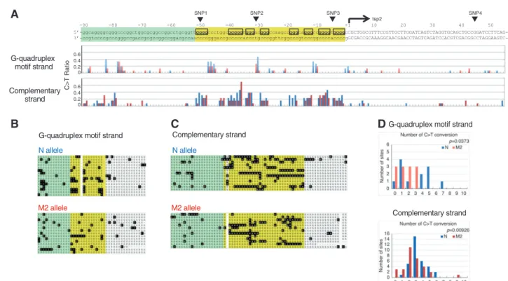
region in vitro.aA square planar structure of a G-quartet (left), pos- sible form of an intramolecular G-quadruplex structure (middle), and possible G-quadruplex formation at a gene promoter. b Genomic structure of the ANXA5 gene promoter region in terms of its G- quadruplex structure. Eight runs of three or four guanines are indicated by the boxed regions. Transcription start points (tsp) are indicated by arrows. The nucleotide at tsp2 is numbered+1, and the nucleotides within the transcript are capitalized. Polymorphisms are indicated by arrowheads. c Conservation of the ANXA5 gene promoter region among mammalian species. Dots indicate conserved nucleotides, and
conserved guanine runs are underlined. Dashes indicate the absence of nucleotides. dOligonucleotides used for CD spectra analysis.eThe propensity for G-quadruplex structure formation at the ANXA5gene promoter region in vitro evaluated by CD spectra. Each polymorphic allele was analyzed. The vertical bars indicate the levels of ellipticity estimated by CD spectra. The ellipticity was measured four times and the curves indicate the mean data values. f Propensity for G- quadruplex structure formation at various concentrations of potas- sium chloride evaluated by CD spectra. The ellipticity was measured at 260 nm, and the error bars represent the S.D. (n=3)
strand Synthesis System for RT-PCR (Invitrogen) with random primers was used to produce single-stranded cDNA templates. TaqMan probes and primers for theANXA5gene (Hs00134054_m1) were obtained commercially (Applied Biosystems). A housekeeping gene, 18S RNA (Hs99999901_s1), was used to normalize the mRNA levels because the expression levels among the samples were stable. All qRT-PCR reactions were performed in triplicate in afinal volume of 25 µl. The cycling conditions used were 2 min at 50 °C, 30 min at 60 °C, and 1 min at 95 °C for RT, followed by 40 cycles of 15 s at 95 °C and 1 min at 60 °C for PCR amplification.
Allele-specific qRT-PCR was carried out as previously described [11]. The transcript from N allele was amplified with the primers 5′-CAGTCTAGGTGCAGCTGCCG-3′ and 5′-GGTGAAGCAGGACCAGACTGT-3′, and that from the M2 allele was amplified with 5′-CAGTC- TAGGTGCAGCTGCCA-3′ and 5′-GGTGAAGCAG- GACCAGACTGT-3′. The product levels were quantified using SYBR Premix Ex Taq II (Takara BIO) and the 7300 real-time PCR system (Applied Biosystems). TheTBPgene was used as an internal control.
Promoter assay
Luciferase reporter constructs were kindly provided by Dr.
Arseni Markoff (University of Muenster, Germany), and the assay was performed as previously described [3]. Briefly, the promoter region and exon 1 encompassing SNP1 to 4 was amplified by PCR and the ~450 bp products were cloned immediately upstream of luciferase initiation codon in the pGL3-Basic Vector (Promega) using the MluI and XhoI sites. Each polymorphic nucleotide change was introduced to the vector by means of site-directed muta- genesis. The resulting constructs were co-transfected with pRL-TK into the HeLa cell line using Lipofectamine 2000 (Invitrogen). Luciferase activity was measured at 48 h after transfection using a Dual-Luciferase Reporter Assay Sys- tem (Promega).
Statistical analysis
Statistical significance was determined using the Studentt- test and one-way analysis of variance (ANOVA).P-values of <0.05 were considered statistically significant. Data are reported as the mean ± SD for each group.
Results
We identified eight runs of three or four guanines with 1–7 nucleotide intervals upstream of theANXA5gene that cor- responded to a consensus sequence of a potential
G-quadruplex forming motif, G3+-N1–7-G3+-N1–7-G3+-N1–7- G3+ (Fig.1a, b) [15]. TheANXA5gene has multiple tran- scription start points (tsp) [23], and although transcripts from tsp1 include 5 runs of guanines at the 5′ region, all eight runs of guanines are located 5′ upstream of the non- transcribed region in cases of transcription starting from tsp2 and tsp3. Thefirst run of guanines is unique to humans, but all of other seven runs are highly conserved among primates. Other mammalian species also carry at least six runs of guanines, suggesting that this G-rich region plays an important role in gene regulation (Fig. 1c).
To evaluate the G-quadruplex structure forming pro- pensity of this region in vitro, we performed a CD spec- troscopy experiment using synthesized oligonucleotides that included the consensus sequence of the potential G-quadruplex forming motif. Typically, parallel form G-quadruplexes display a characteristic positive peak at 260 nm and a negative peak at 240 nm, whereas the anti- parallel form of these structures displays a positive peak at 295 nm and a negative peak at 265 nm on the CD spectra [24]. The oligonucleotide with the N allele sequence pro- duced a positive peak at 260 nm and a trough at 240 nm with a small additional positive peak at 290 nm. This indi- cates that the N allele DNA forms a G-quadruplex structure in vitro, adopting a mixture of parallel and anti-parallel forms. When SNP1, SNP2, and SNP3 were separately introduced into the N allele, no remarkable change was observed in the CD spectra. However, when these variations were combined to form the M2 allele, the CD spectra showed a reduction in the positive peak at 260 nm, sug- gesting that the potential for G-quadruplex formation had been decreased (t-test,P< 0.01) (Fig.1d, e). To evaluate the propensity for G-quadruplex structure formation in vitro, we analyzed the CD spectra for the oligonucleotides under various potassium ion concentrations (Fig. 1e). The M2 allele showed lower positive peaks at 260 nm in 100 mM, and especially in 20 mM, of potassium chloride (P< 0.01), again suggesting that the potential for G-quadruplex for- mation had been decreased.
It was of interest to us to determine how the M2 haplotype, which appears to possess less potential for G-quadruplex structure formation than the N haplotype, impacted on the promoter activity of theANXA5gene. The restricted methy- lation of a G-quadruplex structured DNA region was reported previously [25]. On the assumption thatANXA5polymorph- isms would affect the methylation status of the gene promoter via G-quadruplex structures, and thereby alter gene expres- sion, we performed bisulfite sequencing to locate methylated cytosines in this region. We found that the CpG islands of the ANXA5 gene upstream region were hypomethylated in pla- cental DNA. However, we did not observe any allele-specific alteration of the ANXA5 promoter methylation status (Sup- plementary Fig. S1).