新規二機能性ポリカプロラクトン : 合成、表面特性 および徐放性
張, 宇澄
https://doi.org/10.15017/1931870
出版情報:Kyushu University, 2017, 博士(工学), 課程博士 バージョン:
権利関係:
A Novel Dual-Functionalized Polycaprolactone:
Synthesis, Surface Properties, and Its Controlled Release
Yucheng Zhang
1 -Contents-
Chapter 1 Introduction ... 1
1.1. Polycaprolactone and its functionalization ... 2
1.2. Anti-fouling properties and phosphocholine modification ... 4
1.3. Surface functionalization and catechol groups ... 6
1.4. Surface reorganization and crystallization ... 9
1.5. Cardiovascular disease and drug-eluting stent ... 10
1.6. Suggestion and content ... 12
1.7. Reference ... 14
Chapter 2 Synthesis of Dual-Functionalized Polycaprolactone ... 21
2.1. Introduction ... 22
2.2. Experiment ... 23
2.2.1. Materials ... 23
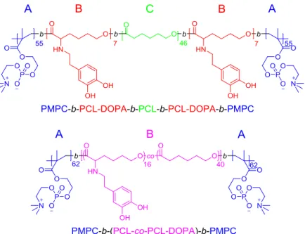
2.2.2. Synthesis of PCL-b-(PCL-DOPA-b-PMPC)2 ... 24
2.2.3. Synthesis of (PCL-co-PCL-DOPA)-b-(PMPC)2 ... 28
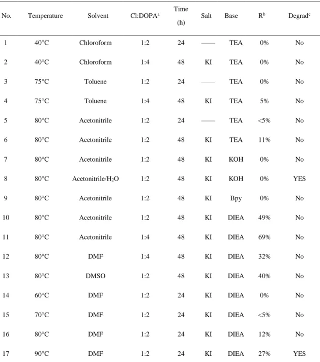
2.2.4. Exploration of dopamine modification ... 30
2.2.5. Degradation test of PCL-b-(PCL-DOPA-b-PMPC)2 ... 31
2.2.6. Characterizations ... 32
2.3. Results and Discussion ... 32
2.3.1. Synthesis of PCL-b-(PCL-Cl)2 and PCL-co-PCL-Cl ... 32
2.3.2. End-functionalization ... 35
2.3.3. Exploration of dopamine modification ... 38
2.3.4. Degradation test of PCL-b-(PCL-DOPA-b-PMPC)2 ... 41
2
2.4. Conclusion ... 42
2.5. Reference ... 43
Chapter 3 Adsorption Behavior of Dual-Functionalized Polycaprolactone... 45
3.1. Introduction ... 46
3.2. Experiment ... 47
3.2.1. Materials ... 47
3.2.2. Adsorption behavior of PCL-b-(PCL-DOPA-b-PMPC)2 on SUS ... 47
3.2.3. Durability of PCL-b-(PCL-DOPA-b-PMPC)2 film on SUS ... 48
3.2.4. Evaluation of protein adsorption ... 48
3.2.5. Characterizations ... 49
3.3. Results and Discussion ... 50
3.3.1. Adsorption behavior of PCL-b-(PCL-DOPA-b-PMPC)2 on SUS ... 50
3.3.2. Durability of PCL-b-(PCL-DOPA-b-PMPC)2 film on SUS ... 56
3.3.3. Evaluation of protein adsorption ... 57
3.4. Conclusion ... 60
3.5. Reference ... 61
Chapter 4 Crystallization-Induced Suppression of Surface Reorganization ... 63
4.1. Introduction ... 64
4.2. Experiment ... 64
4.2.1. Analysis of crystallization state ... 64
4.2.2. Preparation of polymer films ... 65
4.2.3. Evaluation of surface reorganization ... 65
4.2.4. Characterizations ... 66
3
4.3. Results and Discussion ... 66
4.3.1. Analysis of crystallization state ... 66
4.3.2. Evaluation of surface reorganization ... 68
4.3.3. Crystallization-induced suppression of surface reorganization ... 72
4.4. Conclusion ... 73
4.5. References ... 74
Chapter 5 Preparation of Drug-Loading Film and Its Release Properties ... 76
5.1. Introduction ... 77
5.2. Experiment ... 78
5.2.1. Materials ... 78
5.2.2. Preparation of PCL and PCL-b-(PCL-DOPA)2 ... 78
5.2.3. Surface properties of polymer films ... 79
5.2.4. Protein adsorption tests... 80
5.2.5. Preparation of drug-loaded polymer films ... 80
5.2.6. In vitro drug release ... 82
5.2.7. Characterizations ... 82
5.3. Results and discussion ... 83
5.3.1. Preparation of polymer films ... 83
5.3.2. Protein adsorption tests... 85
5.3.3. Preparation of drug-loaded polymer films ... 87
5.3.4. In vitro drug release ... 91
5.4. Conclusion ... 95
5.5. References ... 96
4
Chapter 6 Conclusion and Perspective ... 98
6.1. Additional ideas and application ... 99
6.2. Conclusion ... 100
6.3. References ... 102
Acknowledgement ... 104
1
Chapter 1
Introduction
2 1.1. Polycaprolactone and its functionalization
Polycaprolactone (PCL) is one of the earliest kinds of polyester which was synthesized by Carothers’ group in the early 1930s.1 The ester groups in PCL endow it with a complete degradability.2 PCL is a semi-crystalline polymer, and its crystallinity tends to decrease as the molecular weight increases. In recent years, attentions were drawn to PCL in tissue engineering, owing to their numerous advantages over other biopolymers.3 These advantages include tailorable degradation kinetics, ideal mechanical properties, ease of shaping and manufacture, and drug capacity.2 In addition, a number of drug delivery devices consisting of PCL already have FDA approval and CE Mark registration, which enables a faster avenue to market.4
Figure 1.1. End-functionalization and graft-modification for PCL.
Although PCL is already applied in different biomedical applications, it has several drawbacks due to its undesirable inherent properties including slow degradation rate, poor blood compatibility, and hydrophobicity. The modification of PCL is an appealing approach for this material to obtain particular properties. Generally, the modification of PCL contains end-functionalization and graft-modification.5 As shown in Figure 1.1, the terminal groups of PCL are hydroxyl or carboxyl group, which are determined by the polymerization methods. The end-functionalization could achieve via acylation (for hydroxyl groups) and esterification (for carboxyl groups). Two examples are shown in Figure 1.2, the macroinitiators of atom transfer radical polymerization (ATRP) and reversible addition-fragmentation chain transfer
3
polymerization (RAFT) are prepared from hydroxyl-terminated PCL via acylation and esterification, respectively. The further polymerization is able to obtain a well-designed copolymer.
The graft-modification refers to the modification of PCL main chain, which is more desirable, since this approach is able to tailor the properties, including crystallinity, hydrophilicity, biodegradation rate, adhesion, and mechanical properties.6 The common strategy of PCL graft-modification contains atom transfer radical addition (ATRA) and “click” reaction (Figure 1.3).7 Both of them need a chloride substituted PCL segment which can be prepared by the random/block copolymerization with the monomer of -chloride--caprolactone (-Cl--CL).8
Figure 1.2. Synthesis of PCL-based ATRP macroinitiator and RAFT macroinitiator.
ATRA is based on the haloalkane-methacrylate addition. The reaction is performed with the presence of copper/ligand catalyst system which is similar to ATRP catalyst system (Figure 1.3).9 This reaction can modify PCL with an acceptable conversion rate. However, the undesirable reduction of chloride units is regarded as drawbacks of this method.7 Since ATRA is based on radical mechanism, the termination of radical including cross-linking and quenching induces the reduction of chloride units and increases the polydispersity index (PDI) of the resultant polymer (Figure 1.4).9 “Click” reaction is based on copper-catalyzed 1,3-Huisgens cycloaddition between azides and alkynes. This method has several advantages, such as fast kinetics, mild condition, low
4
toxicity catalysis, and quantificational reaction.8 However, the nitrine-based polymer or material is quite unstable and dangerous, and the reduction of azides units easily occurs during the storage.
Figure 1.3. Methods of graft-modification for -substituted PCL.
Figure 1.4. Potential side reaction of ATRA.
1.2. Anti-fouling properties and phosphocholine modification
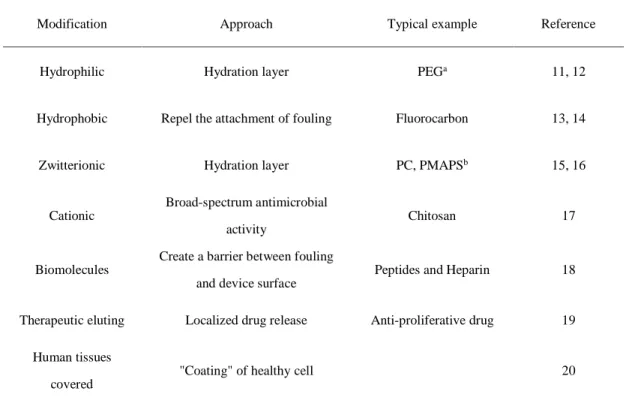
Fouling is used to describe the phenomenon that biological molecules non-specifically adsorb onto the surface of biomaterials on their surfaces, resulting in the reduced performance of such material. Hence, a lot of research has been put forward to realize an anti-fouling surface, and summarized in Table 1.1.10
The human tissue- and fluid-covered surface are considered as the best method since the implanted materials with this coating are recognized as normal tissues without any further pathological changes.20 However, the complicated procedure and long manufacturing period make
5
it difficult for the extensive application. Although hydrophobic surface shows favorable performance in vitro test, the in vivo effectiveness still remains doubtful.13,14 The therapeutic eluting surface is a temporary method for the anti-fouling property.19 After the complete drug release, the surface undergoes fouling again. The denaturation of biomolecules is the main shortage for biomolecules functionalization method.18 Although the use of chitosan which is cationic charged natural polymer exhibits good anti-fouling properties, however, other materials give even worse results.17 The hydrophilic surface benefits from the strong hydration effect which increases the energetic penalty of removing surface binding water for protein attachments.
Although zwitterionic-charged surface shares similar principle with a hydrophilic surface, a stronger hydrophilicity and water-binding effect suggested that this method is more promising.15,16
Table 1.1. Different Methods to Obtain Anti-fouling Surface
Modification Approach Typical example Reference
Hydrophilic Hydration layer PEGa 11, 12
Hydrophobic Repel the attachment of fouling Fluorocarbon 13, 14
Zwitterionic Hydration layer PC, PMAPSb 15, 16
Cationic
Broad-spectrum antimicrobial activity
Chitosan 17
Biomolecules
Create a barrier between fouling and device surface
Peptides and Heparin 18
Therapeutic eluting Localized drug release Anti-proliferative drug 19 Human tissues
covered
"Coating" of healthy cell 20
aPEG is the abbreviation of poly(ethylene oxide); bPC is phosphorylcholine and PMAPS is poly(3-[dimethyl(2′- methacryloyloxyethyl)ammonio]propanesulfonate).
During the last two decades, the excellent performance of phosphorylcholine (PC) modification is gradually recognized by researchers.21 PC presents at the end of phospholipids and
6
the exterior surfaces of cell membranes.21,22 The PC-conjugations have been widely accepted as an effective mean to improve the anti-fouling property for different polymers and surfaces.23,24 Notably, the commercialization of polymerizable PC-based monomer with high purity and polymerization activities, 2-methacryloyloxyethyl phosphorylcholine (MPC), has stimulated the expansive research of this material.24 Surface modification using PMPC brush showed incredible anti-fouling property and biocompatibility. Brash et al. reported the most significant values: less than 5 ng/cm2 for fibrinogen adsorption from the plasma and whole blood using a high density
“graft-from” PMPC-based polymer brush.25
1.3. Surface functionalization and catechol groups
With rapid medical technological advances, a lot of implant devices were introduced to different surgeries including sensory, neurological, cardiovascular, orthopedic, contraception, cosmetic, and organs.17 Since they directly contact with human tissue and body fluid, the biocompatibility of such material is extremely important.26 Although the preparation of biocompatible materials with suitable mechanical properties, shape, and size is difficult, new method focusing on the tailoring the surface properties without changing of mechanical properties has received increasing attention. This method can minimize the impact on the mechanical properties of different devices and endow the modified device with desirable surface properties.27
Figure 1.5. “Graft-from” method for surface modification.
7
The surface functionalization contains “graft-to” method (Figure 1.5) and “graft-from”
method (Figure 1.6).28 In “grafting-from” method, a molecule with initiator was firstly immobilized onto the surface. The further polymerization modifies the surface by selective functional monomers. This method is preferential to get high density and/or high thickness layer.28,29 Since this method involves in-situ polymerization, the size, and shape of the samples are restricted, the chemical inertness is necessary to avoid the undesirable changes, and the
experiment condition and solution selection should be careful to avoid the introduction of toxicity compounds.30
Figure 1.6. “Graft-to” method for surface modification.
The “grafting-to” method is the functionalization of surfaces using a well-designed polymer with anchor groups. This approach involves a simple dip-and-risen procedure, which is the cost-effective and ease of size. Moreover, the molecular weight distributions, special structure, and functionalities can be easily controlled by a well-designed synthesis strategy of the polymer with anchor groups. However, the grafting density and thickness of resultant films are lower than that obtained from “graft-from” method.29
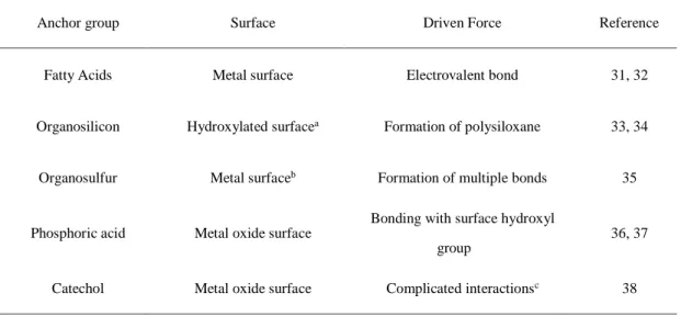
Anchor groups which immobilize the surface modifier onto the substrate, play an important role in both “graft-from” and “graft-to” methods. Table 1.2 summarized some anchor groups utilized to immobilize the molecule or polymer onto varied substrates.
8
Table 1.2. Anchor Groups in Surface Modification
Anchor group Surface Driven Force Reference
Fatty Acids Metal surface Electrovalent bond 31, 32
Organosilicon Hydroxylated surfacea Formation of polysiloxane 33, 34
Organosulfur Metal surfaceb Formation of multiple bonds 35
Phosphoric acid Metal oxide surface
Bonding with surface hydroxyl group
36, 37
Catechol Metal oxide surface Complicated interactionsc 38
aHydroxylated surface includes SiO2, Al2O3, quartz, glass, mica, etc.; bMatal surface includes Ag, Cu, Pt, Hg, Au, etc.; cCatechol groups show strong adhesion properties including hydrogen bonding, metal/metal oxide coordination, and oxidative cross-linking.
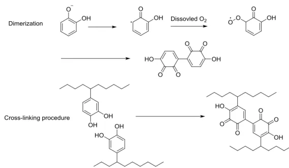
Organosilicon and organosulfur are available for silicon-based material and precious metal, respectively.33-35 Other methods are suited for the stainless steel, titanium, iron, and aluminum which are widely used in the biomedical device.31,36-37 Some comprehensive research showed phosphoric acid possesses the strongest adhesion, but the internet acidity for phosphoric acid leads to the corrosion of the metal surface.37 The fatty acids with the weakest adhesion is only applied in few fields.8 Catechol modification with non-acid, flexible preparation, and good solubility has received increasing attention.39 Catechol groups which exist in holdfast proteins of marine mussels, are the reason why such animals can adhere to the wet rock or ships.40 The most adhesive proteins isolated from mussel (Mytilus sp.) holdfasts (byssus), mfp-3 and mfp-5 are catechol-rich.40 A growing evidence implies that the catechol modification endows various natural and synthetic polymers with wet adhesion.41 Some theory has been put forward to explain the mechanism of catechol adhesion, such as catechol-mediated bidentate hydrogen bonding, metal/metal oxide coordination, or oxidative cross-linking (Figure 1.7 showed the mechanisms of crosslinking of
9
catechol groups).42 Although the mechanism of these adhesion properties still remains controversial, broadly applicable research has sufficiently supported its excellent adhesion property of this material.
Figure 1.7. Dimerization and the cross-linking between catechol groups in different segments.42 1.4. Surface reorganization and crystallization
The interfacial structure of PC-contained amphiphilic surface modifier frequently reorganizes under hydrophobic/hydrophilic conditions, and shows a great impact on its surface properties.21,22 PMPC-contained amphiphilic surface modifier with PC-segregated outmost surface in water results from the inherent strong hydrophilic interaction and hydration effect of PC groups.
However, during the storage, the hydrophobic chain will move out and cover the outmost surface gradually since they possess much lower surface free energy than PC groups.43 In another word, the surface of PMPC-contained amphiphilic polymeric films is switched between the hydrophobic part and hydrophilic PC under different conditions (Figure 1.8). In one research, the U937 macrophage cell was used to evaluate the surface PC density since this cell can adhere to PC surface selectively. There is an obvious reduction of the cell number which is adhering to coated films when changing the surface from fresh coated to the stored under normal ambient condition.
10
This research provides convincing evidence that the surface reorganization might induce the irreversible migration of the PC groups toward bulk state (Figure 1.8).25
Crystalline side chain has been utilized to address this phenomenon. Crystallization is a process that polymer chains fold together and form ordered regions.44 Relatively strong intermolecular forces in crystalline polymer prevent softening even above the glass transition temperature, which indicated the mobility of polymer chain in the semi-crystalline polymer is low.45 The predictable chain fold structure, relatively low mobility, and highly ordered orientation lead to a wide application in the surface orientated structure.46, 47
Figure 1.8. Surface reorganization in PC-based amphiphilic copolymers.43 1.5. Cardiovascular disease and drug-eluting stent
Cardiovascular disease which is also known as heart disease, influences life quality severely.
This disease is not found relating to atherosclerosis in the blood vascular until 1856.
Atherosclerosis is a condition that develops when plaque builds up in the walls of the arteries.
Obviously, atherosclerosis makes blood more difficult to flow through. In the worst case, the heart attack and heart stroke take place with very high mortality.48 During the last decades, percutaneous transluminal angioplasty (PTA) has emerged as an alternative treatment. This surgery uses a small, flexible plastic tube or catheter, with a "balloon" at the end of it. When the tube arrives at the lesion, it inflates to open the blood vessel, and normal blood flow is restored. This treatment
11
benefits from the minimally invasive property, immediate restoration of blood flow, and symptoms
dissipation.49
Despite the success of coronary stenting surgery, in-stent restenosis continues to limit its long-term effectiveness.50 In-stent restenosis refers to re-narrowing of a blood vessel after stent implantation.13 The proliferation and migration of smooth muscle cells are considered as the main mechanisms of in-stent restenosis. To address this issues, the concept of using coronary stents for localized delivery of anti-proliferative drugs with programmed pharmacokinetics is an appealing solution.51 Stent with localized drug delivery properties is also known as drug-eluting stents (DESs).52 The DESs contain one special drug reservoir which ensures a programmed drug release.
Since they contact with the coronary artery wall, anti-proliferation drugs can be directly delivered to the target site, resulting in a therapeutically effective drug concentration in the surrounding tissues. This approach minimizes the systemic drug concertation, which reduces the risk of systemic toxicity. Despite its relatively short history, DESs have already made significant impacts in the PTA treatment, whereby 8 kinds of different FDA-approved DESs are already clinically available, and more DESs are expected to be developed in the near future.53
The driving force of the drug-eluting contains diffusion, degradation/dissolution, and ion-exchange. The diffusion-controlled release uses a polymer layer functions as a barrier to reduce the diffusion of the drug and achieve a controlled release.54-55 The drug release rate of diffusion-controlled drug release at steady state remains constant (zero-order release). While the drug releases from the matrix, the drug is away from the surface, resulting in non-zero-order release.52 Although the non-degradable polymer is utilized in this approach, the use of degradable polymer can reduce the late adverse events and pathologic reactions.56 Moreover, a thin polymer layer which is so-called rate-controlling membrane, can be prepared onto the surface of the
12
drug/polymer matrix. This layer can tailor the drug release kinetics.57 The use of this layer reduces the initial burst release significantly and achieve the long-term drug release.58 Dissolution/degradation-controlled release is based on dissolution or degradation of a polymer/drug matrix membrane.59 Since the water-soluble polymer dissolved rather quickly, the hydrophobic biodegradable polyester or amphiphilic polymer is the most common material. The drug release of this mechanism is significantly influenced by the degradation rate or dissolution rate of polymer/drug matrix membrane.58 Ion-exchange controlled release refers to the release of ionized drugs which are bonded to the polymer layer through the electrovalent bond in advance.
After implantation, the surface ion exchange takes place between bonded drug and the ion in the blood (i.e. Na+, K+, and Cl-). Although ion exchange is a promising approach for DNA-controlled release, the neutral drug is not available for this notion.52
1.6. Suggestion and content
In this thesis, a novel PCL-based drug eluting coating was developed, including the top PC-based rate-controlling membrane, middle drug-loading polymer layer, and bottom PC thin film (Figure 1.9). The top PC-based rate-controlling membrane can control the diffusivity of the drug loaded in the polymer film and improve the surface biocompatibility. The middle drug-loaded layer consists of PCL and dopamine-modified PCL to stabilize drugs. The bottom PC thin film endow stainless steel sheet with a good biocompatibility, after the drug-eluting layer is fully degraded. The preparation of dopamine and PC dual-functionalized PCL designed as PMPC-block-poly(-dopamine--caprolactone)-block-PCL-block-poly(-dopamine--caprolacton e)-block-PMPC [PCL-b-(PCL-DOPA-b-PMPC)2] and (PCL-co-PCL-DOPA)-b-(PMPC)2 was described in Chapter 2. Although they show similar structure, the crystallinity of PCL segments in PCL-b-(PCL-DOPA-b-PMPC)2 and (PCL-co-PCL-DOPA)-b-(PMPC)2 is different, which might have influence on the surface reorganization. The adsorption behavior of
13
PCL-b-(PCL-DOPA-b-PMPC)2 was clarified using SUS as the template and described in Chapter 3. The interfacial structure changes under hydrophobic/hydrophilic condition was evaluated in Chapter 4. In Chapter 5, drug-loaded polymer films were prepared, and the role of catechol-modified PCL and PCL-b-(PCL-DOPA-b-PMPC)2 rate-controlling membranes were verified by in vitro drug release experiment.
Figure 1.9. Design of tri-layer drug-eluting coating
14 1.7. Reference
1. van Natta, F. J.; Hill, J. W.; Carothers, W. H. Studies of Polymerization and Ring Formation.
XXIII.1 -Caprolactone and its Polymers. Journal of the American Chemical Society 1934, 56, 455-457.
2. Jagur-Grodzinski, J. Polymers for Tissue Engineering, Medical Devices, and Regenerative Medicine. Concise General Review of Recent Studies. Polymers for Advanced Technologies 2006, 17 (6), 395-418.
3. Grant, R.; Hay, D.; Callanan, A. A Novel Drug Induced Hybrid Electrospun PCL-Cell Derived ECM Scaffold for Liver Tissue Engineering. Tissue Engineering Part A 2017, 23 (13-14), 650-662.
4. Gilmartin, D. J.; Soon, A.; Thrasivoulou, C.; Phillips, A. R.; Jayasinghe, S. N.; Becker, D. L.
Sustained Release of Cx43 Antisense Oligodeoxynucleotides from Coated Collagen Scaffolds Promotes Wound Healing. Advanced Healthcare Materials 2016, 5 (14), 1786-1799.
5. Du, J.; Armes, S. P. Preparation of Biocompatible Zwitterionic Block Copolymer Vesicles by Direct Dissolution in Water and Subsequent Silicification within Their Membranes. Langmuir 2009, 25 (16), 9564-9570.
6. McFadden, E. P.; Stabile, E.; Regar, E.; Cheneau, E.; Ong, A. T. L.; Kinnaird, T.; Suddath, W.
O.; Weissman, N. J.; Torguson, R.; Kent, K. M.; Pichard, A. D.; Satler, L. F.; Waksman, R.;
Serruys, P. W. Late Thrombosis in Drug-Eluting Coronary Stents after Discontinuation of Antiplatelet Therapy. The Lancet 2004, 364 (9444), 1519-1521.
7. Riva, R.; Lenoir, S.; Jérôme, R.; Lecomte, P. Functionalization of Poly(ε-Caprolactone) by Pendant Hydroxyl, Carboxylic Acid and Epoxide Groups by Atom Transfer Radical Addition.
Polymer 2005, 46 (19), 8511-8518.
8. Zeininger, L.; Portilla, L.; Halik, M.; Hirsch, A. Quantitative Determination and Comparison of the Surface Binding of Phosphonic Acid, Carboxylic Acid, and Catechol Ligands on TiO2
Nanoparticles. Chemistry 2016, 22 (38), 13506-13512.
15
9. Nguyen, J. D.; Tucker, J. W.; Konieczynska, M. D.; Stephenson, C. R. Intermolecular Atom Transfer Radical Addition to Olefins Mediated by Oxidative Quenching of Photoredox Catalysts.
Journal of the American Chemical Society 2011, 133 (12), 4160-4163.
10. O’Brien, B.; Carroll, W. The Evolution of Cardiovascular Stent Materials and Surfaces in Response to Clinical Drivers: A Review. Acta Biomaterialia 2009, 5 (4), 945-958.
11. Liu, Z.; Dong, K.; Liu, J.; Han, X.; Ren, J.; Qu, X. Anti-Biofouling Polymer-Decorated Lutetium-Based Nanoparticulate Contrast Agents for In Vivo High-Resolution Trimodal Imaging.
Small 2014, 10 (12), 2429-2438.
12. Spasojevic, M.; Paredes-Juarez, G. A.; Vorenkamp, J.; de Haan, B. J.; Schouten, A. J.; de Vos, P. Reduction of the Inflammatory Responses against Alginate-Poly-L-Lysine Microcapsules by Anti-Biofouling Surfaces of PEG-b-PLL Diblock Copolymers. PloS one 2014, 9 (10), e109837 (1-11).
13. Pechook, S.; Sudakov, K.; Polishchuk, I.; Ostrov, I.; Zakin, V.; Pokroy, B.; Shemesh, M.
Bioinspired Passive Anti-Biofouling Surfaces Preventing Biofilm Formation. Journal of Materials Chemistry B 2015, 3 (7), 1371-1378.
14. Mohamed, A. M. A.; Abdullah, A. M.; Younan, N. A. Corrosion Behavior of Superhydrophobic Surfaces: A Review. Arabian Journal of Chemistry 2015, 8 (6), 749-765.
15. Yin, H.; Akasaki, T.; Sun, T. L.; Nakajima, T.; Kurokawa, T.; Nonoyama, T.; Taira, T.;
Saruwatari, Y.; Gong, J. P. Double Network Hydrogels from Polyzwitterions: High Mechanical Strength and Excellent Anti-Biofouling Properties. Journal of Materials Chemistry B 2013, 1 (30), 3685-3693.
16. Liu, P.; Chen, Q.; Li, L.; Lin, S.; Shen, J. Anti-biofouling ability and cytocompatibility of the zwitterionic brushes-modified cellulose membrane. Journal of Materials Chemistry B 2014, 2 (41), 7222-7231.
17. Elshaarawy, R. F. M.; Mustafa, F. H. A.; van Geelen, L.; Abou-Taleb, A. E. A.; Tadros, H. R.
Z.; Kalscheuer, R.; Janiak, C. Mining Marine Shell Wastes for Polyelectrolyte Chitosan
16
Anti-Biofoulants: Fabrication of High-Performance Economic and Ecofriendly Anti-Biofouling Coatings. Carbohydrate Polymers 2017, 172, 352-364.
18. Credi, C.; De Marco, C.; Molena, E.; Pla Roca, M.; Samitier Marti, J.; Marques, J.;
Fernandez-Busquets, X.; Levi, M.; Turri, S. Heparin Micropatterning onto Fouling-Release Perfluoropolyether-Based Polymers via Photobiotin Activation. Colloids and surfaces. B, Biointerfaces 2016, 146, 250-259.
19. Pan, C. J.; Tang, J. J.; Weng, Y. J.; Wang, J.; Huang, N. Preparation, Characterization and Anticoagulation of Curcumin-Eluting Controlled Biodegradable Coating Stents. Journal of Controlled Release 2006, 116 (1), 42-49.
20. Liberio, M. S.; Sadowski, M. C.; Soekmadji, C.; Davis, R. A.; Nelson, C. C. Differential Effects of Tissue Culture Coating Substrates on Prostate Cancer Cell Adherence, Morphology and Behavior. PloS one 2014, 9 (11), e112122 (1-13).
21. Matsuura, R.; Tawa, K.; Kitayama, Y.; Takeuchi, T. A Plasmonic Chip-Based Bio/chemical Hybrid Sensing System for the Highly Sensitive Detection of C-reactive Protein. Chemical Communications 2016, 52 (20), 3883-3886.
22. Zwall, F. A. R.; Hemker, C. H. Blood Cell Menbranes and Haemostasis. Haemostasis 1982, 11, 12-39.
23. Whelan, D. M.; Van der Giessen, W. J.; Krabbendam, S. C.; Van Vliet, E. A.; Verdouw, P. D.;
Serruys, P. W.; Van Beusekom, H. M. M. Biocompatibility of Phosphorylcholine Coated Stents in Normal Porcine Coronary Arteries. Heart 2000, 83 (3), 338-345.
24. Ishihara, K.; Mu, M.; Konno, T.; Inoue, Y.; Fukazawa, K. The Unique Hydration State of Poly(2-Methacryloyloxyethyl Phosphorylcholine). Journal of Biomaterials Science. Polymer Edition 2017, 1-16.
25. Feng, W.; Brash, J. L.; Zhu, S. Non-Biofouling Materials Prepared by Atom Transfer Radical Polymerization Grafting of 2-Methacryloloxyethyl Phosphorylcholine: Separate Effects of Graft Density and Chain Length on Protein Repulsion. Biomaterials 2006, 27 (6), 847-855.
17
26. Krishnan, S.; Weinman, C. J.; Ober, C. K. Advances in Polymers for Anti-Biofouling Surfaces.
Journal of Materials Chemistry 2008, 18 (29), 3405-3413.
27. Seetho, K.; Zhang, S.; Pollack, K. A.; Zou, J.; Raymond, J. E.; Martinez, E.; Wooley, K. L.
Facile Synthesis of a Phosphorylcholine-Based Zwitterionic Amphiphilic Copolymer for Anti-Biofouling Coatings. ACS Macro Letters 2015, 4 (5), 505-510.
28. Murakami, D.; Kobayashi, M.; Moriwaki, T.; Ikemoto, Y.; Jinnai, H.; Takahara, A. Spreading and Structuring of Water on Superhydrophilic Polyelectrolyte Brush Surfaces. Langmuir 2013, 29 (4), 1148-1151.
29. Kobayashi, M.; Terayama, Y.; Kikuchi, M.; Takahara, A. Chain Dimensions and Surface Characterization of Superhydrophilic Polymer Brushes with Zwitterion Side Groups. Soft Matter 2013, 9 (21), 5138-5148.
30. Higaki, Y.; Nishida, J.; Takenaka, A.; Yoshimatsu, R.; Kobayashi, M.; Takahara, A. Versatile Inhibition of Marine Organism Settlement by Zwitterionic Polymer Brushes. Polymer Journal 2015, 47 (12), 811-818.
31. Haar, S.; Ciesielski, A.; Clough, J.; Yang, H.; Mazzaro, R.; Richard, F.; Conti, S.; Merstorf, N.;
Cecchini, M.; Morandi, V.; Casiraghi, C.; Samori, P. A Supramolecular Strategy to Leverage the Liquid-Phase Exfoliation of Graphene in the Presence of Surfactants: Unraveling the Role of the Length of Fatty Acids. Small 2015, 11 (14), 1691-1702.
32. Hajdari, Z.; Ćurković, H. O.; Čadež, V.; Šegota, S. Corrosion Protection of Cupronickel Alloy by Self-Assembled Films of Fatty Acids. Journal of The Electrochemical Society 2016, 163 (5), C145-C155.
33. Kämäräinen, T.; Arcot, L. R.; Johansson, L. S.; Campbell, J.; Tammelin, T.; Franssila, S.;
Laine, J.; Rojas, O. J. UV-Ozone Patterning of Micro-Nano Fibrillated Cellulose (MNFC) with alkylsilane self-assembled monolayers. Cellulose 2016, 23 (3), 1847-1857.
34. Castillo, J. M.; Klos, M.; Jacobs, K.; Horsch, M.; Hasse, H. Characterization of Alkylsilane Self-Assembled Monolayers by Molecular Simulation. Langmuir 2015, 31 (9), 2630-2638.
18
35. Mazloum-Ardakani, M.; Dehghani-Firouzabadi, A.; Sheikh-Mohseni, M. A.; Benvidi, A.;
Mirjalili, B. B. F.; Zare, R. A Self-Assembled Monolayer on Gold Nanoparticles Modified Electrode for simultaneous determination of isoproterenol and uric acid. Measurement 2015, 62, 88-96.
36. Metoki, N.; Liu, L.; Beilis, E.; Eliaz, N.; Mandler, D. Preparation and Characterization of Alkylphosphonic Acid Self-Assembled Monolayers on Titanium Alloy by Chemisorption and Electrochemical Deposition. Langmuir 2014, 30 (23), 6791-6799.
37. Cheng, H.; Huai, J.; Cao, L.; Li, Z. Novel Self-Assembled Phosphonic Acids Monolayers Applied in N-Channel Perylene Diimide (PDI) Organic Field Effect Transistors. Applied Surface Science 2016, 378, 545-551.
38. Jeon, I.; Ogumi, K.; Nakagawa, T.; Matsuo, Y. Enhancement of Fill Factor in Air-Processed Inverted Organic Solar Cells Using Self-Assembled Monolayer of Fullerene Catechol. Japanese Journal of Applied Physics 2016, 55 (8), 082301.
39. Waite, J. H. Nature's Underwater Adhesive Specialist. International Journal of Adhesion and Adhesives 1987, 7 (1), 9-14.
40. Lee, B. P.; Messersmith, P. B.; Israelachvili, J. N.; Waite, J. H. Mussel-Inspired Adhesives and Coatings. Annual review of materials research 2011, 41, 99-132.
41. Nicklisch, S. C.; Waite, J. H. Mini-Review: the Role of Redox in Dopa-Mediated Marine Adhesion. Biofouling 2012, 28 (8), 865-877.
42. Moulay, S. Dopa/Catechol-Tethered Polymers: Bioadhesives and Biomimetic Adhesive Materials. Polymer Reviews 2014, 54 (3), 436-513.
43. Yang, S.; Zhang, S. P.; Winnik, F. M.; Mwale, F.; Gong, Y. K. Group Reorientation and Migration of Amphiphilic Polymer Bearing Phosphorylcholine Functionalities on Surface of Cellular Membrane Mimicking Coating. Journal of biomedical materials research. Part A 2008, 84 (3), 837-841.
44. Bittiger, H.; Marchessault, R. H. Crystal Structure of Poly-Caprolactone. Acta Crystallographica 1970, B26 1923-1927.
19
45. Muller, A. J.; Balsamo, V.; Arnal, M. L.; Jakob, T.; Schmalz, H.; Abetz, V. Homogeneous Nucleation and Fractionated Crystallization in Block Copolymers. Macromolecules 2002, 35, 3048-3058.
46. Woodruff, M. A.; Hutmacher, D. W. The Return of A Forgotten Polymer—Polycaprolactone in the 21st Century. Progress in Polymer Science 2010, 35 (10), 1217-1256.
47. Ho, R.-M. Crystallization-Induced Orientation for Microstructures of Poly(L-lactide)-b-poly(-caprolactone) Diblock Copolymers. Macromolecules 2003, 36, 9085-9092.
48. Kaplan, G. A.; Keil, J. E. Socioeconomic Factors and Cardiovascular Disease: A Review of the Literature. Circulation 1993, 88 (4), 1973-1998.
49. Knutsson, A.; Boggild, H. Shiftwork and Cardiovascular Disease: Review of Disease Mechanisms. Reviews On Environmental Health 2000, 15 (4), 359-372.
50. Eberhart, R. C.; Su, S.-H.; Nguyen, K. T.; Zilberman, M.; Tang, L.; Nelson, K. D.; Frenkel, P.
Review: Bioresorbable Polymeric Stents: Current Status and Future Promise. Journal of Biomaterials Science, Polymer Edition 2003, 14 (4), 299-312.
51. Tung, R.; Kaul, S.; Diamond, G. A.; Shah, P. K. Narrative Review: Drug-Eluting Stents for the Management of Restenosis: A Critical Appraisal of the Evidence. Annals of Internal Medicine 2006, 144 (12), 913-919.
52. Acharya, G.; Park, K. Mechanisms of Controlled Drug Release From Drug-Eluting Stents.
Advanced drug delivery reviews 2006, 58 (3), 387-401.
53. Pires, N. M.; van der Hoeven, B. L.; de Vries, M. R.; Havekes, L. M.; van Vlijmen, B. J.;
Hennink, W. E.; Quax, P. H.; Jukema, J. W. Local Perivascular Delivery of Anti-Restenotic Agents from a Drug-Eluting Poly(epsilon-caprolactone) Stent Cuff. Biomaterials 2005, 26 (26), 5386-5394.
54. Siepmann, J.; Lecomte, F.; Bodmeier, R. Diffusion-Controlled Drug Delivery Systems:
Calculation of the Required Composition to Achieve Desired Release Profiles. Journal of Controlled Release 1999, 60, 379–389.
20
55. Peng, T.; Gibula, P.; Yao, K.-D.; Goosen, M. F. A. Role of polymers in improving the results of stenting in coronary arteries. Biomotetiols 1996, 17, 685-694.
56. Alhadad, A.; Ahle, M.; Ivancev, K.; Gottsater, A.; Lindblad, B. Percutaneous Transluminal Renal Angioplasty (PTRA) and Surgical Revascularisation in Renovascular Diseasea Retrospective Comparison of Results, Complications, and Mortality. European Journal of Vascular and Endovascular Surgery : the Official Journal of the European Society for Vascular Surgery 2004, 27 (2), 151-156.
57. Bertrand, O. F.; Sipehia, R.; Mongrain, R.; Rodés, J.; Tardif, J.-C.; Bilodeau, L.; Côté, G.;
Bourassa, M. G. Biocompatibility Aspects of New Stent Technology. Journal of the American College of Cardiology 1998, 32 (3), 562-571.
58. Kaliaa, Y. N.; Guya, R. H. Modeling Transdermal Drug Release. Advanced drug delivery reviews 2001, 48, 159–172.
59. Rao, R.-P.; Ramakrishna, S.; Diwan, P. V. Drug Release Kinetics from Polymeric Films Containing Propranolol Hydrochloride for Transdermal Use. Pharmaceutical Development and Technology 2000, 5 (4), 465–472.
21
Chapter 2
Synthesis of Dual-Functionalized Polycaprolactone
22 2.1. Introduction
Poly(-caprolactone) (PCL) is a kind of aliphatic polyester which composes of hexanoate repeat units.1 Due to its good biocompatibility, drug capacity, and degradability, it has been used for different applications such as tissue engineering, long-term drug delivery systems, microelectronics, adhesives, and packaging.2-4 However, some drawbacks including fouling, lack of the functional groups, and slow degradation rate have restricted its application.5 The contemporary PCL modifications focus on the end-functionalization and graft-modification.6-7 The end-functionalization is easy to achieve by the acylation and further polymerization.8 However, graft-modification with high conversion rate and non-degradation still remains a challenge.9 To our knowledge, both “click” reaction and atom transfer radical addition (ATRA) with drawbacks affected their performance in the functionalization of PCL.10-11
Figure 2.1. Design of two kinds of dual-functionalized block copolymer
In this chapter, the end-functionalization and graft-modification were performed to prepare dual-functionalized PCL. The end-functionalization of MPC was performed using ATRP method.12 And the graft-modification of dopamine was carried out by a nucleophilic substitution reaction.
23
Before the graft-modification, chloride units are introduced to PCL on the basis of ring opening polymerization using -chloride-ε-caprolactone (-Cl--CL).11 Resultant polymers were designated as poly(-chloride-ε-caprolactone)-block-poly(ε-caprolactone)-block-poly(-chloride -ε-caprolactone) [PCL-b-(PCL-Cl)2] and PCL-co-PCL-Cl. The further graft-modification were performed on the basis of nucleophilic substitution. The reaction condition is investigated carefully to achieve an acceptable conversion rate and avoid the degradation of PCL main chain.
At last, the end-functionalization was performed to obtain final target which is shown in Figure 2.1.
2.2. Experiment
2.2.1. Materials
Potassium bicarbonate, potassium iodide (KI),
2-(methacryloyloxy)ethyl-2-(trimethylammonio) ethyl phosphate (MPC), tris[2-(dimethylamino)ethyl]amine (Me6Tren), 3-chloroperoxybenzoic acid, sodium sulfite, imidazole, dopamine hydrochloride, tetrabutylammonium fluoride (TBAF) tetrahydrofuran solution (1mol/mL), tert-butyldimethylsilyl chloride (TBDMS-Cl), and N,N-ethyldiisopropylamine (DIPEA) were purchased from Tokyo Chemical Inc. (TCI), Japan, and
used as received. CuBr(I), stannous octoate (Sn(OCT)2), 2-bromine butyryl bromine, 2,2'-bipyridine (Bpy), α-chloride-cyclohexanone, 1,4-butanediol and ε-caprolactone (ε-CL) were obtained from TCI (Japan) and dried over MgSO4 (Sigma-Aldrich, Japan) for 48 h at room temperature, distilled under reduced pressure before use. Anhydrous toluene and dichloromethane (DCM) were obtained by the ultimate solvent system (Nikko Hansen Corporation, Japan).
Methanol, dimethyl formamide (DMF), acetonitrile, and tetrahydrofuran (THF) were purchased from Wako Pure Chemicals Co. (Japan) and directly used without any further purification.
24
Anhydrous triethylamine (TEA, Sigma-Aldrich, Japan) was prepared by refluxing over CaH2
(Sigma-Aldrich, Japan) and distilled under a nitrogen atmosphere.
2.2.2. Synthesis of PCL-b-(PCL-DOPA-b-PMPC)2
The synthesis strategy is presented in Figure 2.2. Copolymer designated PCL-b-(PCL-Cl)2
was synthesized via 2-steps ring-opening polymerization (ROP). Sn(OCT)2-mediated polymerization of ε-CL (27 g, 0.24 mol) was carried out using 1,4-butanediol (0.45 g, 4.5 mmol) as an initiator in toluene (15 mL) at 75°C for 24 h to obtain PCL. PCL was purified by reprecipitation method using methanol and dried under vacuum oven, resulting in 25 g polymer (yield: 92 %). Mn was 4,500 and polydispersity index (PDI) was 1.3. 1H NMR (400 MHz, CDCl3-d1,): 1.15-1.35 (m, -CO-CH2-CH2-CH2), 1.75-1.61 (m, -CO-CH2-CH2, -O-CH2-CH2), 2.32 (t, O-C-CH2), 3.66 (t, HO-CH2 -O-CH2).
α-Chloride-ε-caprolactone (α-Cl-ε-CL) was obtained through the oxidization of α-chloride-cyclohexanone (7.5 g, 0.056 mol) in DCM (25 mL) at room temperature via Baeyer-Villiger oxidation using 3-chloroperoxybenzoic acid (16 g, 0.061 mol).12 After 24 h, the mixture was quenched by sodium sulfite solution and neutralized by bicarbonate solution.
Subsequently, chloroform was used to extract product. The colorless α-Cl-ε-CL was obtained by distilling under vacuum condition with 74% yield.7 Sn(OCT)2-mediated ROP for α-Cl-ε-CL (5.8 g, 0.038 mol) was carried out using PCL (4.9 g, 0.82 mmol) as a macroinitiator in toluene at 75°C for 24 h. The reaction mixture was poured into methanol to obtain PCL-b-(PCL-Cl)2 triblock copolymer, and resultant sample was dried in a vacuum oven, resulting in 7.3 g. Yield: 65%.
Mn(SEC) was 8,100 and PDI was 1.6. 1H NMR (400 MHz, CDCl3-d1,): 1.15-1.35 (m, -CO-CH2-CH2-CH2-), 1.75-1.61 (m, -CO-CH2-CH2-, -O-CH2-CH2-), 2.02 (m, Cl-CH2-CH2-,
25
Cl-CH2-CH2-CH2-), 2.32 (m, O-CH2-CH2-), 3.66 (m, OH-CH2- -O-CH2-), 4.27 (m, Cl-CH-CO-O-CH2-), 4.32 (m, Cl-CH-).
The dopamine functionalization was performed via substitution reaction. To avoid the oxidation of catechol groups, TBDMS-Cl (15.5 g, 0.1 mol) anhydrous DCM solution with imidazole (8.84 g, 0.13 mol) was slowly added to dopamine hydrochloride solution (7.5 g, 0.040 mol, in 10 mL anhydrous DCM) to obtain TBDMS protected-dopamine. The reaction was performed at room temperature for 6 h and subsequently quenched by saturated potassium bicarbonate solution. After being extracted by chloroform, products were further purified by column chromatography using 3:2 hexane and ethyl acetate mixture as an eluent. A white solid (TBDMS protected dopamine) was obtained in 92% yield.
Subsequently, PCL-b-(PCL-Cl)2 (3.0 g, 0.4 mmol), TBDMS-protected dopamine (4.9 g, 12.8 mmol), DIPEA (2.6 g, 20.1 mmol), KI (0.12 g, 0.7 mmol), and acetonitrile (7 mL) were added into a glass reactor. The mixture was stirred at 80°C under nitrogen environment for 48 h and subsequently poured into MeOH. The product was further purified by column chromatography using chloroform as an eluent. A yellow solid (PCL-b-(PCL-DOPATBDMS)2) was obtained in 75%
yield. Mn(SEC) of 11,000 and polydispersity (Mw/Mn) of 1.6. 1H NMR (400 MHz, CDCl3-d1,): 0.19 (s, -Si-CH3), 0.99 (s, -Si-C-CH3), 1.15- -CO-CH2-CH2-CH2-), 1.75-1.61 (m, -CO-CH2-CH2-, -O-CH2-CH2-), 2.32 (t, -O-CH2-CH2-), 2.63 (m, -NH-CH2-CH2-), 2.78 (m, -NH-CH2-), 3.22 (m, -NH-CH-), 3.66 (t, OH-CH2-), 4.08 (t, -CO-O-CH2-), 6.79-6.59 (m, Ar-H).
The PMPC modification was performed using atom transfer radical polymerization (ATRP).
Firstly, PCL-b-(PCL-DOPATBDMS)2 (1.2 g, 0.071 mmol), 2-bromine butyryl bromine (0.016 g, 0.085 mmol) and anhydrous trimethylamine (0.01 g, 0.10 mmol) in anhydrous DCM were stirred
26
at 45°C for 24 h to obtain block-macroinitiator. The reaction was quenched with water and extracted using chloroform several times. The obtained block-macroinitiatorwas purified by reprecipitation in methanol. Yield: 85%. 1H NMR (400 MHz, CDCl3-d1,): 0.19 (s, -Si-CH3), 0.99 (s, -Si-C-CH3), 1.15-1.35 (m, -CO-CH2-CH2-CH2-), 1.75-1.61 (m, -CO-CH2-CH2, -O-CH2-CH2-), 1.91 (s, Br-CH2-CH3), 2.32 (t, -O-CH2-CH2-), 2.63(s, -NH-CH2-CH2-), 2.78(s, -NH-CH2-), 3.15 (q, -NH-CH-), 4.08 (t, -CO-O-CH2-), 6.79-6.59 (m, Ar-H).
A mixture of block-macroinitiator (1.2 g, 0.07 mmol), MPC (2.95 g, 0.01 mol), Me6Tren (0.046 g, 0.20 mmol), toluene (5 mL), and methanol (3 mL) was deoxygenated by freeze-thaw for 3 cycles, then backfilled with nitrogen. CuBr (0.028 g, 0.20 mmol) was subsequently added to the reaction mixture and kept in oil bath thermostat at 40°C for 18 h to promote ATRP. The mixture was poured into diethyl ether and purified by dialysis against deionized water, and resultant sample was freeze-dried (Yield: 83%). 1H NMR (400 MHz, 1:1 CDCl3-d1 and methonal-d4,):
0.12 (s, -Si-CH3), 0.99 (s, -Si-C-CH3), 1.15-1.35 (m, -CO-CH2-CH2-CH2-), 1.75-1.61 (m, -CO-CH2-CH2-, -O-CH2-CH2-), 1,91 (s, Br-C-CH3), 2.32 (t, O-CH2-CH3), 2.78 (m, -NH-CH2-, -NH-CH2-CH2-), 3.71 (s, -N-CH2-), 4.08 (m, -CO-O-CH2-), 4.23 (m, -P-O-CH2-CH2-O-CO-), 4.33 (s, -P-O-CH2-), 6.67 (m, Ar-H).
The dopamine units in PCL-b-(PCL-DOPATBDMS-b-PMPC)2 were deprotected using 1 mol/L TBAF THF solution for 6 h. The mixture was reprecipitated in diethyl ether for 3 times and dried in the vacuum oven at room temperature overnight to obtain PCL-b-(PCL-DOPA-b-PMPC)2. 1H NMR (400 MHz, 1:1 CDCl3-d1 and methanol-d4: 1.15-1.35 (m, -CO-CH2-CH2-CH2-), 1.75-1.61 (m, -CO-CH2-CH2-, -O-CH2-CH2-), 1,91 (s, Br-C-CH3), 2.32 (t, O-CH2-CH3), 2.78 (m, -NH-CH2-, -NH-CH2-CH2-), 3.71 (s, -N-CH2-), 4.08 (m, -CO-O-CH2-), 4.23 (m, -P-O-CH2-CH2-O-CO-), 4.33 (s, -P-O-CH2-), 6.67 (m, Ar-H).
27
Figure 2.2. Synthesis strategy of PCL-b-(PCL-DOPA-b-PMPC)2.
28 2.2.3. Synthesis of (PCL-co-PCL-DOPA)-b-(PMPC)2
The synthesis strategy of (PCL-co-PCL-DOPA)-b-(PMPC)2 was shown in Figure 2.3.
Copolymer designated PCL-co-PCL-Cl was synthesized on the basis of one-step ROP. The Sn(OCT)2-mediated polymerization of ε-CL (13.5 g, 0.12 mol) and α-Cl-ε-CL (14.7 g, 0.12 mol) was carried out using 1,4-butanediol (0.45 g, 4.5 mmol) as an initiator in toluene (15 mL) at 75°C for 24 h to obtain PCL-co-PCL-Cl. PCL-co-PCL-Cl was purified by reprecipitation method using methanol and dried under vacuum oven, resulting in 25 g polymer (yield: 92 %). Mn was 8,720 and polydispersity (Mw/Mn) was 1.3. 1H NMR (400 MHz, CDCl3-d1,): 1.15-1.35 (m, -CO-CH2-CH2-CH2), 1.75-1.61 (m, -CO-CH2-CH2, -O-CH2-CH2), 2.32 (t, O-C-CH2), 3.66 (t,
HO-CH2 -O-CH2).
Subsequently, PCL-co-PCL-Cl (3.0 g, 0.4 mmol), TBDMS protected dopamine (4.9 g, 12.8 mmol), DIPEA (2.6 g, 20.1 mmol), KI (0.12 g, 0.7 mmol), and acetonitrile (7 mL) were added into a glass reactor. The mixture was stirred at 80°C under nitrogen environment for 48 h and subsequently poured into MeOH. The product was further purified by column chromatography using chloroform as an eluent, resulted in yellow solid with 79% yield. Mn is 11,200 and polydispersity (Mw/Mn) is 1.3. 1H NMR (400 MHz, CDCl3-d1,): 0.19 (s, -Si-CH3), 0.99 (s, -Si-C-CH3), 1.15- -CO-CH2-CH2-CH2-), 1.75-1.61 (m, -CO-CH2-CH2-, -O-CH2-CH2-), 2.32 (t, -O-CH2-CH2-), 2.63 (m, -NH-CH2-CH2-), 2.78 (m, -NH-CH2-), 3.22 (m, -NH-CH-), 3.66 (t, OH-CH2-), 4.08 (t, -CO-O-CH2-), 6.79-6.59 (m, Ar-H).
The PMPC modification was performed using ATRP. Firstly, PCL-co-PCL-DOPATBDMS (1.2 g, 0.071 mmol), 2-bromine butyryl bromine (0.016 g, 0.085 mmol) and anhydrous trimethylamine (0.01 g, 0.10 mmol) in anhydrous DCM was stirred at 45°C for 24 h to obtain random-macroinitiator. The reaction was quenched with water and extracted using chloroform
29
several times. The obtained random-macroinitiatorwas purified by reprecipitation in methanol.
Yield 92%. 1H NMR (400 MHz, CDCl3-d1,): 0.19 (s, -Si-CH3), 0.99 (s, -Si-C-CH3), 1.15-1.35 (m, -CO-CH2-CH2-CH2-), 1.75-1.61 (m, -CO-CH2-CH2, -O-CH2-CH2-), 1.91 (s, Br-CH2-CH3), 2.32 (t, -O-CH2-CH2-), 2.63(s, -NH-CH2-CH2-), 2.78(s, -NH-CH2-), 3.15 (q, -NH-CH-), 4.08 (t, -CO-O-CH2-), 6.79-6.59 (m, Ar-H).
A mixture of random-macroinitiator (1.2 g, 0.07 mmol), MPC (2.95 g, 0.01 mol), Me6Tren (0.046 g, 0.20 mmol), toluene (5 mL), and methanol (3 mL) was deoxygenated by freeze-thaw for 3 cycles, then backfilled with nitrogen. CuBr (0.028 g, 0.20 mmol) was subsequently added to the reaction mixture and kept in oil bath thermostat at 40°C for 18 h to promote ATRP. The mixture was poured into diethyl ether and purified by dialysis against deionized water, and the resultant solution was freeze-dried (Yield: 79%). 1H NMR (400 MHz, 1:1 CDCl3-d1 and methonal-d4,):
0.12 (s, -Si-CH3), 0.99 (s, -Si-C-CH3), 1.15-1.35 (m, -CO-CH2-CH2-CH2-), 1.75-1.61 (m, -CO-CH2-CH2-, -O-CH2-CH2-), 1,91 (s, Br-C-CH3), 2.32 (t, O-CH2-CH3), 2.78 (m, -NH-CH2-, -NH-CH2-CH2-), 3.71 (s, -N-CH2-), 4.08 (m, -CO-O-CH2-), 4.23 (m, -P-O-CH2-CH2-O-CO-), 4.33 (s, -P-O-CH2-), 6.67 (m, Ar-H).
The dopamine units in (PCL-co-PCL-DOPATBDMS)-b-(PMPC)2 were deprotected using 1 mol/L TBAF THF solution for 6 h. The mixture was reprecipitated in diethyl ether for 3 times and dried in the vacuum oven at room temperature overnight to obtain (PCL-co-PCL-DOPA)-b-(PMPC)2. 1H NMR (400 MHz, 1:1 CDCl3-d1 and methanol-d4:
1.15-1.35 (m, -CO-CH2-CH2-CH2-), 1.75-1.61 (m, -CO-CH2-CH2-, -O-CH2-CH2-), 1,91 (s, Br-C-CH3), 2.32 (t, O-CH2-CH3), 2.78 (m, -NH-CH2-,-NH-CH2-CH2-), 3.71 (s, -N-CH2-), 4.08 (m, -CO-O-CH2-), 4.23 (m, -P-O-CH2-CH2-O-CO-), 4.33 (s, -P-O-CH2-), 6.67 (m, Ar-H).
30
Figure 2.3. Synthesis strategy of (PCL-co-PCL-DOPA)-b-(PMPC)2. 2.2.4. Exploration of dopamine modification
Since the nucleophilic substitution is the first time applied for the dopamine modification, the reaction condition was investigated carefully. Generally, the equilibrium constant of nucleophilic substitution reaction relates to the nucleophile, substrate, solvent and leaving groups. In the dopamine graft-modification, the nucleophile (dopamine) and substrate (PCL-b-(PCL-Cl)2 or
31
PCL-co-PCL-Cl) are determined. We didn’t discuss these factors here. Polar protic solvents are not preferred, because nucleophile might hydrogen bonded with the solvent, hindering it from the attacking to carbon. A polar aprotic solvent with low dielectric constant or a hindered dipole end is favored. Furthermore, the conjugate base stability of the leaving group showed a significant effect on the reaction. Since the chloride is not as good leaving group as other halogen units such as iodine or bromine, the halogen exchange reaction was introduced in to achieve better conversion rate. In addition, the base is important for the reaction to absorb the produced hydrochloric during the reaction, and improve the nucleophilicity of amino group in dopamine. However, the use of strong base may influence the polyester main chain due to the hydrolysis of the ester group. The high temperature is able to accurate the reaction rate but induces the degradation of polymer main chain. Because of this, both of conversion rate and degradation were evaluated. Conversion rate was calculated by the 1H NMR and the degradation was evaluated by the combination of molecular weight changes and conversion rate.
2.2.5. Degradation test of PCL-b-(PCL-DOPA-b-PMPC)2
The degradation tests of PCL, PCL-b-(PCL-DOPA)2, and PCL-b-(PCL-DOPA-b-PMPC)2
under enzyme condition were evaluated on the basis of quartz crystal microbalance (QCM, AFFINIX QN, ULVAC, Inc. Japan) measurement. The initial frequency (F0) of different QCM sensors (QCM27C-SUS, AFFINIX QN, ULVAC, Inc. Japan, the quartz crystal resonator with SUS film) was measured in the open air. The QCM sensors were coated by PCL (80 L 50 mg/mL),
PCL-b-(PCL-DOPA)2 (80 L, 50 mg/mL) and PCL-b-(PCL-DOPA-b-PMPC)2 (80 L 25 mg/mL) using spin-coating method (900 rpm, 30 s). Though PCL and PCL-b-(PCL-DOPA)2 are not soluble in water, the unattached PCL-b-(PCL-DOPA-b-PMPC)2 could dissolve in water. To remove those polymers, the obtained sensors were immersed in phosphate buffer saline (PBS, pH= 7.0) for 24 h and washed with pure water sufficiently. After being fully dried under vacuum state, the frequency
32
of coated QCM sensors (F1) was measured for the calculation of coating mass (1 Hz frequency shift implies 30 pg polymer on QCM sensor). Then, polymer-coated sensors were immersed in 1 mL of 0.2 mg/mL lipase (from Pseudomonas cepacia 30 U/mg) PBS solution with sodium azide (0.02%) at 37 °C for 12 h.37,38 Subsequently, the sensors were washed by the deionized water and dried for 12 h under vacuum state. The frequency after degradation test (F2) was measured to calculate the weight loss by Equation 2.1.
WL(%) =FF0−F1
0−F2 (2.1) 2.2.6. Characterizations
The primary structure of obtained polymers was characterized using nuclear magnetic resonance spectroscopy (NMR, AVANCE III 400, Bruker Corporation, Germany). The number-average molecular weight (Mn) and polydispersity index (PDI) were evaluated by size exclusion chromatography (SEC system, JASCO Corporation, Japan). SEC measurements were carried out at a rate of 0.5 ml/min at 313 K using DMF as an eluent. The degradation tests of PCL,
PCL-b-(PCL-DOPA)2, and PCL-b-(PCL-DOPA-b-PMPC)2 under enzyme condition were evaluated on the basis of quartz crystal microbalance (QCM, AFFINIX QN, ULVAC, Inc. Japan) measurement.
2.3. Results and Discussion
2.3.1. Synthesis of PCL-b-(PCL-Cl)2 and PCL-co-PCL-Cl
Sn(OCT)2-mediated polymerization of ε-CL was performed to obtain PCL. The primary structure was evaluated on the basis of 1H NMR spectrum (Figure 2.4). All signals are clearly assigned and they are in good accord with the previous report.13 The number of PCL repeat unit (n(CL)) is calculated by Equation 2.2
33
n
(CL)=
I2.32I ×23.66 (2.2)
where I2.32 and I3.66 are the integral intensity of the signals at 2.32 and 3.66 ppm, respectively, assigned to methylene protons beside the carbonyl group and methylene protons beside the PCL hydroxyl terminal groups. Furthermore, Mn can be calculated by the Equation 2.3
𝑀n(PCL) = 90 + 114 × DP(CL) (2.3)
where the values of 90 and 114 are the formula weight of 1,4-butyl glycol, CL repeat units. The primary structure of polymers is summarized in Table 2.1.
5.0 4.5 4.0 3.5 3.0 2.5 2.0 1.5 1.0
e
HO O
O O
OH O
O
n nO
d d c
b a
e d c
b a
ppm
Figure 2.4. 1H NMR spectrum of PCL.
To prepare PCL-b-(PCL-Cl)2,Sn(OCT)2-mediated ROP of α-Cl-ε-CL was performed using obtained PCL as a macroinitiator. The primary structure and chemical composition of PCL-b-(PCL-Cl)2 were evaluated using 1H NMR measurements (Figure 2.5.a). The number of PCL-Cl repeat unit (n(CL-Cl)) are determined using Equation 2.4
n(CL−Cl)=(I(4.35~3.97)−I2.31)×2
I3.66×3 (2.4) where I(4.35~3.97) and I2.31 are the integral intensity of the signals at the range from 4.35 to 3.97 ppm and at 2.31 ppm. The signal at 4.31 ppm and 4.23 ppm are assigned to methenyl and methylene protons beside both sides of the ester groups in PCL-Cl segment. Meanwhile, the 4.03 and 2.31
34
ppm are assigned to two methylene protons beside both sides of ester groups in PCL segment. Mn
can be calculated by the equation 2.5
𝑀n= 𝑀n(PCL) + 148 × DP(CL−Cl) (2.5)
where the value 148 is the formula weight of CL-Cl repeat unit.
Meanwhile, the 1H NMR spectrum of PCL-co-PCL-Cl was shown in Figure 2.5.b, n(CL) and n(CL-Cl) are obtained using the 1H NMR spectrum of PCL-co-PCL-Cl by Equation 2.2 and 2.4, and the Mn is calculated using Equation 2.5.
5.0 4.5 4.0 3.5 3.0 2.5 2.0 1.5 1.0
a g
f e
d
O O h
O O
O O
O
n nO
OH O
O Cl O
Cl
H m e m
d d
c a b
h
e c d
b a
g f
ppm
5.0 4.5 4.0 3.5 3.0 2.5 2.0 1.5 1.0
c e
O O
O O
O O
O H
O
O O Cl
Cl
OH
n d d nO
a f h e
d g
b
g
f h
e d c
b
ppm a
b
Figure 2.5. 1H NMR spectrum of a) PCL-b-(PCL-Cl)2 and b) PCL-co-PCL-Cl.
The results of dopamine functionalization were shown in Figure 2.6, the signal at 4.31 and 4.13 ppm was decreased obviously because the chloride groups were substituted by an amino group. Meanwhile, the very strong signals appeared at 0.12 and 0.99 ppm which are resulted in the TBDMS group. The number of dopamine-modified PCL can be calculated using Equation 2.6 n(CL−DOPA)=I(6.86−6.48)×2
I3.63×3 (2.6) where I(6.86~6.84) and I3.66 are the integral intensity of the signals ranging from 6.86 to 6.84 and at 3.66 ppm, respectively, which are assigned to protons of phenyl groups and at methylene protons beside the PCL hydroxyl terminal groups. Furthermore, the conversion rate (R) was calculated from Equation 2.7.