INTRODUCTION
In 1985, Muhe performed the first laparoscopic cholecystectomy using a modified laparoscope(1), and laparoscopic cholecystec-tomy has since emerged as the gold - standard surgical treatment for cholecystitis and gallbladder attack. In 1997, Navarra reported a single - incision laparoscopic cholecystectomy(2). Since then, re-ports have shown that the procedure is neither less invasive nor more efficacious, but that it offers a good cosmetic outcome com-pared with conventional four - port laparoscopic cholecystectomy (3 - 9). Thus, we should clarify that the indication of this procedure is dependent on patient characteristics and the condition of the gall-bladder.
When performing laparoscopic cholecystectomy in patients with cholecystitis, some cases require an additional port or conversion to open surgery because of severe inflammation or adhesion around the gallbladder. Laparoscopic cholecystectomy carries the poten-tial risk of bile duct injury ; therefore, surgeons should proactively manage cases to avoid this serious complication. As few previous reports exist(10), the aim of the present study was to evaluate the risk factors associated with the need for an additional port in single -incision cholecystectomy in patients with cholecystitis. In this study, we focused on additional port in single - incision laparoscopic cholecystectomy, and evaluated risk factors of additional port in this procedure.
PATIENTS AND METHODS
Patient series and ethical statementSeventy - five patients with acute cholecystitis or after acute cholecystitis underwent single - incision cholecystectomy at the Department of General Surgical Science, Gunma University Hospi-tal, Gunma, Japan, between September 2010 and September 2014. Information on gender, age, physical data, laboratory data, surgical procedures, postoperative complications, outpatient course, and histological factors was extracted from medical records. Each labora-tory data was extracted at the highest value in the periods of cholecystitis. Acute cholecystitis was diagnosed according to the updated Tokyo Guidelines for the management of acute cholangitis and cholecystitis (TG13), and we graded the severity of cholecysti-tis (grade I to III) according to these guidelines(11, 12). All patients underwent either preoperative magnetic resonance cholangiopan-creatography (MRCP) or drip infusion cholangiographic- computed tomography (DIC - CT) for investigation of the cystic duct and common bile duct. Patients with signs of bile duct stones underwent preoperative endoscopic retrograde cholangiopancreatography (ERCP). Exclusion criteria of this single - incision cholecystectomy procedure is that the patient had severe cardiovascular disease or pulmonary disease as the American Society of Anesthesiologists (ASA) Physical Status classification : grade III or more. We also excluded the indication of poly - open surgeries’ history expected severe adhesion.
The protocol for this research project was approved by the institutional review board of Gunma University Hospital (approval No. 699), within which the work was undertaken, and it con-forms to the provisions of the Declaration of Helsinki established in 1995 (as revised in Brazil in 2013). All patients signed informed consent forms according to our institutional guidelines.
ORIGINAL
Risk factors for an additional port in single-incision
laparoscopic cholecystectomy in patients with cholecystitis
Kenichiro Araki1,2, Ken Shirabe1, Akira Watanabe1,2, Norio Kubo1,2, Shigeru Sasaki2, Hideki Suzuki2, Takayuki Asao3,
and Hiroyuki Kuwano2
1
Department of Hepatobiliary and Pancreatic Surgery, Gunma University Graduate School of Medicine, Gunma, Japan,2Department of General
Surgical Science, Gunma University Graduate School of Medicine, Gunma, Japan,3Department of Oncology Clinical Development, Gunma
University Graduate School of Medicine, Gunma, Japan
Abstract : Although single-incision laparoscopic cholecystectomy is now widely performed in patients with cholecystitis, some cases require an additional port to complete the procedure. In this study, we focused on risk factor of additional port in this surgery. We performed single-incision cholecystectomy in 75 patients with acute cholecystitis or after cholecystitis between 2010 and 2014 at Gunma University Hospital. Surgical indications followed the TG13 guidelines. Our standard procedure for single-incision cholecystectomy routinely uses two needlescopic devices. We used logistic regression analysis to identify the risk factors associated with use of an additional full -size port (5 or 10 mm). Surgical outcome was acceptable without biliary injury. Nine patients (12.0%%) required an additional port, and one patient (1.3%%) required conversion to open cholecystectomy because of severe adhesions around the cystic duct and common bile duct. In multivariate analysis, high C-reactive protein (CRP) values ( 7.0 mg/dl) during cholecystitis attacks were significantly correlated with the need for an addi-tional port (P = 0.009), with a sensitivity of 55.6%%, specificity of 98.5%%, and accuracy of 93.3%%. This study indi-cated that the severe inflammation indiindi-cated by high CRP values during cholecystitis attacks predicts the need for an additional port. J. Med. Invest. 64 : 245-249, August, 2017
Keywords : Single-incision laparoscopic cholecystectomy, cholecystitis, additional port
Received for publication March 14, 2017 ; accepted May 22, 2017. Address correspondence and reprint requests to Kenichiro Araki, M.D., Ph.D. Department of Hepatobiliary and Pancreatic Surgery, Gunma University Graduate School of Medicine, 3 - 39 - 22 Showa - machi, Maebashi, Gunma 371 - 8511, Japan and Fax : +81 - 27 - 220 - 8802.
Surgical procedure
Surgical indication was determined according to the TG13 guidelines, depending on the severity grade(13). The operation started with a skin incision at the umbilicus, which was 1.5 cm long for patients with a body mass index (BMI) of less than 25 kg/m2, and 2.0 cm long for those with a higher BMI. Subcutaneous
dissection was not performed in any patient. Subsequently, an Alexis wound retractor (XS size ; Applied Medical, Rancho Santa Margarita, California, USA) was installed in the umbilical wound and used to dilate the wound, creating a single free ovoid hole approximately 2.5 × 2.0 cm in diameter. The tips of a glove’s thumb, middle, and small fingers were cut, and four 5 - mm Yelloports (Amco Inc., Japan) were attached into the echo probe cover and confirmed to be airtight. From April 2011 on, we used the EZ access system (Hakko Co., Ltd., Japan) for the single - access port. We rou-tinely used a mini loop retractor II with a 2.2 - mm diameter shaft (Covidien Co., Ltd., Minnesota, USA), and a needle device to grasp the fornix and neck of gallbladder, which we had previously devel-oped(14). We also used an Endo Relief needle forceps device with a 2.4 - mm diameter shaft (HOPE DENSHI Co., Ltd., Japan), depend-ing on the severity of gallbladder inflammation. Our standard procedure was single - access via the umbilicus with one or two needle devices via the right intra or sub coastal regions (single -incision with one or two punctures). After cholecystectomy, 2 - 0 bladed absorbable thread was used for closure of the fascia to pre-vent umbilical hernia.
Statistical analysis
Logistic regression analysis was used to identify risk factors that were independently associated with conversion to an additional port during surgery. We performed a receiver operating charac-teristic (ROC) curve analysis for correlation of the C - reactive pro-tein (CRP) values and an additional port. We obtained the area under the curve (AUC) to distinguish continuous variables. The Mann!Whitney U test, Pearson’s χ2 test, and Fisher’s exact test were used to compare continuous and categorical variables respec-tively, with two - sided P!0.05 indicating significance. All statistical analyses were performed using SPSS software version 21.0 (IBM SPSS Inc., Chicago, USA).
RESULTS
Preoperative characteristics and clinical findings
Patient characteristics and clinical findings are detailed in Table 1. Preoperatively, four patients (5.3%) had common bile duct stones, and acute pancreatitis occurred in four patients (5.3%). Three pa-tients (3.0%) had undergone previous surgery of the upper abdo-men. Computed tomography findings in acute patients with chole-cystitis revealed enhancement of the surrounding fat in 33 patients (44.0%), and fluid collection around the gallbladder in four patients (5.3%). Histological findings of the gallbladder revealed fibrosis of the muscularis and/or subserosa in 38 cases (53.3%), and gangre-nous cholecystitis in one case (1.3%).
Operative outcome and postoperative course
Surgical outcome and postoperative course are shown in Table 2. The median operative time and blood loss were 126.8"44.4 min and 19.5"38.4 ml, respectively. The median length of hospital stay after surgery was 3.5"2.8 days. No intraoperative massive bleed-ing or intraoperative transfusions occurred. Nine patients required an additional port (12.0%), and one patient required conversion to open cholecystectomy because of strong adhesions around the cystic duct and common bile duct.
Major complications, defined as grade II or higher according to
Clavien - Dindo classification, occurred in three patients (4.0%) after surgery. Bile leakage from the cystic duct occurred in one patient (1.3%), and it was treated with an endoscopic retrograde bile duct stent. One patient had intra- abdominal hemorrhage from the additional port site in the upper abdomen, which was success-fully managed with conservative therapy. Common bile duct stones dropped from the gallbladder were found in one patient (1.3%), and treatment with endoscopic stone retrieval was successful. No pa-tients experienced biliary injury, umbilical hernia, surgical site in-fection, or mortality.
Analysis for risk factors for an additional port
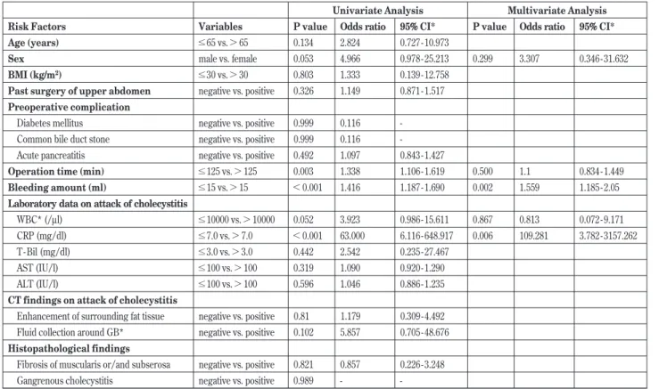
We evaluated the risk factors for an additional port in this series. Logistic regression analysis for the risk of an additional port re-quirement is shown in Table 3. In univariate analysis, operative Table1. Patient characteristics and clinical findings (N = 75)
Age (years) 57 (21 - 87)
Sex ratio (male : female) 37 : 38 BMI (kg/m2) 23.8 (16.5 - 36.0)
Past surgery of upper abdomen 3 (4.0%) Preoperative complication
Diabetes mellitus 2 (2.7%) Common bile duct stone 4 (5.3%) Acute pancreatitis 4 (5.3%) Laboratory data on attack of cholecystitis
WBC (per/l) 7971 (2700 - 18900) CRP (mg/dl) 2.9 (0.01 - 25.9) T - Bil (mg/dl) 1.3 (0.2 - 6.8) AST (IU/l) 125 (15 - 1003) ALT (IU/l) 121 (8 - 874) CT findings on attack of cholecystitis
Enhancement of surrounding fat tissue 33 (44.0%) Fluid collection around GB* 4 (5.3%) Histopathological findings
Fibrosis of muscularis or/and subserosa 38 (50.7%) Gangrenous cholecystitis 1 (1.3%) *GB, Gallbladder
Table2. Operative outcome and postoperative course Operative time (min) 126.8"44.4 Bleeding amount (ml) 19.5"38.4 Hospitalization after surgery (days) 3.5"2.8 Perioperative events
Gallbladder perforation 14 (18.7%) Additional port 9 (12.0%) Conversion to open surgery 1 (1.3%) Postoperative complications
Bile leak 1 (1.3%)
Intra - abdominal bleeding 1 (1.3%) Common bile duct stone dropped from GB* 1 (1.3%) Umbilical hernia 0 (0%) SSI* (Including wound infection) 0 (0%) *SSI, Surgical site infection ; *GB, Gallbladder
time (!125 min), bleeding amount ("15 ml), and CRP values ("7.0 mg/dl) during attacks of cholecystitis were significantly correlated with the requirement of an additional port. In multivariate analysis, bleeding amount ("15 ml) and CRP values ("7.0 mg/dl) were sig-nificantly correlated with an additional port. CT findings in attacks of cholecystitis and histological findings like gangrenous cholecysti-tis were not significantly correlated. Using the Fisher’s test, a sig-nificant correlation between an additional port and CRP values was found (Table 4). Indeed, elevated CRP values correlated with an additional port with a sensitivity of 55.6%, specificity of 98.5%, and accuracy of 93.3%. We then used ROC curve analysis to select for the optimized cutoffs by correlating the values of high CRP, WBC counts, and the need for an additional port (Figure 1).
Only one patient with a high CRP value (23.1 mg/dl) completed the procedure with a single - incision method. This patient had se-vere inflammation in the body and tail of the gallbladder only, so the cystic duct could be divided and dissected without an additional port. Four patients without high CRP values needed an additional port and the reasons included severe inflammation of the cystic Table 3. Logistic regression analysis for the risk of additional port
Univariate Analysis Multivariate Analysis Risk Factors Variables P value Odds ratio 95%% CI* P value Odds ratio 95%% CI* Age (years) !65 vs."65 0.134 2.824 0.727 - 10.973
Sex male vs. female 0.053 4.966 0.978 - 25.213 0.299 3.307 0.346 - 31.632 BMI (kg/m2) !30 vs."30 0.803 1.333 0.139 - 12.758
Past surgery of upper abdomen negative vs. positive 0.326 1.149 0.871 - 1.517 Preoperative complication
Diabetes mellitus negative vs. positive 0.999 0.116 -Common bile duct stone negative vs. positive 0.999 0.116 -Acute pancreatitis negative vs. positive 0.492 1.097 0.843 - 1.427
Operation time (min) !125 vs."125 0.003 1.338 1.106 - 1.619 0.500 1.1 0.834 - 1.449 Bleeding amount (ml) !15 vs."15 !0.001 1.416 1.187 - 1.690 0.002 1.559 1.185 - 2.05 Laboratory data on attack of cholecystitis
WBC* (/μl) !10000 vs."10000 0.052 3.923 0.986 - 15.611 0.867 0.813 0.072 - 9.171 CRP (mg/dl) !7.0 vs."7.0 !0.001 63.000 6.116 - 648.917 0.006 109.281 3.782 - 3157.262 T - Bil (mg/dl) !3.0 vs."3.0 0.442 2.542 0.235 - 27.467
AST (IU/l) !100 vs."100 0.319 1.090 0.920 - 1.290 ALT (IU/l) !100 vs."100 0.596 1.046 0.886 - 1.235 CT findings on attack of cholecystitis
Enhancement of surrounding fat tissue negative vs. positive 0.81 1.179 0.309 - 4.492 Fluid collection around GB* negative vs. positive 0.102 5.857 0.705 - 48.676 Histopathological findings
Fibrosis of muscularis or/and subserosa negative vs. positive 0.821 0.857 0.226 - 3.248 Gangrenous cholecystitis negative vs. positive 0.989 - -*CI, confidence interval ; *WBC, white blood cell ; *GB, Gallbladder
Table 4. Relationship between high CRP values and additional port Port added Single incision
High CRP ("7.0 mg/dl) 5 1 P!0.001 (Fisher’s exact test) Low CRP (!7.0 mg/dl) 4 65
Figure 1.
Receiver operating characteristic (ROC) curve for additional port by high CRP values ("7.0 mg/dl) during a cholecystitis attack. A value of CRP = 7.0 mg/dl predicted the need for an additional port with a sensitivity of 0.556 and a specificity of 0.985. Area under the ROC curve (AUC) was 0.823.
duct and neck of gallbladder (n = 2), requirement for suture clo-sure of a short cystic duct stump (n = 1), and intra- abdominal adhe-sion due to four previous abdominal surgeries (n = 1).
DISCUSSION
This study showed that although single - incision laparoscopic cholecystectomy is feasible and safe with acceptable operative out-comes for patients with cholecystitis, 19% of our patients required an additional full - size port to complete the procedure. High CRP values, reflective of severe inflammation, are an independent risk factor for the requirement of an additional port.
Since single - incision laparoscopic cholecystectomy was first re-ported(2), it has been shown to be a feasible and efficacious proce-dure that is comparable to conventional multi - port laparoscopic cholecystectomy(3 - 6). To date, a few meta- analyses, including several randomized trials of highly variable quality, have compared single - incision cholecystectomy with conventional laparoscopic cholecystectomy(15 - 17). While several reports described no ad-vantage in terms of patient satisfaction after single - incision pro-cedures, they did describe an advantage in terms of cosmetic out-comes. However, postoperative morbidity tended to be higher after single - incision cholecystectomy, whereas postoperative pain and hospital stay were similar between single - access and conventional laparoscopic procedures(17).
Although expected complications include severe inflammation of the gallbladder, Mirizzi syndrome, and confluent stones, few reports have analyzed the difficulties and risk factors associated with an additional port in single - incision laparoscopic cholecystectomy(10). The results of the present study suggest that single -incision cholecystectomy might be safely performed for patients with cholecystitis with acceptable operative outcomes. This study also clarified that high CRP values (!7.0 mg/dl) are indeed a significant risk factor for conversion to an additional port. When a single - access procedure is elected for patients with cholecystitis with high CRP values, the likelihood of the need for an additional port should be considered for the patient’s safety. If the patients with high CRP value are elected single - incision cholecystectomy, these cases should be performed by experienced surgeon. The results of this study may help senior surgeons to decide whether young surgeons or residents perform this surgery.
Another concern associated with single - incision cholecystec-tomy is the appropriate selection of either a pure incision port method or a single - access method using needlescopic devices. In contrast to the single - access technique, the surgical approach of pure single - incision cholecystectomy has been criticized for violat-ing important surgical principles, such as triangulation (co - axial setup) and tissue tension when Calot’s triangle is dissected. These results potentially make it more difficult to identify the cystic artery and cystic duct, and to achieve a ‘critical view’ in order to avoid biliary duct injury(18). In our hospital, single - incision cholecystec-tomy with one or two needlescopic devices is now the standard procedure for patients with cholecystitis, except in cases of severe systemic preoperative complications or large scars of the upper abdomen. This single - incision procedure is feasible with similar outcomes compared with the conventional multiport method. Moreover, since laparoscopic cholecystectomy is a good training procedure for residents, it is easier to adapt this surgery for young candidates. Therefore, we use needlescopic devices in all cases and no longer perform any operations using the pure single - incision method. A mere 2 to 3 - mm incision is necessary using needle-scopic devices, and the resulting scars are almost invisible. Only one randomized trial has compared needlescopic cholecystectomy with pure single - incision cholecystectomy, reporting a positive effect of the single - incision technique in terms of pain and cosmetic
outcomes(19). However, that trial was not blinded.
The limitations of this study include the small number of patients and the use of clinical data from a single center. Further analysis with a larger population and multiple centers should be performed in the future. Additionally, our hospital has more than 15 years of needlescopic laparoscopic cholecystectomy (two ports and two puncture method) experience. Naturally, surgeons should have adequate experience with single - incision surgery, needlescopic surgery, and conventional laparoscopic cholecystectomy before attempting this surgery.
In conclusion, this study suggests that single - incision laparo-scopic cholecystectomy is adoptable for patients with cholecystitis, but that the severe inflammation as indicated by elevated CRP val-ues during cholecystitis attacks may require consideration of con-version to an additional port.
ACKNOWLEDGEMENT
The authors have no conflict of interest.
REFERENCES LIST
1. Reynolds W, Jr : The first laparoscopic cholecystectomy. JSLS. 5 : 89 - 94, 2001
2. Navarra G, Pozza E, Occhionorelli S, Carcoforo P, Donini I : One - wound laparoscopic cholecystectomy. Br J Surg 84 : 695, 1997
3. Romanelli JR, Roshek TB 3rd, Lynn DC, Earle DB : Single -port laparoscopic cholecystectomy : initial experience. Surg Endosc 24 : 1374 - 1379, 2010
4. Yu WB, Zhang GY, Li F, Yang QY, Hu SY : Transumbilical single port laparoscopic cholecystectomy with a simple tech-nique : initial experience of 33 cases. Minim Invasive Ther Allied Technol 19 : 340 - 344, 2010
5. Rivas H, Varela E, Scott D : Single - incision laparoscopic cholecystectomy : initial evaluation of a large series of patients. Surg Endosc 24 : 1403 - 1412, 2010
6. Elsey JK, Feliciano DV : Initial experience with single - incision laparoscopic cholecystectomy. J Am Coll Surg 210 : 624 -626, 2010
7. Phillips MS, Marks JM, Roberts K, Tacchino R, Onders R, DeNoto G, Rivas H, Islam A, Soper N, Gecelter G, Rubach E, Paraskeva P, Shah S : Intermediate results of a prospective randomized controlled trial of traditional four - port laparo-scopic cholecystectomy versus single - incision laparolaparo-scopic cholecystectomy. Surg Endosc 26 : 1296 - 1303 2012 8. Ma J, Cassera MA, Spaun GO, Hammill CW, Hansen PD,
Aliabadi - Wahle S : Randomized controlled trial comparing single - port laparoscopic cholecystectomy and four - port la-paroscopic cholecystectomy. Ann Surg 254 : 22 - 27, 2011 9. Sasaki A, Ogawa M, Tono C, Obara S, Hosoi N, Wakabayashi
G : Single - port versus multiport laparoscopic cholecystec-tomy : a prospective randomized clinical trial. Surg Laparosc Endosc Percutan Tech 22 : 396 - 399, 2012
10. Chuang SH, Yang WJ, Chang CM, Lin CS, Yeh MC : Is routine single - incision laparoscopic cholecystectomy feasible? A retrospective observational study. Am J Surg 210 : 315 - 321, 2015
11. Kimura Y, Takada T, Strasberg SM, Pitt HA, Gouma DJ, Garden OJ, Büchler MW, Windsor JA, Mayumi T, Yoshida M, Miura F, HiguchiR, Gabata T, Hata J, Gomi H, Dervenis C, Lau WY, Belli G, Kim MH, Hilvano SC, Yamashita Y : TG13 current terminology, etiology, and epidemiology of acute cho-langitis and cholecystitis. J Hepatobiliary Pancreat Sci 20 : 8 - 23,
2013
12. Yokoe M, Takada T, Strasberg SM, Solomkin JS, Mayumi T, Gomi H, Pitt HA, Garden OJ, Kiriyama S, Hata J, Gabata T, Yoshida M, Miura F, Okamoto K, Tsuyuguchi T, Itoi T, Yamashita Y, Dervenis C, Chan AC, Lau WY, Supe AN, Belli G, Hilvano SC, Liau KH, Kim MH, Kim SW, Ker CG ; Tokyo Guidelines Revision Committee : TG13 diagnostic criteria and severity grading of acute cholecystitis (with videos). J Hepato-biliary Pancreat Sci 20 : 35 - 46, 2013
13. Yamashita Y, Takada T, Strasberg SM, Pitt HA, Gouma DJ, Garden OJ, Büchler MW, Gomi H, Dervenis C, Windsor JA, Kim SW, de Santibanes E, Padbury R, Chen XP, Chan AC, Fan ST, Jagannath P, Mayumi T, Yoshida M, Miura F, Tsuyuguchi T, Itoi T, Supe AN : Tokyo Guideline Revision Committee : TG13 surgical management of acute cholecystitis. J Hepato-biliary Pancreat Sci 20 : 89 - 96, 2013
14. Asao T, Yanagita Y, Nakamura J, Hosouchi Y, Takenoshita S, Nagamachi Y : Usefulness of a visceral mini - retractor accessi-ble without trocar port during laparoscopic surgery. Surg Endosc 13 : 91, 1999
15. Markar SR, Karthikesalingam A, Thrumurthy S, Muirhead L,
Kinross J, Paraskeva P : Single - incision laparoscopic surgery (SILS) vs. conventional multiport cholecystectomy : system-atic review and meta- analysis. Surg Endosc 26 : 1205 - 1213, 2012
16. Pisanu A, Reccia I, Porceddu G, Uccheddu A : Meta- analysis of prospective randomized studies comparing single - incision la-paroscopic cholecystectomy (SILC) and conventional mul-tiport laparoscopic cholecystectomy (CMLC) : J Gastrointest Surg 16 : 1790 - 1801, 2012
17. Garg P, Thakur JD, Garg M, Menon GR : Single - incision laparoscopic cholecystectomy vs. conventional laparoscopic cholecystectomy : a meta- analysis of randomized controlled trials. J Gastrointest Surg 16 : 1618 - 1628, 2012
18. Strasberg SM, Hertl M, Soper NJ : An analysis of the prob-lem of biliary injury during laparoscopic cholecystectomy. J Am Coll Surg 180 : 101 - 125, 1995
19. Lee PC, Lo C, Lai PS, Chang JJ, Huang SJ, Lin MT, Lee PH : Randomized clinical trial of single - incision laparoscopic cholecystectomy versus minilaparoscopic cholecystectomy. Br J Surg 97 : 1007 - 1012, 2010