STUDIES ON THE MICROBIAL DEGRADATION OF HOMOCHOLINE
(~g: ~ IC: ct Q 7.1\ =E ::] IJ ~CD~~ IC: ~ -r Q fin~)
ISAM ALI MOHAMED AHMED ALI
Ph.D. Dessertation
Submitted to the United Graduate School of Agricultural Sciences, Tottori University in partial fulfilhnent of the requirements for the degree of
Doctor of Philosophy in Bioresources Sciences
2010
DEDICATION
I wou!dlllie to dedicate this humfite thesis to:
My beloved wife Khalda and my sweet son Elmujtaba My family: Ali, Zeinab, Buthina, Fatihia, Mohamed Ahmed, Hajer, N awal, Eghbal, Entisar and Ekhlass
for their generous Love antfszg;yort
TABLE OF CONTENTS
TABLE OF CONTENTS ... i
LIST OF FIGURES ... v
LIST OF TABLES ... vii
ABBREVIATIONS ... viii
CHAPTERl:GENERALINTRODUCTION 1.1 INTRODUCTION ... I 1.2 SYNTHETIC LONG CHAIN QUATERNARY AMMONIUM COMPOUNDS (sQACs) ... 4
1.3 NATURALLY OCCURRING QUATERNARY AMMONIUM COMPOUNDS (nQACs) ... 5
1.3.1 Choline ... . 1.3.2 Homocholine 1.3.3 Glycine Betaine 1.3.4 B-Alanine Betaine. 1.3.5 L-Carnitine 1.4 MICROBIAL DEGRADATION OF QUATERNARY AMMONIUM ... 9
. ... 9
11 COMPOUNDS ... 13
1.4.1 Biodegradation of Long Chain QAC by Microorganisms 13 1.4.2 Biodegradation of Naturally Occurring QAC by Microorganisms ... . 16
1.4.2.1 Bacterial metabolism of choline 1.4.2.2 Bacterial metabolism of 4-N-trimethylamino-1-butanol... ... 19
1.4.2.3 Bacterial metabolism ofL-carnitine 1.5 RESEARCH OBJECTIVES ... 21
CHAPTER 2: SCREENING, ISOLATION AND IDENTIFICATION OF HOMOCHOLINE DEGRADING MICROORGANISIMS 2.1 INTRODUCTION ... 23
2.2 MATERIALS AND METHODS ... 24
2.2.1 Materials 2.2.2 General Methods 2.2.3 Chemical Synthesis ... 24
2.2.4 Screening and Isolation of Homocholine Degrading Bacteria ... 27
2.2.5 Morphological, Biochemical and Physiological Characterization ... 28
2.2.6 Sequencing of 16S rRNA Gene 2.2.6.1 Polymerase chain reaction (PCR) 2.2.6.2 Agarose gel electrophoresis of DNA and visualization 2.2.6.3 PCR product purification and sequencing ... 32
2.2.8 Creation of Phylogenetic Trees 2.2.9 Analytical Methods ...
2.2.9.1 Thin layer chromatography (TLC) 2.2.9.2 Capillary electrophoresis
2. 3 RESULTS AND DISCUSSION ... 35 2.3 .1 Screening of Homocholine Degrading Microorganisms ... 35 2.3.2 Isolation and Characterization of Homocholine Degrading Arthrobacter sp. Strain E5
... 37 2.3.2.1 Morphological and physiological characteristics of strain E5 ...
2.3.2.2 16S rR.l\TA gene sequence analysis
2.3.2.3 Utilization of several organic compounds by strain
2.3 .3 Isolation and Characterization of Homocholine Degrading Rhodococcus sp. Strains A2
2.3.3.1 Enrichment and isolation of strain A2
2.2.3.2 Morphological and physiochemical characterization of strain A2 2.3.3.2 Phylogenetic analysis of strain
2.3.3.3 Growth of strain A2 on homocholine and other structurally related compounds ... 47 2.3.4 Isolation and Characterization of Homocholine Degrading Pseudomonas sp. Strains
A9 and B9b ... 50
2.3.4.1 Morphological and physiological characteristics of the isolated strains
2.3.4.2 16S rRNA gene sequence analysis of strains A9 and B9b ... 51 2.3.4.3 Utilization of several C.N. sources by strains A9 and B9b ... 54
2.4 CONCLUSIONS ... 56 CHAPTER 3: DEGRADATION OF HOMOCHOLINE BY RESTING CELL
SUSPENSIONS AND GROWING CELL CULUTRES OF THE ISOLATED STRAINS
3.1 INTRODUCTION ... 57 3.2 MATERIALS AND METHODS ... 59
3. 2.1 Materials 3.2.2 General Methods 3.2.3 Chemical Synthesis
3.2.4 Growth and Homocholine Degradation by the Isolated Strains ... .
3.2.5 Homocholine Degradation by Resting Cells of the Isolated Strains ... 62 3.2.6 Isolation and Quantification of the Metabolites ofHomocholine ... 62
3.2.6.1 Homocholine
3.2.6.2 Trimethylaminopropionaldehyde (TMAPaldehyde)
3.2.6.3 P-alanine betaine and trimethylamine (TMA) ... 63 3.2.6.4 Trimethylamine
3.2.7 Analytical Methods
3. 2. 7.1 Capillary electrophoresis
3.2.7.2 Gas chromatography-mass spectrometry (GC-MS) ... 65
3 .2. 7.3 Fast atom bombardment- mass spectrometry (F AB-MS) 3.3 RESULTS AND DISCUSSION ... 65
3. 3.1 Identification of Homocholine Degrading Metabolites ... 65
3.3.2 Degradation of Homocholine by Resting Cells of the Isolated Strains ... 69
3.4 CONCLUSIONS ... 74
CHAPTER 4: ENZIMATIC ACTIVITIES IN THE DEGRADATION PATHWAY OF HOMOCHOLINE BY THE ISOLATED STRAINS 4.1 INTRODUCTION ... 77
4.2 MATERIALS AND METHODS ... 79
4.2.1 Materials .... 79
4.2.2 General Methods 4.2.3 Screening for Homocholine degrading Enzymatic Activities ... 80
4.2.3.1 Plate replica staining method 4.2.3.2 Cell free extract preparation ... 81
4.2.3.3 NAD+-dependent homocholine dehydrogenase 4.2.3.4 NAD+ -dependent TMAPaldehyde dehydrogenase ... 82
4.2.3.5 Membrane-bound homocholine dehydrogenase 4.2.3.6 Homocholine oxidase 4.2.3.7 NAD+ -dependent 3-hydroxypropionate dehydrogenase ... 83
4.2.4 Time Course of the Growth and Enzyme Formation of Pseudomonas sp. strains A9 .84 4.2.5 Stability of Homocholine Dehydrogenase Activity of Strain A9 4.2.6 Formation ofHomocholine Dehydrogenase on Various Media 4.2.7 Measurement of Protein Content 4.2.8 Measurement of Molecular ... 84
... 85
.86 4.2.9 Polyacrylamide Gel Electrophoresis ... 86
4.2.1 0 Intact and Dry Cell Reaction of Pseudomonas sp. Strain A9 4.2.11 Detection of 3-Hydroxypropionate by LC/MS/MS 4.3 RESULTS AND DISCUSSION ... 89
4.3.1 Screening for Homocholine degrading Activity ... 89 4.3.2 Time Course of the Growth and Enzyme Formation of Pseudomonas sp. Strains A9 90 4.3.3 Formation ofHomocholine Dehydrogenase on Various Media
4.3.4 Stability ofHomocholine Dehydrogenase of Pseudomonas sp. Strain A9 4.3.5 Intact and Dry Cell Reaction of Pseudomonas sp. Strain A9
4.3.6 Substrate Specificity of Homocholine Dehydrogenase
.94
4.3. 7 Measurement of the Native Molecular Mass of Homocholine Dehydrogenase ... 10 1 4. 3. 8 Detection of 3 -hydroxypropionate Dehydrogenase Activity 102
CHAPTER 5: GENERAL CONCLUSIONS ... 107
REFERENCES ... 111
ACKNOWLEDGMENT ... 127
ABSTRACT ... 129
iii~
...
133LIST OF ORIGINAL PUBLICATIONS ... 135
Fig. 1. 1 Metabolic fate of choline Fig. 1. 2 Metabolism of choline
LIST OF FIGURES
Fig. 1. 3 The synthetic pathway to ~-alanine betaine in plant.... 10 Fig. 1. 4 Function of L-camitine in fatty acid metabolism ... 11 Fig. 1. 5 General degradation pathway of cationic surfactants ... 15
Fig. 1. 6 Choline oxidation pathway 18
Fig. 1. 7 Degradation pathway of 4-N-trimethylamino-1-butanol in Pseudomonas sp. 13 CM ... 20
Fig. 2. 11 H NMR spectrum of chemically synthesized 3 -N-trimethylamino-1-propanol ... 26 Fig. 2. 2 Histogram showed the cell growth of isolated microorganisms.
Fig. 2. 3 Morphology of Arthrobacter sp. strain E5 as seen by light microscopy under optical and phase contrast conditions ... .
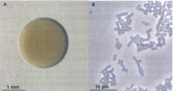
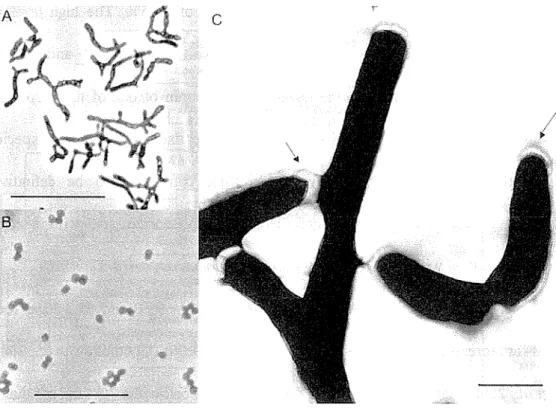
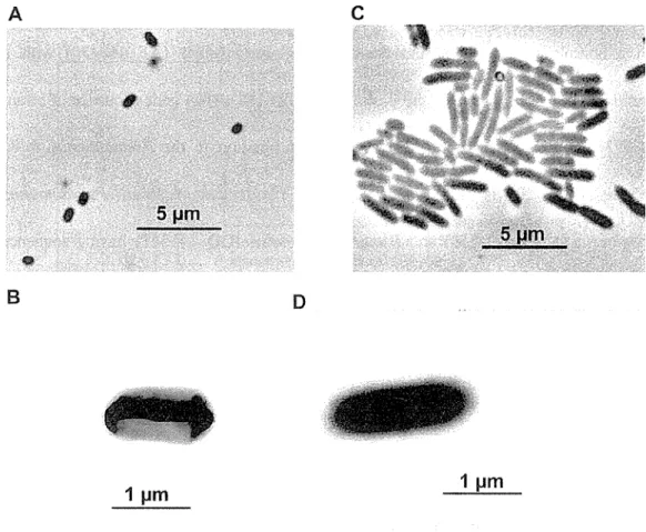
Fig. 2. 4 Phylogenetic tree based on 16S rRNA sequence showing the relationship between strain E5 and most closely related species of the genusArthrobacter ... 41 Fig. 2. 5 Morphology of Rhodococcus sp. strain A2 as seen by phase contrast microscopy and
transmission electron microscopy
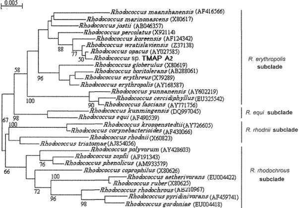
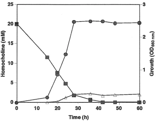
Fig. 2. 6 Phylogenetic tree based on 16S rRNA sequence showing the relationship between strain A2 and most closely related species of the genus Rhodococcus . ... .47 Fig. 2. 7 Time course study of the growth and metabolism of homocholine by Rhodococcus sp.
strain A2 ... 49 Fig. 2. 8 Morphology of isolated strains, Pseudomonas sp. strain A9 and B9b as seen by phase contrast microscopy and transmission electron microscopy ... 51 Fig. 2. 9 Phylogenetic tree based on 16S rRNA sequence showing the relationship between the isolated strains A9 and B9b and most of the closely related P~eudomonas species ... 53
Fig. 3. 11 H NMR spectrum of chemically synthesized 3 -N-trimethylamino-1-propionic acid (~
Alanine betaine)
Fig. 3. 2 Capillary electrophoresis chromatogram showing the generated metabolite peaks from the degraded homocholine by the isolated strains alongside with authentic standards peaks of trimethylamine and ~-alanine betaine
Fig. 3. 3 GC-MS spectra of the metabolite trimethylamine formed during the degradation of homocholine by the isolated strains ... 68 Fig. 3. 4 F AB-MS spectra of metabolites in the degradation pathway of homocholine by the isolated strains ... 69
Fig. 3. 5 Time course degradation of homocholine and the generation of the metabolites, trimethylamine and beta-alanine betaine by strains E5, A2 and A9
Fig. 3. 6 Proposed pathway for the aerobic biodegradation of homocholine by the isolated strains
Fig. 4. 1 Bovine serum albumin standard curve as determined by Lowry method ... 86
Fig. 4. 2 Screening of NAD+ -dependent dehydrogenase activity in the isolated strains by (A) replica staining and (B) spectrophotometric assays ... . Fig. 4. 3 Time course of the growth and homocholine dehydrogenase activity formation of Pseudomonas sp. strain Fig. 4. 4 Effect of stabilizers on the stability of homocholine dehydrogenase of Pseudomonas sp. strain A9 ... 96
Fig. 4. 5 Evaluation of the stability ofhomocholine dehydrogenase during storage at 4°C ... 96
Fig. 4. 6 Degradation ofhomocholine by resting cells of Pseudomonas sp. strain A9 ... 97
Fig. 4. 7 Degradation ofhomocholine by dried cells of Pseudomonas sp. strain A9 ... 98
Fig. 4. 8 Native-PAGE analysis of crude homo choline dehydrogenase ... 10 1 Fig. 4. 9 Estimation of the native molecular mass of homocholine dehydrogenase by size exclusion chromatography on TSK-gel G3000 SW column ... 102
Fig. 4. 10 Native-PAGE analysis of crude 3-hydroxypropionate dehydrogenase ... 103
Fig. 4. 11 LC/MS/MS analysis of 3 -hydroxypropionate in the degradation pathway of homocholine by the intacted cells of Pseudomonas sp. strain A9 ... 104 Fig. 4. 12 Proposed degradation pathway of homocholine by Pseudomonas sp. strain A9 ... 1 06
LIST OF TABLES
Table 1. 1 Structural features of synthetic sQACs and their industrial usage
Table 2. 1 Basal-homocholine (basal-HC) liquid medium
Table 2. 2 Basal-homocholine (basal-HC) agar medium ... 28 Table 2. 3 Nutrient broth (meat extract) media
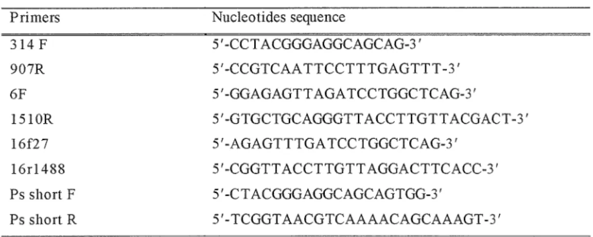
Table 2. 4 Primers used in PCR amplification
Table 2. 5 Morphological and physiological characteristics of Arthrobacter sp. strain E5 ... .42 Table 2. 6 Morphological and physiological characteristics of Rhodococcus sp. strain A2 ... .44 Table 2. 7 Utilization of homo choline and other structurally related compounds by
Rhodococcus sp. strain A2 ... .49 Table 2. 8 Phenotypic characteristics of Pseudomonas sp. strains A9 and B9b ... 55
Table 4. 1 Protease inhibitor cocktail
Table 4. 2 Reaction mixture for activity staining on native-PAGE gels Table 4. 3 Formation of Homo choline dehydrogenase on various media
Table 4. 4 Substrate specificity of homocholine dehydrogenase from strain A9 ... 1 00 Table 4. 5 NAD+-dependent 3-hydroxypropionate dehydrogenase activity in the cell extracts of
homocholine-, choline- and glucose-growing cells 103
~-AB
Basal-He CTAB DCIP
DMA-Propanol DNPH
DTT FAB-MS GC-MS GMC MLA nAChR nQACs NTB PMS sQACs TMA TMABA TMA-Butanol TMAP
TMAPaldehyde QACs
ABBREVIATIONS
~-Alanine betaine
Basal-homocholine medium
Cetyl trimethyl ammonium bromide 2,6-dichlorophenol-indophenol 3-N-dimethy larnino-1-propanol 2, 4-dinitropheny hy drazine Dithiothreitol
Fast atom bombardment- mass spectrometry Gas chromatography-mass spectrometry Glucose-methanol-choline oxidoreductases Methyllycaconitine
Nicotinic acetylcholine receptor
Naturally occurring quaternary ammonium compounds Nitro tetrazolium blue
Phenazine methosulfate
Synthetic quaternary ammonium compounds Trimethy I amine
4-N-trimethylamino-1-butyraldehyde 4-N-trimethylarnino-1-butanol
3-N-trimethylarnino-1-propanol (homocholine) Trimethylarninopropionaldehyde
Quaternary ammonium compounds
CHAPTERl
GENERAL INTRODUCTION
1.1 INTRODUCTION
Quaternary ammonium compounds (QACs) are organic compounds that contain four functional groups attached covalently to a positively charged central nitrogen atom (~N+). These functional groups (R) include either long chain alkyl, benzyl or methyl groups. The term quaternary ammonium compounds refer to either chemically synthesized long-chain quaternary ammonium compounds (sQACs) or naturally occurring quaternary ammonium compounds (nQACs). Long-chain sQACs have been important since their bactericidal properties were recognized in the 1930s. Since then, they are extensively used in domestic and industrial applications as surfactants, emulsifiers, fabric softeners, disinfectants and corrosion inhibitors (Garcia et al., 2004;
Patrauchan and Oriel, 2003). Such extensive use of sQACs in various branches of industry has resulted in increased presence of these compounds in wastewater, waterways, lakes, sludges and soils (Gerike et al., 1978). These compounds are toxic to aquatic plant and animal organisms. With their surface-active properties, they facilitate solubility of many dangerous rnicropollutants, such as pesticides, of which toxins can easily penetrate living organisms (Grabinska-Sota 2004). On the other hand, nQACs are widely distributed in the biosphere: there are more than 100 reported examples including well-known representatives such as choline, glycine betaine and ~-alanine
betaine (Anthoni et al., 1991) which have different biological functions, such as adaptation of organisms to environmental stress and transportation of chemical groups in many metabolic processes (Anthoni et al., 1991). Choline is an essential nutrient that is widely distributed in foods, principally in form of phosphatidylcholine. Choline or its metabolites are required for synthesis of phospholipids in cell membranes, methyl group metabolism and cholinergic neurotransrnission and transmembrane signaling, as well as for lipid-cholesterol transport and metabolism (Fig. 1.1). This quaternary amme IS
present in tissues predominantly as one of the choline-phospholipids such as phosphatidylcholine and sphingomyelin (Zeisel et al., 1994). The osmoprotective role of glycine betaine, which derived from choline via an oxidation reaction, is evident in a number of diverse microbial systems, including enteric bacteria (Andresen et al., 1988), soil bacteria (Smith et al., 1988), halophilic bacteria (Galinski et al., 1982), cyanobacteria (Mackay et al., 1984) and methanogenic archaebacteria (Robertson et al., 1990). Besides being an osmoprotectant, glycine betaine also has a role in general metabolism where one methyl group of glycine betaine is incorporated into methionine in mammals (Finkelstein et al., 1972, Millian et al., 1998) and microorganisms (White et al., 1971), or into cobalamin (Vitamin B12) in microorganisms (White et al., 1971).
Recently, glycine betaine was proven to be an effective cryoprotectant for a wide range of prokaryotic organisms during freeze-drying, liquid-drying and liquid nitrogen freezing (Cleland et al., 2004). L-Camitine a trimethylated amino acid that is similar in structure to choline is highly polar zwitterionic quaternary amine carboxylic acid and present in some prokaryotes and all eukaryotes (Ramsay et al., 2001). In prokaryotic cells, L-carnitine serves either as a nutrient, such as a carbon and nitrogen source (Kleber, 1997), or as an osmoprotectant (Jung et al., 1990; Robert et al., 2000).
However, in eukaryotic cells, it serves exclusively as a carrier of acyl moieties through
various subcellular compartments (Ramsay et al., 1993). Moreover, it has an important role in the transport of activated long-chain fatty acids across the inner mitochondrial membrane (Bremer, 1983). 3-N-Trimethylamino-1-propanol (homocholine) an analogue of choline, in which the amino alcohol group is lengthened by one CH2-group, has been shown to resemble choline in many aspects of cholinergic metabolisms (Boksa and Collier, 1980). It has been reported that when homocholine transported into the rat brain synaptosome, it acetylated and released as acetylhomocholine from a superior cervical ganglion and minces of mouse forebrain by a calcium-dependent process during depolarization (Collier et al., 1977; Carroll and Aspry, 1980). Moreover, it was found to be effective in preventing fat infiltration both in fat and cholesterol fatty livers (Channon et al., 1937). From the choline-like structure, one would expect that homocholine is degraded similarly and at a rate comparable to that of choline. To date, no microorganism degrading homocholine as a sole source of carbon and nitrogen has been isolated and consequently, this catabolic pathway remains unclear.
~
<,<:• H,•: --0·---C --r.~ CH,:OPO,CHPfl;t"{CH~'
I I ~--
-<1-(,-.t..o-tH <) ,' ''<, in
! !I ! 1~ ! NH
phospha;;~~~dl~~~~"~-,Hz-t,•-·. -~
o
f
S=uctural membrane phospholipidsHO,_ ,' H, ' I_,CH;
• ·p~ C. '.,..N~
phosphochohne,jl . o· -~ 'c><,.
\~.c;)
···t/
Neurotransmitterlr----1&w.aw=~?--~ethyl
group donor1 'm • ·- m1 o
0
1• Crls , II / i
'y~ ~~ ~ I
_.-CHs 10Sif1 ' H,c.._";"~\
~
ll C , N'+ , I \1' ·c : 0acetylcholine
II
HO/ -..._c"('~GH,
/Jl "-" ,/
>;U·
~ ch~li~e
11 6etaineplatelet-activating fac~pr
;'"!l
Signaling phospholipids
sphingosylphosphorylcholine
Fig. 1. 1 Metabolic fate of choline (Degani et al., 2006)
1.2 SYNTHETIC LONG CHAIN QUATERNARY AMMONIUM COMPOUNDS (sQACs)
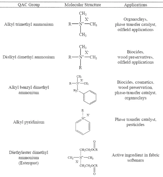
Synthetic quaternary ammonium compounds (sQACs) containing a long chain alkyl group or a benzyl group are cationic surfactants (Table 1.1) that play an important role in many industrial fields due to their versatile physico-chemical properties. Such widespread uses as disinfectants, fabric softening agents, foam depressants, and antistatic agents lead to massive discharge into the environment with its associated concerns (Wee and Kennedy, 1982).
Potential risks have been reported in many previous studies that repeated exposure to long-chain sQACs can induce microbial resistance against antibiotics in many pathogenic microorganisms (McDonnell and Russell, 1999). In addition, discharge of long-chain sQACs can disturb the purifying activities of natural aquatic systems or public wastewater treatment plants because of their toxicity to microbial life (Laopaiboon et al., 2002; Tubbing and Admiraal, 1991). The possibility to eliminate sQACs-related pollution effectively has been investigated in many ways. As one of the most economical means, biological treatment found to be an effective way to remove sQACs. In particular, aerobic treatment processes can provide rapid biodegradation via a consortium of bacteria (Scott and Jones, 2000).
Table 1. 1 Structural features of synthetic sQACs and their industrial usage QAC Group
Alkyl trimethyl ammonium
Dialkyl dimethyl ammonium
Alkyl benzyl dimethyl ammonium
Alkyl pylidinium
Dierhylester dimethyl ammonium (Esterqn::1t)
Molecular Structure CH,
1
x-
R-~....,.-cH3
CH
I
3CH,
I ~-
R-l'r-cH,
I -
R
CH-
I
~---
R-::~--cri;
H''D I
R
x·
0 CH,cH,OCR
II
I - -
CE,-N""-CH,
. x·l -- -
CH,CH,OCR
- - II
0
ApplicatiollS
Orgnnoclays, phase tran;;fer catalyst,
oilfield application>.
Biocides.
wood preservatives.
oilfield applications
Biocide::,. cosmetics.
wood preservation.
phase-transfer catalyst.
organoclnys
Plw~e transfer cataly,t.
pesticides
Active ingredient in falnic softeners
1.3 NATURALLY OCCURRING QUATERNARY AMMONIUM COMPOUNDS (nQACs)
Naturally occurring quaternary ammonium compounds constitute a class of metabolites with more than 100 reported examples, such as choline, glycine betaine, ~-alanine
betaine, y-butyrobetaine and L-camitine (Anthoni et al., 1991). They form a structurally heterogeneous class of compounds with a unifYing character of a polar and fully methyl substituted nitrogen atom, creating a permanent positive charge on the N moiety
(Rhodes and Hanson, 1993). Their occurrence apparently reflects several independent evolutionary patterns ranging from highly specialized functions for each nQAC to a general strategy for adaptation of the organisms to fluctuating or stressing environmental conditions (Anthoni et al., 1991). Moreover, it has been suggested that nQACs may be involved in transporting various chemical moieties within plants, acts as macromolecular components, and have role in overcoming water and salt stresses (Blunden and Gordon, 1986). In addition, the presence or absence of these compounds could be used as taxonomic indicator as well as they could used in the pharmacology to prevent artherosclerosis (Hoppe, 1979), to reduce plasma cholesterol levels (Kaneda and Abe, 1984), smooth muscle relaxant (Baker and Murphy, 1976) and to inhibit mammalian neuromuscular transmission (Hosein and McLennan, 1959).
1.3.1 Choline
Choline is an essential nutrient that is widely distributed in foods, principally in the form of phosphatidylcholine. Organ meats, eggs, soybeans, nuts, fish and broccoli are particularly good sources (Howe et al., 2004). The only source of choline other than diet is de novo biosynthesis of phosphatidylcholine from phosphatidyl-ethanolamine (Zeisel, 1990). Choline is required to make the phospholipids such as phosphatidylcholine, lysophosphatidylcholine, choline plasmalogen and sphingomyelin- essential components of all cell membranes. It is a precursor for the biosynthesis of the neurotransmitter acetylcholine and in biological methylation reactions. Choline phospholipids in cell membranes play a role in generating various second messengers during signal transduction (Zeisel, 1990; Zeisel and Blusztajn, 1994). Choline can be acetylated, phosphorylated and oxidized (Fig. 1.2). Only a small fraction of dietary choline is acetylated by choline acetyltransferase (EC.2.3.1.6) (Zeisel, 1990; Zeisel and
Blusztajn, 1994). This enzyme is highly concentrated in the terminals of cholinergic neurons, but also present in non-nervous tissue such as the placenta. Phosphorylation of choline is the first step in the mqjor pathway for phosphotidylcholine synthesis, catalyzed by choline kinase (EC. 2.7.1.32), using
Mi+
and ATP. This enzyme is widely distributed in mammalian tissues, including liver, brain, kidney and lung.Choline deficiency results in liver dysfunction, fatty liver, decreased in growth, infertility, Alzheimer's disease, abnormal kidney function, decreased red blood cell synthesis, high blood pressure and liver cancer (Zeisel, 1990; Zeisel and Canty, 1993;
Zeisel and Blusztajn, 1994).
S-adenosyl-homocysteine
Dimethylglycine Phosphatidyl-
ethanolamine Methionine __..,. S-adenosylmethionine Homocysteine
Betaine
..
Betaine aldehyde dehydroge:;Jase
Betaine aldehyde Trimethylamine
!
....
Choline dehydrogenase
Phosphatidylcholine
i
CDP-choline :diacylglycerol cholinephosphotransfeaseCDP-choline
i
CTP :phosphocholine cytidy!ytram:.ferasePhosphocholine
i
Choline
Choline .l.jnase
--IIl-li- Acetylcholine
Choline acetyltrar1.lerase
Fig. 1. 2 Metabolic pathway of choline (Hassan, 2008)
1.3.2 Homocholine
Homocholine (3-N-trimethylamino-1-propanol) an analogue of choline, in which the amino alcohol group is lengthened by one CH2-group, has been shown to resemble
1980). After it is transported into the rat brain synaptosome, it is acetylated and released as acetylhomocholine from a superior cervical ganglion and minces of mouse forebrain by a calcium-dependent process during depolarization (Collier et al., 1977;
Carroll and As pry, 1980). Homocholine, however, differs dramatically from choline in a very important aspect. That is not acetylated by solubilized cholineacetyltransferase, but is acetylated by intact rat brain synaptosomes (Collier et al., 1977), possibly because homocholine is acetylated by cholineacetyltransferase that is associated with vesicular and/or neuronal membranes (Carroll and Aspry, 1980). Nelson et al. (1980) assumed that the extracellular precursors of choline and homocholine may be directly accumulated by a crude vesicular fraction of mouse forebrain, independently of the cytoplasm, and may be utilized to replace the loss in acetylcholine. Additionally, they suggested that the extracellular products, acetylcholine and acetylhomocholine, may not be capable of replacing acetylcholine lost from a crude vesicular fraction. Moreover, acetylhomocholine is released from brain slices both spontaneously and in response to stimulation via mechanism similar to those that released acetylcholine, but some differences in specificity of the acetylcholine storage and/or release processes might be present (Nelson et al., 1980). Previous studies on acetylhomocholine synthesis and release have partly classified these false transmitter criteria by showing that homocholine is acetylated in tissues containing cholinergic nerve terminals (Collier et al., 1977), that acetylhomocholine is released by a calcium-dependent mechanisms during nerve stimulation (Collier et al., 1977), and that acetylhomocholine has actions qualitatively similar to but quantitatively different from those of acetylcholine on both muscarinic and nicotinic receptors (Hunt and Renshaw, 1934; Curtis and Ryall, 1966;
Barrass et al., 1970). In pharmacology, choline analogues such as homocholine, methylcholine and ethylcholine are used to study the chemical specificity of the various
processes involved in neurotransmission and in particular it has been used to investigate the subcelular origin of the transmitter released by stimulation.
1.3.3 Glycine Betaine
Glycine betaine is found in microorganisms, plants and animals, and is a significant component of many foods including wheat, shellfish, spinach and sugar beets (Craig, 2004). Glycine betaine is compatible osmolyte that increases the water retention of cells, replaces inorganic salts and protect intracellular enzymes against osmotically induced or temperature induced inactivation. In human, glycine betaine is catabolized via a series of enzyme reactions that occur mainly in the mitochondria of liver and kidney cells. Glycine betaine also acts as a catabolic source of methyl groups via transmethylation for use in many biochemical pathways. The formation of methionine from homocysteine can occur either via glycine betaine or 5-methyltetrahydrofolate.
The conversion of homocysteine to methionine is important to conserve methionine, detoxify homocysteine and produce S-adenosylmethionine (Millian et al., 1998).
Glycine betaine is synthesized by a two-step oxidation of choline or by methylation reaction from glycine. Two N-methyltransferases genes from halotolerant cyanobacteriumAphanothece halophytica have been isolated (Wadi tee et al., 2003).
1.3.4 P-Alanine Betaine
Many plants, bacteria, and marine algae accumulate quaternary ammonium compounds (QACs) in response to abiotic stresses such as drought and salinity (Rhodes and Hanson, 1993; Gorham, 1995). Zwitterionic compounds such as glycine betaine, ~
alanine betaine and proline betaine, are known to be the most effective osmoprotectants
(Yancey, 2005). In most members of the highly stress-tolerant plant family Plumbaginaceae, B-alanine betaine was reported to replace glycine betaine (Hanson et al., 1991, 1994). B-alanine betaine synthesis is not constrained by choline availability, because it is derived by theN-methylation of the non-protein amino acid, B-alanine (Fig.
1.3). Unlike glycine betaine synthesis, B-alanine betaine synthesis does not require oxygen, and hence it was suggested to be suitable for osmoprotection under saline and hypoxic conditions (Hanson et al., 1994; Rathinasabapathi, 2000). Accordingly,
B-
alanine betaine was distributed among species of the Plumbaginaceae, adapted to a wide range of adverse stress environments including saline and hypoxic conditions (Hanson et al., 1994). Although B-alanine betaine accumulation has been intensely studied for its role in osmoprotection (Hanson et al., 1994; Rathinasabapathi et al., 2000, 2001 ), early work on this compound in marine algae also suggested that it had a cholesterol-reducing effect in animal feeding experiments (Abe and Kaneda, 1973).
1~:\Tan.ineJ
'\; -M~Ihyl P-alaninc
N.N-Dimclhyl P-alanine
N. N,N-Tnm<:thyl P-alaninc:
=~-Alanine betaine
H.N~COOII
l
.\' ·<l<it'IW\ l'f .f. ·IIJ('flltrJJII/1<' dt'f'<'l!ci<.•tlf\'-mcFhl·lrrarrs[f'rc/sc
II C ' 's~lUOH H
l
S ·mkmHrl·l. ·mcrhwmnc dt'f'll'ndem S-merhlltnm.l{i:ra.wII,C, ~COUll
H1C/N
j S·adt•Jiold·l.·lllc'tlltmll/1<.' dc'f)Cfld<'lll
+
.\'-mctln·lcraru{erascII,C
tr;e.=::N~coo H,C/
Fig. 1. 3 The synthetic pathway to B-alanine betaine in plant (Duhaze et al., 2003)
,.
1.3.5 L-Camitine
A trimethylated amino acid roughly similar in structure to choline, L-carnitine is a highly polar zwitterionic quaternary amine carboxylic acid present in some prokaryotes and all eukaryotes (Ramsay et al., 2001). In prokaryotic cells, L-carnitine serves either as a nutrient, such as a carbon and nitrogen source (Kleber, 1997), or as an osmoprotectant (Jung et al., 1990; Robert et al., 2000). However, in eukaryotic cells, it serves exclusively as a carrier of acyl moieties through various subcellular compartments (Ramsay et al., 1993). It has an important role in the transport of activated long-chain fatty acids across the inner mitochondrial membrane (Bremer, 1983). Cytosolic long- chain fatty acids, which are present as CoA esters, are transesterified to L-carnitine at the mitochondrial outer membrane (Fig. 1.4).
0
II S-CoA
R-C-
Fattyacyl ~~
Co A
cooe
CHI ,
I -
HS-CoA
cooe
0 CH, I
II I -
HO-C-H R-C- 0 - C -H C:H, I
I -
<i!>N(CH,J, L-Camitine
I
CH, I -
<XlN(CHJ3 Acylcarnitine
" l r- )
( LC1 ~3L'''"'J
0
II
~- Curnitine ~
J/' =-~yltransft>rn.~ "'-.
R-C- S-CoA [! HS-CoA
Fnlly acyl CoA
!NTERMEMBRANE SPACE
MATRIX
Fig. 1. 4 Function ofL-carnitine in fatty acid metabolism (Nelson and Cox, 2004)
In this reaction, the acyl moiety of the long chain fatty acids is transferred from CoA to the hydroxyl group of carnitine. The resulting long chain acylcarnitine esters are transported over the inner mitochondrial membrane via a specific carrier, camitine- acylcamitine translocase. In the mitochondrial matrix, the enzyme carnitine acetyltransferase is able to reconvert short and medium chain acyl-CoAs into acylcamitines using intrarnitochondrial carnitine. Through this mechanism of reversible acylation, carnitine is able to modulate the intracellular concentrations of free CoA and acyl-CoA. Furthermore, it is involved in the transfer of the products of peroxisomal
B -
oxidation, including acetyl-CoA, to the mitochondria for oxidation to C02 and H20 in the Krebs cycle (Jakobs and Wanders, 1995).
In humans, approximately 75% of body L-carnitine sources comes from the diet and 25% from de novo biosynthesis (Tein et al., 1996). In general, L- carnitine is low in foods of plant origin and high in animal foods (Rebouche, 1992). L-Carnitine ultimately is synthesized from lysine and methionine. L-Lysine provides the carbon chain and nitrogen atom of carnitine, and L-methionine provides the methyl group. In the pathway of L-camitine in mammals, 8-N-trimethyl-L-lysine is synthesized only as a post-translational modification of protein synthesis and is released for L-carnitine biosynthesis by normal processes of protein turnover. A number of proteins contain one or more 8-N-trimethyl-L-lysine residues, including histones, actin, myosin, and calmodulin. The majority of 8-N-trimethyl-L-lysine synthesized in the body probably is formed in skeletal muscle (Rebouche, 1992).
1.4 MICROBIAL DEGRADATION OF QUATERNARY AMMONIUM COMPOUNDS
1.4.1 Biodegradation of Long Chain QAC by Microorganisms
The susceptibility of cationic surfactants to biodegradation was first reported in the 1950s. Gerike (1982) confirmed the study done before using activated sludge and cetyl trimethyl ammonium bromide (CTAB), dodecyl benzyl dimethyl ammonium chloride and didecyl dimethyl ammonium chloride.
Studies on the biodegradation pathways have been conducted for various sQACs.
The utilization of tetramethyl ammonium chloride by Pseudomonas sp. was described by Hampton and Zatman (1973). According to their work, the oxidation of this compound is initiated by splitting the C-N bond, therefore producing methanal and trimethylamine. The intermediate trimethylamine is oxidized to trimethylamine N-oxide (Large et al., 1972) or transformed to methanal and dimethylamine (Colby and Zatman, 1973; Meiberg and Harder, 1978). The trimethylamine N-oxide is further converted to dimethylamine (Large, 1971; Myers and Zatman, 1971), followed by the generation of methanal and methylamine (Colby and Zatman, 1973). Methylamine is then converted to methanal and ammonium. Based on an investigation of the utilization of alkyl trimethyl ammonium bromides by microorganisms derived from sewage and soil, Dean- Raymond and Alexander (1977) demonstrated that the first step of degradation occurs with hydroxylation of the terminal carbon of the alkyl group and the resulting carboxylic acid undergoes P-oxidation. Another study dealing with hexadecyl trimethyl ammonium chloride suggested a central fission of Calky1-N bond as the first step of degradation (Van Ginkel et al., 1992). Not only hexadecyl trimethyl ammonium chloride, but also
hexadecanal and hexadecanoate produced as metabolites, were all utilized by Pseudomonas species. It is believed that the central Calkyi-N bond fission is first carried out by an oxygen dependent dehydrogenase as suggested for alkyl trimethyl ammonium compounds (Van Ginkel et al., 1992) and then intermediates are formed consecutively by N-demethylation reactions.
In contrast to the previous findings, a recent study proposed the degradation of dodecyl trimethyl ammonium chloride by Pseudomonas sp. strain 7-6, isolated from a wastewater treatment plant, via dual pathways, which besides an initial attack on the Caikyi-N bond also initiates degradation via cleavage of a Cmethy1-N bond, hence producing dodecyl dimethylarnine as an intermediate product (Takenaka et al., 2007).
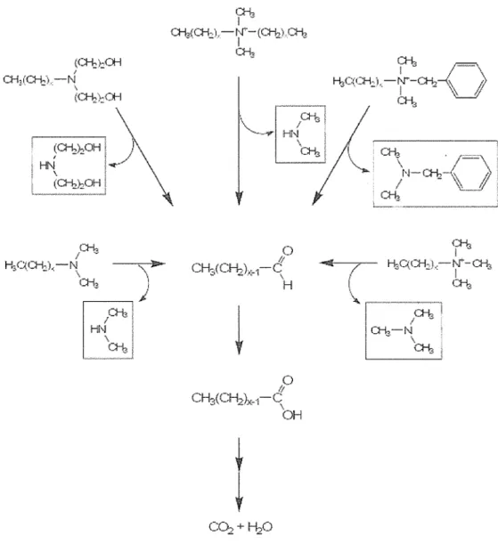
Overall, general degradation pathways of cationic sw-factants shown in Fig. 1.5 indicated that biodegradation is mostly commenced with cleavage of the Calkyi-N bond irrespective of the type of sQACs and the degradation of the produced alkanals proceeds via B-oxidation for complete mineralization (Van Ginkel, 2004).
On the other hand, because of the sw-factants character and the limited enzymatic ability of individual microorganisms, only a few known surfactants, alkane sulphonates, alkyl sulphates, and alkyl arnines, are completely degraded by a single microorganism (Van Ginkel, 1996). For this reason, consortia of microorganisms are highly efficient, as well as necessary for the complete degradation of surfactants (Scott and Jones, 2000;
Van Ginkel, 1996). The lack of the isolation of microbes degrading quaternary ammonium surfactant to completion may be due to the fact that in all those studies nitrogen was provided in form of ammonium in addition to the nitrogen-containing
surfactants. In this way, no selective pressure to use nitrogen deriving from the surfactant itself was imposed during enrichment and isolation.
(CI-b}~OH
CH,(CI-J.,)c-N I
- ' - . \
(C:I-b)...OH
(0-l,.):,OH '
(O-I£1:PH
l:!-b
H;C~CH.::),-f\j
\
CH3
;;,~
\
,)
CH., I ~
%(CH.::l. -~.t-(Cl-h)CH3 CH3
/f 0 CH3(C~t:-1-C\
H
~
/f 0 CH3(CH2)x-1-C~
(JH
CC~+~O
t
CH., ,.=-...
I ~ I \
1--1f''(1-H-N'"-Cl-h-<x\ /;
, '3 -·\- - . I \L__5f
CI-t,
CH.,
\4-CH,J~·) c~
...'\__j
{:::1-1:3 I
Clll( / ~C(CH.:;),-r-Cf-1.;
l"E:J·. a-~z-rJ::ris \ :.
a-lz
C!:-!:,1
Fig. 1. 5 General degradation pathway of cationic surfactants.
The alkanals formed through the oxidation of the a.-carbon of the alkyl chain are further metabolized via ~-oxidation (Van Ginkel, 2004).
1.4.2 Biodegradation of Natur-ally Occurring QAC by Microorganisms 1.4.2.1 Bacterial metabolism of choline
Choline is found ubiquitously in nature and the ability to degrade choline is widespread amongst microorganisms. The aerobic degradation of choline was firstly reported in
.
Achromobacter cholinophagum that metabolized choline via betaine as first oxidation products (Shieh 1964). Subsequently, Kortstee (1970) showed that many microorganisms of the genera Agrobacterium, Arthrobacter, Micrococcus, Pseudomonas, Rhizobium and Streptomyces were found to grow with choline as a sole source of carbon and nitrogen. He also found that all the tested choline-utilizing bacteria were able to grow with betaine, N, N-dimethylglycine or sarcosine as a sole carbon and nitrogen source.
In microorganisms, the degradation of choline proceeds by stepwise oxidation of the ethanol-group yielding betaine aldehyde and glycine betaine (Fig. 1.6). Thereafter, glycine betaine is degraded by subsequent splitting off the methy 1-groups, leading to the metabolites dimethylglycine, sarcosine and finally glycine (Shieh, 1964; Kortstee, 1970;
Mori, 1981). In plants such as spinach (Rathinasabapathi et al., 1997), sugar beet and amaranth (Russell et al., 1998), the first step reaction is catalyzed by choline monooxygenase (EC.l.14.15. 7), a ferredoxin-dependent soluble Rieske-type protein, whereas in animals and microorganisms, it is catalyzed by either membrane-bound choline dehydrogenase (EC.l.l.99.1) (Nagasawa et al., 1976; Landfald and Strom, 1986;
Russell and scope, 1994; Tsuge et al., 1980) or soluble flavoprotein, choline oxidase (EC.l.l.3.17) (lkuta et al., 1977; Yamada et al., 1977; Rozwadowski et al., 1991).
Choline oxidase catalyzes the four-electron oxidation of choline to glycine betaine via betaine aldehyde as intermediate (Ikuta et al., 1977). Based on amino acid sequence
compansons, the enzyme can be grouped in the glucose-methanol-choline flavin- dependent (GMC) oxidoreductase enzyme superfamily, which comprises enzymes like glucose oxidase, cholesterol oxidase, or cellobiose dehydrogenase that utilizes FAD as a cofactor for catalysis and non-activated alcohols as a substrate. The highly GC rich codA gene encoding for choline oxidase was cloned from genomic DNA of Arthrobacter globiformis and expressed to high yield in Escherichia coli (Fan et al., 2004). The resulting recombinant enzyme was highly purified and showed to be a dimmer of identical subunits containing covalently bound FAD.
In the second step, the oxidation of betaine aldehyde is catalyzed by a soluble, NAD(Pt-dependent betaine aldehyde dehydrogenase (EC.l.2.1.8) that has been purified to homogeneity from Pseudomonas aeruginosa (Nagasawa et al., 1976), E. coli (Falkenberg et al., 1990), Xanthomonas translucens (Mori et al., 1992), Arthrobacter globiformis (Mori et al., 2001) and Cylindrocarpon didymum (Mori et al., 1980).
Betaine-homocysteine methyltransferase is an enzyme catalyzing methyl transfer reaction from glycine betaine to homocysteine, forming dimethylglycine and methinonine, respectively. Betaine-homocysteine methyltransferase (EC.l.2.1.8) has been purified to homogeneity from rat (Lee et al., 1992), pig (Garrow, 1996), human (Skiba et al., 1982) and cyanobacteria (Waditee and Incharoensakdi, 2001). It is a zinc metalloenzyme consisting of a hexamer of 45 kDa subunits and is active in the livers and kidneys of humans and pigs, but only in the livers of rats (Mckeever et al., 1991;
Millian and Garrow, 1998). Following the betaine-homocysteine methyltransferase reaction, the last two reactions of choline oxidation convert dimethylglycine to glycine via sarcosine as an intermediate. The oxidative demethylation of dimethylglycine to
form sarcosine is catalyzed by dimethylglycine oxidase (Mori et al., 1980) or membrane bound dimethylglycine dehydrogenase (Porter et al., 1985). Subsequently, the formation of glycine from sarcosine is catalyzed by the mitochondrial enzyme sarcosine dehydrogenase (Porter et al., 1985) or sarcosine oxidase (Frisell, 1971; Mori et al., 1980).
( CH3)::.t.,-~ CH2CH.::OH Choline
1
Choline deilydrogcuos·eCholillt' OYidL"t.st
(CH3)3"!'(-CH:;:CHO Betaine aldehyde
l Bttotnc aldeilydc deilnirogena''"
(CH3}3~-CH2COOH
Glydnebeta ine SH( CH.::)~?'\H:1CHCOOH
Homocystei11e
--~"
""'-,\
J
B~.'taiiiC-hoi!!Oc:1·.sr(i•w il!erin·Tn·(l'i.Sfera.sc CH,S( CH2)2~H2CHCOOH~'Iethionine
k / /
(CH3)2~CH2CC)OH
Dimethylglydne
i
CH.,);'CH::COOH
Snrco::,ine
H2::-.JCH.~COOH
l
Glycine
Dif!J(•rfi.i·1gf1·cf.iif.' d._:·ll] ·d1·ogt11ase
Dimerli.1·1g1~n'iue o.Yfd(l ~e
Sm·cosin~.' ·drogena.s e Sarcosi;J(' o-.:fda se
Fig. 1. 6 Choline oxidation pathway (Hassan, 2008)
1.4.2.2 Bacterial metabolism of 4-N-trimethylamino-1-butanol
4-N-trimethylamino-1-butanol (TMA-Butanol) is an analogue of choline, in which the amino alcohol group is lengthened by two CH2-groups, has been shown to resemble choline structurally. In the screening for TMA-Butanol degrading microorganisms from soil, many bacterial strains were isolated. One of these strains, Pseudomonas sp. 13CM was found to degrade TMA-Butanol rapidly and showed high and stable NAD+- dependent alcohol dehydrogenase activity was selected as a TMA-Butanol degrading strain (Hassan, 2008). In Pseudomonas sp. 13CM, and similar to choline, TMA-Butanol was oxidized to 4-N-trimethylamino-1-butyraldehyde (TMABaldehyde) by a NAD+- dependent TMA-Butanol dehydrogenase (Fig. 1.7), and consequently TMABaldehyde was oxidized to y-butyrobetaine by a NAD+ -dependent TMABaldehyde dehydrogenase (Hassan et al., 2007 & 2008). Kinetic studies on substrates and substrate analogs of both enzymes showed that positively charged trimethylammoniun1 or trimethylammonium groups of the substrates have critical effect on the catalytic activity of the enzymes (Hassan, 2008). Similaly, Gadda et al., (2004) reported that the trimethylammonium moiety is critically important for the binding of ligands at the active site of choline oxidase.
(C:!13)3~+(~!12)4()!1 .:1-N-Trimethylamino-1-butanol
~AD-
D1A-Butanol dehydrogenase
"\TADII
+
t i( Cli3)3 "\T-(~li2)3Cli()
-4-N-Trimethylmninobutyralclehyde
~AD-
-)\1 T~\1A.Baldehyde
dehydrogena'Se :..JADli+
IIl
(Cli3)3~(Cli~)3C()Oli
: -Butyrobetaine
Fig. 1. 7 Degradation pathway of 4-N-trimethylamino-1-butanol in Pseudomonas sp.
13CM (Hassan, 2008)
1.4.2.3 Bacterial metabolism of L-camitine
Different Pseudomonas and Agrobacterium species are able to grow aerobically using L-camitine as a sole source of carbon and nitrogen. Under aerobic conditions, oxidation of L-camitine is catalyzed by NAD+ -dependent L-camitine dehydrogenase (EC.
1.1.1.108) (Goulas, 1988; Mori et al., 1988, 1994). The L-camitine dehydrogenase gene from Xanthomonas translucens was cloned and expressed in E. coli (Mori et al., 1988). 3-Dehydrocamitine formed by the L-camitine dehydrogenase could be degraded to glycine betaine by the addition of NAD+, ATP, and CoA (Lindstedt et al, 1967). A NAD+ -dependent D-camitine dehydrogenase (EC.l.l.l.254) has been purified and characterized from various Agrobacterium species, which were able to utilize D- camitine as a sole source of carbon and nitrogen (Hanschmann et al., 1994; Setyahadi et
al., 1997). During growth on D-carnitine, a carnitine racemase and L-carnitine dehydrogenase were induced in Pseudomonas sp. AK1 (Monnich et al., 1995).
Enterobacteriacea such as E. coli, Salmonella typhimurium and Proteus vulgaris are able to convert L-carnitine via crotonobetaine to y-butyrobetaine in the presence of carbon and nitrogen sources under anaerobic conditions (Seim et al., 1982).
A two-step pathway, including L-carnitine dehydratase (EC. 4.2.1.89) and a crotonobetaine reductase (EC. 1.3.99), was demonstrated in E. coli (Eichler et al., 1994) and Proteus sp. (Engemann et al., 2005). The isolation and characterization of the cai operon encoding the structural genes of the anaerobic L-carnitine pathway in E. coli and Proteus sp. were reported (Bernal et al., 2007; Eichler et al., 1994; Engemann et al., 2005).
Acinetobacter calcoaceticus 69N is able to metabolize L-carnitine, L-0- acylcarnitines and y-butyrobetaine as a sole carbon source. A. calcoaceticus is able to split the C-N bond of L-and D-carnitine to yield trimethylamine (Englard et al., 1983) and a monoxyenase catalyzed cleavage has been postulated (Ditullio et al., 1994).
1.5 RESEARCH OBJECTIVES
Based on the above-mentioned literature review, research on the biodegradation of homocholine by soil microorganisms is not yet carried out and consequently, the biodegradation pathway of this compound remains unclear. Furthermore, from the choline-like structure, one would expect that homocholine could be degraded similarly and at a rate comparable to that of choline. Consequently, an osmoprotectant
p
-alanine betaine could be an intermediate product in the degradation pathway of homocholine.Therefore, the study ofhomocholine degradation pathways and the enzymes involved in this degradation pathway is of considerable interest. From an application standpoint, the
recent findings that many marine bacterial and plant species accumulate ~-alanine
betaine in response to salt stress or water deficit (Hanson et al., 1994; Rathinasabapathi et al., 2000) have spurred considerable interest and investigation on ~-alanine betaine biosynthesis, with the goal of genetically engineering water and osmotic stress resistance in beneficial bacteria and crop plants (Rathinasabapathi et al., 2000).
Based on the aforementioned assumptions and concepts, the overall objective of this thesis was to obtain information to help in understanding homocholine degradation ability by soil bacteria as well as to elucidate the degradation pathway of this compound by the isolated strains.
The specific aims of the study were:
1- Screening of microorganisms that able to grow with homocholine as a sole source of carbon and nitrogen.
2- Isolation, characterization and identification of microorganisms capable of degrading homocholine.
3- Postulation of the degradation pathway ofhomocholine by the isolated strains.
4- Screening for the enzymatic activities in the degradation pathway of homocholine by the isolated strains.
CHAPTER2
SCREENING, ISOLATION AND IDENTIFICATION OF HOMOCHOLINE DEGRADING MICROORGANISMS
2.1 INTRODUCTION
Homocholine (3-N-trimethylamino-1-propanol) an analogue of choline, in which the amino alcohol group is lengthened by one CHrgroup, has been shown to resemble choline in many aspects of cholinergic metabolisms (Boksa and Collier, 1980). It is transported into rat brain synaptosome, and is acetylated and released as acetylhomocholine from a superior cervical ganglion and minces of mouse forebrain by a calcium-dependent process during depolarization (Collier et al., 1977; Carroll and As pry, 1980). It is also effective in preventing fat infiltration both in fat and cholesterol fatty livers (Channon el al., 1937). It is well kno·wn that choline is an essential nutrient that is widely distributed in foods, principally in form of phosphatidylcholine. It is also a precursor of membrane and lipoprotein phospholipids and the neurotransmitter acetylcholine; thus it is important for the integrity of cell membranes, lipid metabolism and cholinergic nerve function (Zeisel et al., 1994; Zeisel 200). From the simple and choline-like structure, one would expect that homocholine is degraded in a similar way and rate comparable to that of choline. To date, no microorganism degrading homocholine as a sole source of carbon and nitrogen has been isolated and consequently
the catabolic pathway is not yet elucidated. Considering the importance of this compound and the design and development of similar compounds, it is important to know the microbial strategies and the biochemical pathway of its degradation.
2.2 MATERIALS AND METHODS
2.2.1 Mater·ials
3-N-dimethy lamino-1-propanol (D MA-Propanol), 3-N-dimethylaminopropionic acid (DMA-Propionic acid) and 3-aminopropionaldehyde diethylacetal were purchased from Tokyo Kasai (Tokyo, Japan). Yeast extract was from Nacalai Tesque (Kyoto, Japan).
Peptone was from Nihon Seiyaku (Tokyo, Japan). Gram staining kit and methyl iodide were purchased from Wako Chemicals (Tokyo, Japan). Primers used in this work were obtained from Sigma Genosys (Sigma, Japan). Unless otherwise specified, all other reagents used in this study were of analytical grade.
2.2.2 General Methods
General weight measurements were made using IB-200H electronic balance (Shimadzu corp. Kyoto, Japan) and smaller quantities and measurements for the preparation of standards were made using Mettler AE240 analytical balance (Mettler, Toledo, AG, Switzerland). pH was determined using an Horiba F-22 pH meter (Horiba Ltd, Kyoto, Japan). Cultures and extracts were centrifuged using either a medium size centrifuge Himac CF16RX2 or big size centrifuges Himac SCR 18B and RC 20B (Hitachi Koki Co Ltd., Tokyo, Japan). Inoculated liquid media were incubated either in at 25°C using a NR-300 double shaker (Taitec corporation, Cupertino, CA, USA) or at 30°C and low temperature in LTI-601SD EYALA low temperature incubator equipped with NR-30
double shaker (EY ALA, Tokyo, Japan). Incubation of plates occurred in Taitec M- 260F temperature controlled incubate box. Media were autoclaved at 121°C for 20 minutes using BS-235 high pressure steam sterilizer (Tomy Seiko Co., Ltd., Tokyo, Japan). Filter sterilization of solutions was carried out using 0.22 !ill1 disposable filters MILLEX®-GA (Millipore, Molshiem, France). Samples were vortex using a Vortex Genie 2 (Scientific Industries, Inc., USA). Benzoylation of samples and standards was carried out at 90°C using Eyala MG-200 hot plate (Tokyo Rikakika Co., Ltd., Japan).
PCR Thermal Cycler Dice model TP 600 of TaKaRa was used for PCR reactions (TaKaRa Bio Inc., Shiga, Japan).
2.2.3 Chemical Synthesis
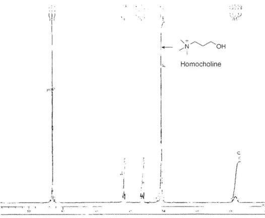
3-N-trimethylarnino-1-propanol iodide (homocholine) was prepared by N-methylation ofDMA-Propanol following the method of Hassan et al. (2007). Briefly, Methyl iodide (160 mmol) was added dropwise to a solution ofDMA-Propanol (100 mmol) in diethyl ether (200 rnl) at room temperature. After stirring for 1 h, the mixture was filtered. The precipitate was dried in a vacurnm dessiccator for overnight and afforded white powder (85 %). The structure and purity of homocholine were confirmed according to its 1H NMR spectrum (ECP 500 MHz, NMR spectrometer; JEOL). Approximately 5 mg homocholine was dissolved in D20 (Merck, Co. Inc., Darmstadt, Germany) and analyzed using 1H NMR. The strong signal of NMR spectrum at 2.9 ppm (Fig. 2.1) reflected the presence of trimethyl group with 99% purity.
3-N-trimethylarninopropionaldehyde was prepared from 3-arninopropion- aldehyde diethylacetal by methylation with methyl iodide. Large excess amount of
methyl iodide (42 ml) was added to a suspension of 3-aminopropionaldehyde diethylacetal (5.0 g, 34.0 mmol) and K2C03 (11.7 g, 35 mmol) in 50 ml ethanol at room temperature. After stirring at 40°C for 2 h, the rrllx.ture was cooled, and evaporated in vacuo. The residue was dissolved in 50 ml water and then extracted with CH2Cl2 (50 ml x 3 times). Combined organic extracts were dried over sodium sulfate, filtrated and concentrated. The residue was crystallized from methanol-ethylacetate to give colorless solid (8.5 g, 77%) of 3-N- trimethylaminopropionaldehyde diethylacetal iodide.
Thereafter, it was hydrolyzed by 0.1 M HCl for 3 h at room temperature to produce TMAPaldehyde iodide as a 30 mM solution. The structure of TMAPaldehyde iodide was determined by 1H-NMR spectroscopy and capillary electrophoresis .
(I ..
I '
r
"'
-· ... ----·"""-.
~--.,:--:,
~,
"2:~~-~-·" -.
I;
(
"N
""''./"''oH. /
lv_ Homocholine
c
I r
ll
~.
1 );...- - - ~r ~\._ _ _.._._,___ ~---·---·______/' ·~--- .!
- - - · , ...
Fig. 2. 11H NMR spectrum of chemically synthesized 3-N-trimethy lamino-1-propanol (Homocholine)
2.2.4 Screening and Isolation of Homocholine Degrading Bacteria
Enrichment cultures of soil samples from different location in Tottori University and around Tottori City, Tottori, Japan, were used to obtain homocholine degrading strains.
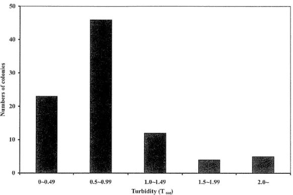
Approximately 1 00 mg of each soil sample was inoculated into 5 rnl basal medium (basal-homocholine media; Table 2.1) containing 5 giL homocholine as sole source of carbon and nitrogen; 2 giL KH2P04; 2 giL K2HP04 ; 0.5 giL MgS04 • 7H20; 0.5 giL yeast extract; and 1 giL polypeptone (pH 7.0). Cultivation was done at 30°C for 2 to 7 days in reciprocal shaker at 144 strokes/min. Subsequently, 200 ~ of the culture was transferred to fresh basal-homocholine (basal-HC) media for another 2 days incubation.
After enrichment culture, an appropriate amount (1 rnl) of culture solution was taken and serial 10-fold dilutions were prepared with physiological saline. Then, 100-~
samples of the 107 to 109 dilutions were plated on agar medium of basal-HC (Table 2.2) and/or meat extract (Table 2.3) and then incubated at 30°C for 2 days. After successive transfers to new plates, individual, distinguishable colonies were selected and stroked into slant media. Single colonies were reinoculated in basal-HC liquid media, and the cell growth was estimated by measuring the turbidity at 660 nm with Novaspec II spectrophotometer (Amersham Pharmacia Biotech, Uppsala, Sweden). A suitable control was set using basal media without substrate (homocholine). The highly growth isolates were either stroked into basal-HC slant media and stored at
soc
in a cold room or transferred to 50% glycerol and stored in a refrigerator at -80°C. Highly growth isolates were further screened for the removal of homocholine from the growth media using TLC plates and capillary electrophoresis as described bellow.Table 2. 1 Basal-homocholine (basal-HC) liquid medium TMA-Propanol iodide (Homocholine)
K2HP04 KH2P04 MgS04.7H20 Yeast extract Peptone Total volume
5.0 g 2.0 g 2.0 g 0.5 g 0.5 g 1.0 g 1000 mL
Table 2. 2 Basal-homocholine (basal-HC) agar medium
TMA-Propanol iodide (Homocholine) 5.0 g
K2HP04 2.0 g
KH2P04 MgS04.7H20 Yeast extract Peptone Agar
Total volume
Meat extract Peptone NaCl Agar
Table 2. 3 Nutrient broth (meat extract) media
2.0 g 0.5 g 0.5 g 1.0 g 15.0 g 1000 mL
5.0 g 10.0 g
5.0 g 15.0 g
Total volume 1000 mL
The pH of all media was adjusted to pH 7.0 with either 1M NaOH or 1M HCl.
2.2.5 Morphological, Biochemical and Physiological Characterization
The morphological characterization of the cells was examined using optical microscope (Olympus BX 41; Olympus Corp., Tokyo, Japan) under light and phase contrast conditions, and by using a transmission electron microscope (JEOL 100 CX II Tokyo,
Japan). For observation of cell morphology by transmission electron llllcroscopy (TEM), cells were grown on basal-HC liquid media to both exponential and stationary phases, collected and suspended in physiological saline solution. A small drop of the suspension was placed on carbon-coated copper grid (Nisshin EM Co. Ltd, Tokyo, Japan) and negatively stained with 0.1% uranyl acetate. After air-drying, the grids were examined by the electron microscope. The Gram staining was tested using a Gram staining kit (Wako Pure Chemical Industries, Ltd. Tokyo, Japan) according to the manufacture's instructions, and using KOH method (Buck 1982). Gram stained bacterial cells were observed using optical microscope under both light and phase contrast conditions. Catalase activity was determined by bubble formation in a 3% hydrogen peroxide (H202) solution. A colony was picked from the agar plate and mixed with the solution of 3% H202 . Frothing indicated the catalase activity. Motility was checked by water hanging drop method. Utilization of different carbon sources and selected metabolic activities were investigated by using API 20 NE commercially available test kit (Biomerieux SA, Geneva, Switzerland) following the manufacture's instructions. The API stripes tests were visually read after 24h and 48h. Utilization of selected carbon compounds and combined carbon and nitrogen sources were tested by measuring turbidity in liquid culture (5 ml) at pH 7.0 using basal medium supplemented with 5 gil (w/v) of the compound of choice. The compounds used were: choline, homocholine, 4- N-trimethylarnino-1-butanol, glycine betaine, ~-alanine betaine, y-butyrobetaine,
dimethyl glycine, dimethylarnino-1-propionic acid, sarcosine, L-carnitine, D-carnitine, propionate, acrylate, 3-hydroxypropionate, malonate, trimethylamine, dimethylarnine and monomethylamine. All tests were preformed with cells taken from the colonies pre-