• 検索結果がありません。

Diagnostic Ability of Diffusion-weighted Magnetic Resonance Imaging to Discriminate Ampullary Neoplasms : A Preliminary Study of 15 Cases

N/A
N/A
Protected

Academic year: 2021

シェア "Diagnostic Ability of Diffusion-weighted Magnetic Resonance Imaging to Discriminate Ampullary Neoplasms : A Preliminary Study of 15 Cases"

Copied!
10
0
0

読み込み中.... (全文を見る)

全文

(1)

Diagnostic Ability of Diffusion-weighted Magnetic Resonance Imaging to Discriminate Ampullary Neoplasms : A Preliminary Study of 15 Cases

Masatsugu N AGAHAMA

*1)

, Naotaka M ARUOKA

1)

, Eiichi Y AMAMURA

1)

, Yuichi T AKANO

1)

, Nobuyuki T AKEYAMA

2)

, Toshi H ASHIMOTO

2)

, Takahiro U MEMOTO

3)

, Junichi T ANAKA

3)

and Hiroshi T AKAHASHI

1)

Abstract : We assessed the diagnostic capability of diffusion-weighted magnetic resonance imaging (DWI) to predict the histological diagnosis of ampullary lesions to resolve the diagnostic uncertainty of endoscopic biopsy for ampullary neoplasms.

From January 2009 to August 2011, we performed DWI using b values of 0 and 1000 s / mm

2

for 15 patients with a histological diagnosis of ampullary lesion (ade- nocarcinoma, n= 8 ; adenoma, n =4 ; hyperplasia, n =3) . We compared the signal intensities (determined by comparing signal intensities of ampullary lesions and rating them as markedly hyperintense, hyperintense, or hypo-to-isointense relative to the duodenal wall) and the apparent diffusion coefficient (ADC, 10

­3

mm

2

/ s) values of the ampullary lesions on DWI among the three groups based on the histological diagnosis. Values are expressed as median (range) . The cancer-group lesions showed a significantly higher signal intensity than either adenoma or hyper- plasia (markedly hyperintense / hyperintense / hypo-to-isointense ; adenocarcinoma, 7 / 1 / 0 ; adenoma, 0 / 4 / 0 ; hyperplasia, 0 / 0 / 3 ; P<0.005) . The ADC values were significantly lower in adenocarcinoma at 1.46 (0.83-1.63) than in either adenoma at 2.14 (1.92-2.37) or hyperplasia at 2.06 (1.88-2.53) (P< 0.005) . In addition, the ADC values in the malignant group (adenocarcinoma) were significantly lower than those in the benign groups (adenoma and hyperplasia) (P <0.001) . The findings suggested that DWI could contribute significantly to accurate preprocedural diagnosis of ampullary lesions.

Key words : ampullary neoplasm, diffusion-weighted magnetic resonance imaging, apparent diffusion coefficient (ADC) , endoscopic biopsy, preprocedural diagnosis.

Introduction

 Ampullary neoplasms occur rarely and have a complex anatomy, making it difficult to accomplish accurate preprocedural diagnosis

1-4)

. The primary cause of this difficulty is the diagnostic uncertainty of endoscopic biopsy for ampullary lesions ; i.e., false-negative findings for cancer are frequent in the ampulla of Vater

1-4)

. Consequently, inappropriate treatment Original

1)

Department of Medicine, Division of Gastroenterology, Showa University Fujigaoka Hospital, 1-30 Fujigaoka, Aoba-ku, Yokohama, 227-8501, Japan.

2)

Department of Radiology, Showa University Fujigaoka Hospital.

3)

Department of Gastroenterological and General Surgery, Showa University Fujigaoka Hospital.

To whom corresponding should be addressed.

(2)

procedures are sometimes performed including pancreaticoduodenectomy (PD) for localized adenoma and ampullectomy for hyperplasia or invasive adenocarcinoma

1-4)

. Therefore, a more reliable method of endoscopic biopsy and / or better supplemental diagnostic tools are needed to achieve more accurate preprocedural diagnoses of ampullary lesions.

 Recently, diffusion-weighted magnetic resonance imaging (DWI) was reported to be useful for differentiating the histology of some neoplasms other than ampullary neoplasms, and has since gained wider acceptance

5-8)

. In this monograph, we show preliminary results of the investigation into whether DWI could contribute to accurate preprocedural diagnosis of ampullary lesions.

Patients and methods

 This retrospective study was approved by the institutional review board and was conducted in accordance with the Health Insurance Portability Accountability Act. The institutional review board waived the requirement for patient informed consent.

 From January 2009 to August 2011, we obtained the histological diagnosis of ampullary lesions in 17 patients. Of these, 15 patients (adenocarcinoma in 8 patients, adenoma in 4, and hyperplasia in 3) underwent magnetic resonance imaging (MRI) , while the remaining two patients did not consent to MRI due to financial reasons. We thus included the 15 patients who underwent MRI in this study and reviewed their clinical records to assess the following variables : patient demographics, clinical course, diagnostic imaging findings, methods for determining final histological diagnosis, and final histological diagnosis.

 Our basic management strategy for investigating the ampullary lesion was as follows.

Endoscopic retrograde cholangiopancreaticography (ERCP) and endoscopic ultrasonography

(EUS) was routinely performed for all patients with ampullary lesions to evaluate local extension of the lesion along the pancreatic and / or bile ducts unless the patient refused these examinations. Endoscopic biopsy was almost always performed simultaneously with ERCP. Abdominal computed tomography, MRI, and / or conventional ultrasonography were also always performed before conducting either ERCP or endoscopic biopsy to avoid mistaking postprocedural changes for intrinsic findings. When biopsy results proved the presence of carcinoma or when they were indicative of adenoma, but the lesion was not considered removable endoscopically because of considerable extension along the pancreatic and/or bile ducts, the patient was referred for surgery. If the biopsy results were indicative of adenoma and the lesion was considered removable endoscopically, endoscopic papillectomy (EP) was performed according to the method described elsewhere

9)

. When the papillectomy margin was positive for tumor and the residual lesion was considered removable endoscopically, repeat endoscopic resection was applied. When the papillectomy margin was positive for tumor and repeat endoscopic resection was considered infeasible, or the papillectomy specimen contained a malignant component, the patient was referred for surgery. If the biopsy results showed hyperplasia, endoscopic biopsy was performed every 3 months until 12 months after the initial diagnosis, and biannually thereafter.

 MRI was performed using a 1.5-T MRI unit (SignaHDxt ; GE Healthcare Japan, 4-7-127

(3)

Asahigaoka, Hino-shi, Tokyo, Japan) . T1-weighted images, T2-weighted images, and magnetic resonance cholangiopancreatography were acquired first. Thereafter, DWI using b values of 0 and 1000 s / mm

2

was performed. The MRI images were evaluated with a picture archiving and communication system (PACS) (Impax 4.0 ; Agfa, Mortsel, Belgium) by two experienced physicians (N. T., radiologist with 14 years of experience ; M. N., gastroenterologist with 21 years of experience) . Images were evaluated first qualitatively and then quantitatively. The DWI image was displayed to the two reviewers without any other information concerning the patient.

For qualitative analysis, the signal intensity(SI) of the ampullary lesion on the DWI with a b value of 1000 s / mm

2

was determined by comparing SI between the lesion and the adjacent duodenal wall, and was classified as markedly hyperintense relative to the adjacent duodenal wall markedly hyperintense ;(MHI) , hyperintense (HI) , or hypo-to-isointense (HII)
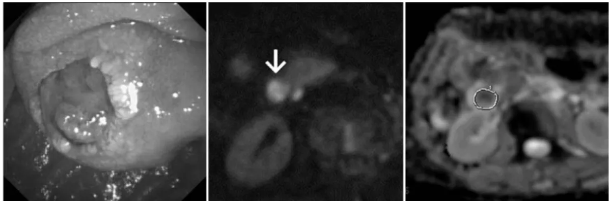
6)

. For quantitative analysis, the apparent diffusion coefficient (ADC) value was calculated by manually placing a region of interest (ROI) over the ampullary lesion. The ROI was drawn freehand along the outline of the lesion identified in the DWI images used for qualitative analysis and was set as large as possible (Fig. 1) . Care was taken to avoid the inclusion of any area outside the tumor in the ROI. SI and ADC values were determined on a single slice of DWI images containing the largest available tumor area in each patient. After independently evaluating the images alone, the two reviewers subsequently discussed the findings to reach a final consensus.

 To assess the usefulness of DWI for differentiating histological diagnoses of ampullary lesions, we compared the SI and ADC values of the lesion among the following three groups based on the final histological diagnosis: adenocarcinoma group (ACG) , adenoma group (AG) , and hyperplasia group (HG) .

 The statistical analyses were performed by a nonparametric method as follows. Fisherʼs exact probability test was used for categorical variables, and the Mann-Whitney U-test was used to analyze numerical variables. Results are shown as percentages and / or rates of patients, or as median (range) . Commercial statistical software (SPSS 13.0 for Windows ; SPSS, Chicago, IL)

was used, and two-tailed P values of less than 0.05 were considered significant.

Results

Clinical course and determination of the final diagnosis

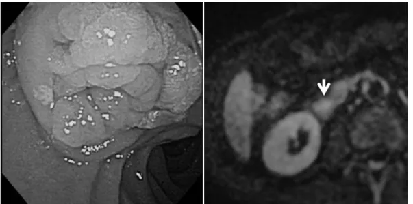
 Table 1 details all patient characteristics. In the ACG, one patient (Case 2) rejected the offer of either surgery or chemotherapy and was therefore treated with endoscopic stenting alone. In Case 3, the initial diagnosis was adenoma diagnosed by biopsy specimen. However, the EP specimen showed that the tumor was mainly composed of cancer. Therefore, the patient was referred for surgery and underwent PD. In Cases 4 and 6 (Fig. 1) , distant metastases were present when diagnosing the ampullary lesion histologically, and because these 2 patients did not wish to receive chemotherapy, they were managed with endoscopic stenting alone. In Case 8

(Fig. 2) , although the endoscopic biopsy specimen showed adenoma, the EUS findings showed

extension of the tumor across the duodenal wall, suggesting a cancerous lesion. Therefore,

the patient did not undergo EP and was referred for surgery to undergo PD after obtaining

(4)

informed consent. The PD specimen revealed that the tumor was mainly composed of adenoma, with some areas of adenocarcinoma.

 In the AG, two patients underwent PD. In Case 9, extension of the lesion along the bile

Table 1.  Details of the 15 patients included in the present study.

Case No

Age

(yr) Gender cholangitis

Dilatation of BD

/ PaD

Presence of jaundice

Tumor size

(mm)

Signal intensity on

the DWI

ADC value of DWI

( 10

­3

mm

2

/s)

Final histological diagnosis

Method or specimen for final diagnosis

Treatment

1 62 Female no yes/yes no 15 MHI

||

1.49 adenocarcinoma Endoscopic biopsy PD

2 88 Female no yes/yes no 15 MHI

||

1.31 adenocarcinoma Endoscopic biopsy ES

**

3 57 Female no no/no no 16 MHI

||

1.48 adenocarcinoma Specimen of EP

††

PD

4 77 Male yes yes/yes yes 29 MHI

||

0.83 adenocarcinoma Endoscopic biopsy ES

**

5 71 Female no no/yes no 12 MHI

||

1.44 adenocarcinoma Endoscopic bopsy PD

6 71 Female no yes/yes no 15 MHI

||

1.51 adenocarcinoma Endoscopic biopsy ES

**

7 67 Male no yes/no no 16 MHI

||

1.40 adenocarcinoma Endoscopic biopsy PD

8 78 Male no yes/yes no 17 HI

‡‡

1.63 cancer in adenoma Specimen of PD PD

9 74 Male yes yes/no no 12 HI

‡‡

2.20 adenoma Specimen of PD PD

10 61 Female no no/no no 10 HI

‡‡

1.92 adenoma Specimen of PD PD

11 68 Female no no/no no 15 HI

‡‡

2.37 adenoma Repeated biopsies

Intensive follow up none

12 82 Male no yes/no yes 15 HI

‡‡

2.07 adenoma Repeated biopsies

Intensive follow up ES

**

13 71 Male no no/no no 9 HII

||||

1.88 hyperplasia Repeated biopsies none

14 67 Male no yes/yes no 14 HII

||||

2.06 hyperplasia Repeated biopsies none

15 62 Male no no/no no 10 HII

||||

2.53 hyperplasia Repeated biopsies none

, bile duct;

, pancreatic duct;

, diffusion-weighted magnetic resonance imaging; , apparent diffusion coefficient;

||

, markedly hyperintense to the adjacent duodenal wall; , pancreaticoduodenectomy;

**

, endoscopic stenting;

††

, endoscopic papillectomy;

‡‡

, hyperintense to the adjacent duodenal wall;

||||

, hypo-to-isointense to the adjacent duodenal wall.

Fig. 1.  Endoscopy and diffusion-weighted magnetic resonance imaging of Case 6 (adenocarcinoma) .

The endoscopy showed an obviously elevated lesion with an erosive depression in the

ampulla of Vater (left) . Diffusion-weighted magnetic resonance imaging revealed that the

tumor, which was markedly hyperintense relative to the adjacent duodenal wall (white

arrow) , was easily identified (center) . The apparent diffusion coefficient (ADC) value was

calculated by manually placing a region of interest (ROI) over the ampullary lesion. The

ROI was drawn by freehand along the outline of the lesion identified in the DWI images

used for qualitative analysis and was set as large as possible (right) .

(5)

duct was considered too long to be removed completely by either EP or transduodenal surgical ampullectomy, and PD was undertaken. In Case 10 (Fig. 3) , the patient firstly underwent EP ; however, although the ampullectomy specimen showed no malignant component, the specimen margin was positive for tumor at the bile duct direction and the residual lesion was undetectable. The patient elected to have the tumor completely removed and therefore underwent PD. The resected PD specimens for these two AG patients (Cases 9 and 10)

revealed only adenoma in the tumor. Cases 11 and 12 rejected the recommendation of either EP or surgery despite the biopsy specimen evidently showing adenoma. Therefore, these 2 patients received intensive follow up consisting of bimonthly or trimonthly endoscopic examination and biannual abdominal CT and / or MRI study. No changes in the size, morphological features, or biopsy results were observed until 28 months in Case 11 and 30 months in Case 12 after the initial diagnosis. According to their clinical courses, we consider it logical that the final diagnosis for these two patients was regarded as adenoma.

 In the HG, repeated biopsy specimens consistently revealed hyperplasia and no morphological changes were observed in Case 13 (Fig. 4) , Case 14, and Case15. Therefore, we concluded that the final diagnosis for these three cases was hyperplasia.

Comparison of variables among the three groups

 There were no significant differences in age, gender, symptoms, occurrence of cholangitis and / or pancreatitis, and size of the lesion among the three patient groups. Size of the lesion was principally measured by EUS, or by other modalities in patients who refused to receive EUS.

 The distribution in classification of the SI according to the DWI findings was significantly dif- ferent among the three groups as follows. The MHI / HI / HII ratio was 7 / 1 / 0 in ACG, 0 / 4 / 0

Fig. 2.  Endoscopy and diffusion-weighted magnetic resonance imaging of Case 8 (cancer in

adenoma) . The endoscopy showed an elevated lesion with granular surface in the ampulla

of Vater (left) . Diffusion-weighted magnetic resonance imaging revealed the tumor, which

was markedly hyperintense relative to the adjacent duodenal wall (white arrow) .

(6)

in AG, and 0 / 0 / 3 in HG, thus ACG lesions showed significantly higher SI than AG or HG.

Representative cases are shown in Fig. 1-4. Meanwhile, the ADC value of ampullary lesions was significantly lower in ACG {1.46(0.83-1.63)} than in either AG {2.12(1.92-2.37)} or HG

{2.06(1.88-2.53)} , and the malignant (ACG) group showed significantly lower ADC values than the benign (AG and HG) groups (Fig. 5) . ADC values of more than 1.88 10

­3

mm

2

/ s were seen only in the benign group, whereas ADC values of less than 1.63 10

­3

mm

2

/ s were seen only in the malignant group. In this respect, ADC values of less than 1.63 10

­3

mm

2

/ s showed 100% sensitivity and specificity in the diagnosis of a malignant lesion.

Discussion

 We recognize that the present study had several drawbacks and thus the findings must be considered preliminary. First of all, the sample size was very small. Secondly, the retrospective nature of the study impeded the integrity of any conclusions that could be drawn. In spite of these problems, however, the fact that the SI and ADC values on DWI enabled a clear distinction between cancerous and benign lesions in the present study suggests that DWI is a very promising modality for the discrimination of ampullary lesions.

 The precise reason why DWI is able to show malignancies as hyperintense is not yet known.

However, the high cellularity of malignant tumors is assumed to be the main reason, with water molecules in the extracellular fluid of cancer tissue possibly more restricted in Brownian motion than water molecules in the surrounding normal tissue

6)

. The mechanism of visualizing body malignancies on DWI is therefore assumed to be basically the same as that of visualizing a fresh brain infarction.

Fig. 3.  Endoscopy and diffusion-weighted magnetic resonance imaging of Case 10. In this patient,

the ampullary lesion was confirmed to be totally composed of adenoma based on

examination of the pancreaticoduodenectomy specimen. The endoscopy showed a distinct

elevation with marked redness in the ampulla of Vater (left) . Diffusion-weighted magnetic

resonance imaging revealed the tumor, to be slightly, but obviously, hyperintense relative to

the adjacent duodenal wall (white arrow) .

(7)

 In terms of the rare occurrence of ampullary lesions, a multicenter prospective study is considered practical for gaining the large sample size necessary to achieve more robust statistical significance and to yield the ADC cut-off values for predicting the malignancy of an ampullary tumor. However, some issues remain to be resolved before a multicenter study can be conducted. Significant variability in absolute ADC values seems inevitable depending on the

Fig. 5.  Scatter plotting of the apparent diffusion coefficient value of diffusion-weighted magnetic resonance imaging. The apparent diffusion coefficient value of diffusion-weighted magnetic resonance imaging drew a clear distinction between malignant and benign lesions.

Fig. 4.  Endoscopy and diffusion-weighted magnetic resonance imaging of Case 13 (hyperplasia) .

The endoscopy showed a thick tortuous elevation with a relatively smooth surface in the

ampulla of Vater (left) . Diffusion-weighted magnetic resonance imaging only visualized the

tumor, which was isointense relative to the adjacent duodenal wall (white arrow) .

(8)

coil systems, imagers, vendors, and field strength

10-12)

, and ADC values may also vary according to the b values used

10-13)

. Furthermore, interobserver variability in ROI size and positioning in a single slice method is reportedly significant

14)

. To eradicate these issues and conduct a multicenter study, inter-institutional validation using a temperature-controlled fluid for quality control

12)

, calculation of ADC value using intravoxel incoherent motion theory using 3 or more b values

13, 15)

, and application of whole-volume ROIs

14)

are currently under consideration in our community.

 We recognize that our current management algorithm for ampullary lesions, which includes EP, should be considered challenging at this stage because the occurrence rate of potentially life-threatening complications in patients receiving EP is reported to be markedly higher than in those who underwent endoscopic resection of gastric or colonic lesion, and the efficacy of EP remains unjustifiable

1-4)

. Furthermore, it is uncertain which of the following should be applied for a salvage procedure when the ampullectomy margin is positive for tumor, i.e., repeat endoscopic resection, transduodenal resection, or PD

1-4)

. Thus it is difficult to state that EP is an established treatment procedure. In addition, it has been reported that preoperative biliary drainage causes significant fibroproliferative inflammatory changes in bile ducts, making dissection difficult on performing PD

16)

and increasing the complication rate after PD

16-18)

. EP is obviously a more complicated procedure and thus considered to adversely affect the adjacent tissues more than biliary stenting alone, suggesting that PD following ampullectomy could be more technically difficult than PD without prior ampullectomy. Conversely, it is obviously excessive if PD is applied for localized adenoma with low-grade dysplasia

1-4)

. In other words, an inappropriate choice of treatment procedure would have a markedly larger impact on the clinical course in patients with ampullary lesions than in patients with early gastric or colonic cancer, both of which are often treated endoscopically

1-4, 16-18)

; thus, accurate preprocedural diagnosis is essential for avoiding such an inappropriate choice. However, the reported false-negative rate of endoscopic biopsy for cancer in ampullary lesions is high at 12〜60%

4, 19-21)

. In fact, two (25%)

of the 8 patients with ampullary cancer in the present study showed a false-negative finding on endoscopic biopsy for cancer. Some authors reported that deeper biopsy after sphincterotomy was useful for ensuring an accurate diagnosis in their retrospective studies

22-24)

. However, a prospective study concerning this subject showed that sensitivity was 21% before and 37% after sphincterotomy, and concluded that endoscopic biopsies do not allow for reliable preoperative diagnosis of ampullary lesions

25)

. Therefore, it is considered necessary to establish a more reliable method for endoscopic biopsy and / or an alternative diagnostic tool that could help to definitively discriminate ampullary neoplasms. We consider that DWI is a promising candidate as such a diagnostic modality to reliably discriminate ampullary lesions based on the present results.

We now plan to conduct a multicenter prospective study to gain more robust statistical power and to yield adequate cut-off ADC values for predicting the malignancy of ampullary lesions.

 In conclusion, DWI showed significantly higher SI and significantly lower ADC values in

patients with ampullary cancer than in patients with either adenoma or hyperplasia. Therefore,

we believe that DWI could contribute significantly to the accurate preprocedural diagnosis of

(9)

ampullary neoplasms.

Conflict of interest  None declared.

References

1) Yoon YS, Kim SW, Park SJ, et al. Clinicopathologic analysis of early ampullary cancers with a focus on the feasi- bility of ampullectomy. Ann Surg. 2005;242:92-100.

2) Winter JM, Cameron JL, Olino K, et al. Clinicopathologic analysis of ampullary neoplasms in 450 patients: Impli- cations for surgical strategy and long-term prognosis. J Gastrointest Surg. 2010;14:379-387.

3) Katsinelos P, Paroutoglou G, Kountouras J, et al. Safety and long-term follow-up of endoscopic snare excision of ampullary adenomas. Surg Endosc. 2006;20:608-613.

4) Kim JH, Kim JH, Han JH, et al. Is endoscopic papillectomy safe for ampullary adenomas with high-grade dyspla- sia? Ann Surg Oncol. 2009;16:2547-2554.

5) Kartalis N, Lindholm TL, Aspelin P, et al. Diffusion-weighted magnetic resonance imaging of pancreas tumors. Eur Radiol. 2009;19:1981-1990.

6) Nasu K, Kuroki Y, Tsukamoto T, et al. Diffusion-weighted imaging of surgically resected hepatocellular carcinoma:

imaging characteristics and relationship among signal intensity, apparent diffusion coefficient, and histopathologic grade. AJR Am J Roentgenol. 2009;193:438-444.

7) Sandrasegaran K, Sundaram CP, Ramaswamy R, et al. Usefulness of Diffusion-weighted imaging in the evaluation of renal masses. AJR Am J Roentgenol. 2010;194:438-445.

8) Paudyal B Paudyal P, Tsushima Y, et al. The role of ADC value in the characterisation of renal carcinoma by diffusion-weighted MRI. Br J Radiol. 2010;83:336-343.

9) Matsusaki S, Okano H, Nishikawa K, et al. Endoscopic diagnosis and treatment for tumor of the papilla of vater.

Gastroenterol Endosc. 2009;51:1738-1747.(in Japanese) .

10) Sasaki M, Yamada K, Watanabe Y, et al. Variability in absolute apparent diffusion coefficient values across differ- ent platforms may be substantial: a multi-vendor, multi-institutional comparison study. Radiology. 2008;249:624-630.

11) Braithwaite AC, Dale BM, Boll DT, et al. Short- and mid-term reproducibility of apparent diffusion coefficient measurements at 3.0-T diffusion-weighted imaging of the abdomen. Radiology. 2009;250:459-465.

12) Chenevert TL, Galban CJ, Ivancevic MK, et al. Diffusion coefficient measurement using a temperature controlled fluid for quality control in multicenter studies. J Magn Reson Imaging. 2011;34:983-987.

13) Le Bihan D, Breton E, Lallemand D, et al. Separation of diffusion and perfusion in intravoxel incoherent motion MR imaging. Radiology. 1988;168:497-505.

14) Lambregts DM, Beets GL, Maas M, et al. Tumour ADC measurements in rectal cancer: effect of ROI methods on ADC values and interobserver variability. Eur Radiol. 2011;21:2567-2574.

15) Guiu B, Cercueil JP. Liver diffusion-weighted MR imaging: the tower of Babel? Eur Radiol. 2011;21:463-467.

16) Wagholikar GD, Sikora SS, Pandey R, et al. Morphological changes in bile ducts following preoperative biliary stenting. Indian J Gastroenterol. 2003;22:166-169.

17) Eshuis WJ, van der Gaag NA, Rouws EA, et al. Therapeutic delay and survival after surgery for cancer of the pancreatic head with or without preoperative biliary drainage. Ann Surg. 2010;252:840-849.

18) Morris-Stiff G, Tamijmarane A, Tan YM, et al. Pre-operative stenting is associated with a higher prevalence of post-operative complications following pancreatoduodenectomy. Int J Surg. 2011;9:145-149.

19) Galandiuk S, Hermann RE, Jagelman DG, et al. Villous tumors of the duodenum. Ann Surg. 1988;207:234-239.

20) Beger HG, Treitschke F, Gansauge F, et al. Tumor of the ampulla of Vater: experience with local or radical resec-

(10)

tion in 171 consecutively treated patients. Arch Surg. 1999;134:526-532.

21) Clary BM, Tyler DS, Dematos P, et al. Local ampullary resection with careful intraoperative frozen section evalua- tion for presumed benign ampullary neoplasms. Surgery. 2000;127:628-633.

22) Bourgeois N, Dunham F, Verhest A, et al. Endoscopic biopsies of the papilla of Vater at the time of endoscopic sphincterotomy: difficulties in interpretation. Gastrointest Endosc. 1984;30:163-166.

23) Huibregtse K, Tytgat GN. Carcinoma of the ampulla of Vater: the endoscopic approach. Endoscopy. 1988;20 suppl 1:

223-226.

24) Ponchon T, Berger F, Chavaillon A, et al. Contribution of endoscopy to diagnosis and treatment of tumors of the ampulla of Vater. Cancer. 1989;64:161-167.

25) Menzel J, Poremba C, Dietl KH, et al. Tumors of the papilla of Vater- inadequate diagnostic impact of endoscopic forceps biopsies taken prior to and following sphincterotomy. Ann Oncol. 1999;10:1227-1231.

[Received September 2, 2013 : Accepted June 24, 2014]

Table 1.  Details  of  the  15  patients  included  in  the  present  study.
Fig. 2.  Endoscopy  and  diffusion-weighted  magnetic  resonance  imaging  of  Case  8  (cancer  in  adenoma)
Fig. 3.  Endoscopy  and  diffusion-weighted  magnetic  resonance  imaging  of  Case  10
Fig. 5.  Scatter  plotting  of  the  apparent  diffusion  coefficient  value  of  diffusion-weighted  magnetic  resonance  imaging

参照

関連したドキュメント

Correspondence should be addressed to Salah Badraoui, [email protected] Received 11 July 2009; Accepted 5 January 2010.. Academic Editor:

7 The current density J z at the center of the channel is higher for a micropolar fluid than that for a Newtonian fluid, and it will decrease as the microrotation parameter

The system evolves from its initial state without being further affected by diffusion until the next pulse appears; Δx i x i nτ − x i nτ, and x i nτ represents the density

A class of nonlinear fourth-order telegraph-di ff usion equations TDE for image restoration are proposed based on fourth-order TDE and bilateral filtering.. The proposed model

(In a very recent preprint, Niethammer and Vel´azquez [9] have obtained a remarkable estimate for the effective potential of a single particle in the supercritical case by taking

Theorem 4.8 shows that the addition of the nonlocal term to local diffusion pro- duces similar early pattern results when compared to the pure local case considered in [33].. Lemma

Ruan; Existence and stability of traveling wave fronts in reaction advection diffusion equations with nonlocal delay, J. Ruan; Entire solutions in bistable reaction-diffusion

We formulate the heavy traffic diffusion approximation and explicitly compute the time-dependent probability of the diffusion approxi- mation to the joint queue length process.. We