Japanese Physical Therapy Association
JapanesePhysicalTherapyAssociation
me\twzaeeag16ts
ng
1
53N9ff
0989ij)
Effectof
Hindlimb
Suspension
Gastrocnemius
Muscle
on
Young
and
Adult
in
Mouse*
Nobuhide
HAIDA
andKatsuhiko
TACHINO*"
ABSTRACT
The
purpose
of
this
studywas
to
determine
the
effect
efhypokinesiafhypodynamia
(H/H)
onanthropometric,
histochemical
and
histologic
characteristics of medialgastrocnemius
niuscles
(MG)
in
adult versusimmatuTe
mice.Hindlimb
suspensionfor
two
weeks was usedte
produce
atrophyin
two
groups
of mice, ages4
andI2
weeks, with non-suspendedanimals
serving' as controls.Young
HfH
mice exhibited markeddecreases
in
body
weight and muscle weight.H/H
reducedthe
diameter
ofboth
type
I
andtype
2
fibers,
increased
the
percentage
eftype
I
fibers,
and de-creased thepercentage
oftype
2
fibeTs
in
young
MG.
For
all measuTementsyoung
mice were more affectedthan
adult micc.The
greater
effgcton
young
mice
suggeststhat
most ofthe
atrophy occurTing
with
HrH
wasdue
to
developmental
aTrest,Key
words:Hypoklnesia!Hypodynamia,
Gastrocnemius,
Atrophy
Introduction
Djsuse
atrophy maybe
produced
in
animals
by:
(1)
isolution
of
the
lumbo-sacral
cord orthe
peripheral
innervation,
(2)
limb
immobilization
(joint
fixation
by
casting,
joint
pinning
andbracing),
(3)
muscletenotomy,
(4>
prolonged
gen-eral
anesthesia,(5)
smallcage
restraint,
(6)
space
fiight
hypokinesia,
and(7)
hindclimb
suspensionhypokinesiafhypodynamia
(HIH).
HfH
produced
by
hindlimb
suspension
differs
from
other models ofdisuse
atrophyin
that
the
hindlimb
muscles
are
free
to
undergo
a
full
Tange
of voluntaryisotonic
contractions.
Despite
freedom
ofmove-ment,
hindlimb
musclesin
the
suspended animalhave
reduced
mechanicalIoading
(hypodynamia)
anddecreased
metor
activity
(hypokinesia).
HIH
*
igffenlrc
jz
ig
yv-rwFlvamaotswa\UtJErflt
**
lkmff,pt,
scwrnex
:siFIJit\Kml"immraft\gve\tsra
#*;F-・・・Departmtint-o'f
Physi・cal・Therapy,
School
ofAllied
Medical
Profes$lons,
Kanazawa
University
(Receiyed
12
October
1987)
suspension results
in
the
musclesbecoming
virtu-ally electricallysilent
as
measured
by
electro-myography.
However,
allactivities
are notin-hibited
in
normalyoung,
active animalsZ}-3).AIthough
severalstudies
regardingthe
effect
of
HfH
on
skeletal
muscle
have
been
reported,
most
previous
investigations
have
been
carried
out
in
young
immature
rodentsin
the
age
range
of
three
to
seven
weeks.
One
o,f
the
major
unresolved
probleips
in
those
studies wasthe
useof
growing
animals
in
whichthe
induced
atrophy mightbe
due
to
developmental
arrest ratherthan
to
a wastingaway
oftissu'e.
The
objectives
of
this
study
were:
(a)
t.o
evalu-ate
the
effect ofHIH
ongastrocnemius
musclesof
mice
with
anthropornetric,
histologic
and
histo-chemical
measurements,and
(b)
to
compare
ef-fects
ofHfH
on}'oung
and adult aninials.Methods
'
'Rodents
used
for
thifi
s.tudy
were
4
nc*taza#
strain,
Forty
male mice, age4
orI2
weeks,
ware randomlydivided
into
two
group$,
HIH
apdcontrol
(CON).
HfH
wasinduced
by
using a modifiedMorey
method4),The
hindquarters
were su$pendedfor
aperiod
o{1two
weeks,
usingbody
haTness
and
swivel
in
a
slighthead-down
tilt
that
barely
prevented
the
hindlimb
from
contacting
any
supportive
surface.
Access
to
feod
and
watcr waspermitted
by
use ofthe
forelimbs
on
a
grid
surfaced
floony,
Laboratory
Rat
Chow
and water were available ad
libitum.
AEter
two
ureeks
of
suspension,
at
age
6
or14
weeks,the
micewere
sacrificedby
cervical
dis-location,
Gastrocnemius
muscles were rapidlyexcised
and
weighed,
)Veights
were
expressed
as absolute(mg)
and relative(mg/body
weight)
value.
Medial
gastrecnemius
(MG)
sampleswere
frozen
in
isopentane
cooled
to
freezing
point
withliquid
nitrogen.Tissue
wasthen
sectiolledat
10um
in
a cr>,ostat at-200C,
For
staining, myosinATPase
atpH
IO.3
and4,6,
and
Hema-toxylin
andEosin5}
were used.Staining
for
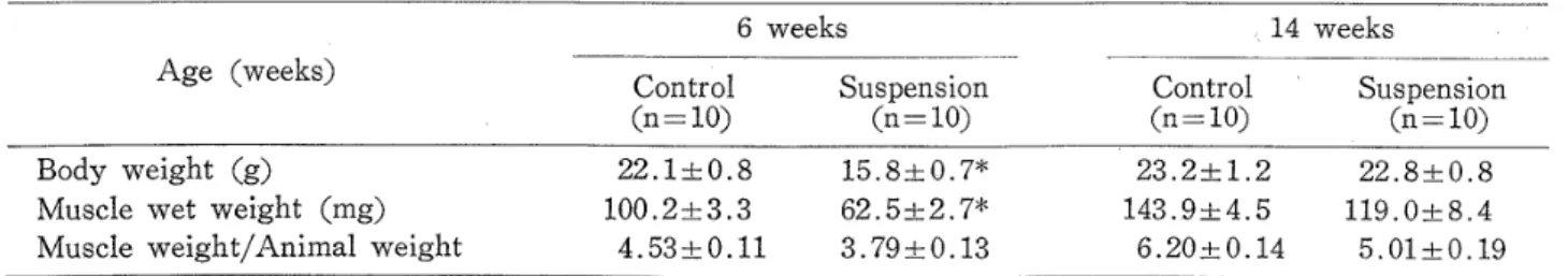
Table
1
Anthropometric
ee16#ag
1
e
measurements
m>rosin
AT'I'ase
identified
fast
twitch,
type
2
fibers
(alkali
stable) andslow
twitch,
type
1
fibers
(acid
stable).The
fiber
aTeasand
the
proportions
of
type
l
and
type2
fibers
were
obtained
with
a
semi-automatic morphometric s>rstem
(Cosmozone
98,
Nilion
Kohgaku).
C}ualitative
analysls ofthe
morpholegylpathology
of
the
muscles
wusper-formed
using
a
modified
Dubowitz's
method6).
Stuclent's
t-test
ivas usedto
examinethe
effectof age,
HIH,
andfiber
types
onthe
histochemical
measurement.
Significance
was accepted atp<
O.05.
Results
Anthropometric
measurements:Suspension
sig-nificantly Teducedthe
body
vreight
ofthe
6
week
old
mice
(22.]
±O.8g
for
the
CON,
I5,8
±O.7g
for
the
NIH),
but
did
not affectthe
14
week old mice(2S,2
±1.2
g
for
tlie
CON,
22.8
±O.8
g
for
the
Hi'H).
There
were
marked
decreases
in
tlie
young
muscle weight and muscleweightfbody
weight.
Older
m.icewere
less
affected
(Tab]e
1),
'
of
young
and
adult
mice6
weeksAge
(weeks)
Centrol(n=10)Suspension
(n=10)
14.weeks
Control
Suspension
(n=IO)
(n=10)
Body
weight
(g)
Muscle
wet weight(mg)
Muscle
weight!Animalweight
22.1
±O.8
100.2
±3,3
4.53
±O.11
15.8
±Q,7*
62.5
±2.7*
3.79
±,
O.13
23,2
±1.2
143.9
±,
4.5
6.20
±O.14
22,8
±O.8
119,O
±8.4
5.01
±O.19
ValuesControlare
means ±S.E.
vs.
suspension,
*P<O.05Table
2
Fiber
size, andfiber
typecomposltlonin
control and suspended muscles6
weeks
Suspension
(n=:1O)
14
Control(n
==1O)
weeks
Age
(weeks)
Control(n=:10)
Suspension
(n=10)
Fiber
size
type
1
type
2
Fiber
type
type
1
type
2
(ptrnE)
composition(%)
1,041
±100
975
±114
13.6
±O.9
86.4
±1.0
566
±50*
548
±39*
19.3
±2.4*
81.7;2.2
1,310
±249
1,161
±244
13,O
±O,7
87.0
±,
O.7
785
±86*
979
±150*
16.7
±2.9
83.3
±2.9
ValuesControlare
means ±S.E,
Japanese Physical Therapy Association
JapanesePhysicalTherapy Association
・i"・:/-.tk
''giii//I/1111///i/l/i:{"t'
"as'sc
Hindlirnb
suspension ongastrocnemius
muscle'y.rTee....l,{,//./j.tfu,ss
/
k.
'
,ee
eeesx
ee
gi/M
ac
ases
ee
sc
ue
60
40
20
./.×
Control
OType
1
eType
2
・×
eN"
Xe
a
t.--o
s
O'Xo5
ptee}
wwps...'W"eskge.tsew&a
tslr,'.';i'I.-li"i""Sli(iff
."
e"S"e//,es・,mas'twes.
tv,t.yg
'
'
・・,.\'max'
9t.
60no"
g
4oq8P-20k
/
.6week
olduHIHx.
XXX,
ox---o.
×
×
.× .
60
40
20
14week
old"HIH./eXX
/o/oxl
×
.× .
x.
×
.Fig.
1Sections
of
gastrocllemius
muscles
stained
for
myosin
ATPase
at
pH
9.4
from
eont-rol(A),
6
week old musele(B)
and14week
eld muscle(C)
with suspension.In
this
pre-paration,
Type
1
fibers
are appearedpale,
andType
2
fibers
aredark
(Type
2B)
orintermediate
(Type
2A),
×115.
Histochemical
characteristics:Representative
histochemical
data
are shownin
Fig.
1.
Figure
1,
A-C,
was stainedfor
myosinalkali-ATPase.
In
contrast
to
Fig.
IA,
which wasfrom
a controlMG
and
showsnormal
fiber
size,Figs.
IB
andFig.
2
Distribution
lations
sented
ATPase
IC,
were
from
H/H
muscles, and showreductiori
of
the
diameter
of
both
type
1
and
type
2
fibers.
Fiber
atrophy
was
greater
at
6
than
at
14
weeks
oE
age
(Table
2).
Type
I
fiber
size
decreased
46%
and
41%,
and
type
2
fiber
sizedecreased
44%
andI6%,
respectively,
for
the
6
andI4
week
rnice,There
was
also a significantincrease
in
the
percentage
oft}rpe
1
fibers
(from
13.6%
to
l9,39{,)
in
the
young
MG
with aparallel
decrease
in
2
fibers
from
suspension.
HIH
did
not
signifi-cantly
alter
the
fiber
type
distribution
in
old
MG.
Fig.
2
$1iows
the
distribution
ef
type
1
andtype
2
fibers
in
MG.
From
these
data
suspended
young
mice show
greater
atrophyof
both
fiber
types
than
CON
er
old
mice.
Histopathological
evaluation:Figure
8A
shows
a
section of aMG
from
aCON
mouse.Figure
8B
4
8
12
16
20
×
lo2
Fiber
size(gem2)
of
type
1
andtype
2
fiber
popu-from
gastrocnemius
museles arepre-as a
function
offiber
slze.Myosin
stainingidentified
fiber
type.
6
pt\ffza*
Table
3
Incidence
offiiber
abnormalitiesin
centroland suspended
rnuscles
6
weeks14
weeks
Age
(weeks)
control
SuEPen'
control
SU.SPen"
ston slon
(n=10)
(n=10)
(n=tlO)
(n=10)
Internal
nucleiNecrosis
Phagocytosis
Basophilia
Fiber
splittingInflamrnation
Fibresis
oooooo3 54215310 oooooo2 o111119
ee
t
es
s
wa
ew
X
ss
ee
g,ee
ew
ueg/
ee
,
ee
1ee
ee
ss
ee
es
lee
tw
///
,ge,
ts
/ee
la
li
,
/ha
Slli
'
ee
y
/
boew'
/
ee
ma
,
ges
ee
ee
es
Fig.
3
Sections
ofgastrocnemius
musclesstained
for
hematoxylin
and eosinfrom
controlmouse
(A).
Fig.
3B.
shows musclefibers
with
internal
nuclei(arrows) ,andgreater
liferation
ofthe
perimysial
connectivetissue
than
in
Fig.
3A.
×175.
gives
evidence ofinternal
nuclei and anincrease
of connectivetissue.
As
canbe
seenin
Table
3,
suspension
resultedin
the
occurrence of severalhistopathological
changes
in
both
agegroups.
The$e
consisted
ofthe
appearance orincrease
in
ng16kse
1
e
internal
nuclei,phagocytosis,
fibrosis,
andfiber
necrosis and splitting.
There
was noevidence
of
neuropathic
change,
In
CON
micethere
werea]so
a
few
fiber
abnormalities
but
only
in
the
6
week
old
MG.
Discussion
Mice
usedin
this
study
were6
and14
weeks ofage
at
the
termination
of atwo
week suspensionperiod.
H!H
had
marked
effects
on
the
6
week,
but
yery
little
effect
on
the
14
week
eld
mice,
Comparison
withtlie
other studies reported usingolder
rodents
is
diracult
since
different
periods
of
suspension
were
used.
XMith
three
or moTeweeks
of
HIH,
muscle
atTophy
clearly
occurred
in
adult
rats2),T}.
LeBlanc
et
al.S}found
the
same
degree
of
atrophy
in
rodents
ranging
in
age
from
13
to
29
weeks withtwo
weeksof
suspen-sion,
but
McNulty
etal.9)
reported
results
similar
to
ours
in
i7
week
eld
rats
with
HIH
periods
as
long
as
four
weeks.
A
major criticism olthe
HfH
modelhas
been
the
use
of
young
growing
animals
since
atrophy canbe
due
to
the
result ofdevelopmental
arrestas
well
as
to
a
decrease
in
bize
or
Iobs
oi
weightie),Although
sexualmaturity
occurrsin
the
ratand mouse
between
seven
and
eight
weeks
of
ageii),i2), muscle
growth
anddevelopment
con-tinue
for
relatively
along
period.
In
the
129
strain
of
mice,
muscle
weight
increases
upto
21
weeks
depending
upon sexalld
muscletypc.
Fiber
diameter
increases
in
a
curvilinear
manner
through
28
weeks
of
age
but
with
most
of
the
growth
occurr;ngby
10
weeksin
the
MGiL')ni4}.
There
is
a unimoclaldistTibution
consisting almost completelyof
small
iibersi3}.
Significant
histo-diemical
fiber
type
clianges
appearto
cease atteT8
weeksi-).
In
this
study,
CON
mice1iad
changes similarto
most ofthose
reportedin
the
IiLerature.
INJhile
agehad
noeffect
on
body
weight
or
muscle
weight,
there
was
afiber
sizedifference
Japanese Physical Therapy Association
JapanesePhysicalTherapy Association
Hindlimb
suspenSion
ono
£
growth
during
suspension whilethe
l4
week
old mice were at
least
approachingfull
maturity.
Therefore,
it
appearsthat
atrophy
occurring
with
HfH
in
6
week old micewas
primarily
due
to
the
result ofdevelopmental
arrest,Such
a
distinction
is
important
since
areduc-tion
in
protein
content
and synthesis capacity,and!or
a
morerapid
protein
degradation
has
been
reported
to
occur
with
HIHi5)'i9).
Loss
of'muscle
mass
in
disuse
is
due,
atleast
in
part,
to
loss
of
muscle
protein20),2i),
andslower
protein
synthesis and
faster
protein
degradation
or con-currentchanges
ofl)oth
processes
candecrease
the
deposition
of
muscle
protein22?.
Since
rates ofboth
protein
synthesis anddegTadation
are
significantly
greater
in
growing
than
in
adult
skeletal
muscle23),the
sig'nifiFanceof
these
clianges
with
HIH
in
growing
-animals
needs
clarification,
Furthermore,
the
factors
that
stimu-late
muscle
growth
in
immature
animals
might
be
affected
by
H/H
or
limb
immobilization
ofgrowing
muscle23).
'
.
H!H
resultedin
a significant reductionin
the
sizesof
type
1
and
type
2
fibers,
an
increase
in
the
percentage
ofthe
type
1
fibers
witha'
parallel
decrease
in
the
type
2
fibers
ascompaTed
to
the
CON
on
young
mice.
This
greater
influence
en
slow
twitch
fibers
is
coincident
with
the
trans-formation
of
aprop'ortion
o,fthe
type
l
fibers
into
type
2,
and
accompanied
both
by
a selectivedecline
in
the
slow
twitch
component of myosin without'a,change,
in
fast
twitch
rnyosin
and
a
shift
in
fiber
type
distribution
as
indicated
by
exantination
QE
myosin
ATPase
stained
muscles.H/H
studies2d)-26)have'shown
greater
atrophy of antigr4vity,Ioad
bearing
muscles such asthe
MC
ankleplanter
flexors.
7the
MG.is
gffected
morein
non-weight-bearingconditi'ons
than
muscles used
for
ankle
dorsal
fiexion,
such
as
tibialis
anterior.Atrophy
ofthe
MG
withHIH
may
indicate
that
it
is
normally used as ananti-gravity
muscle
in
normallocomotion
in
the
gastrocnemius
muscle7
rodent.
'
In
contrastto
otheranimal
models
of
disuse,
HfH
does
not Testrictthe
hindlimb
musclesfrom
full
range
of
voluntary
isotonic
contractions.
foVhile
virtually
electrical
silent
as
measured
by
single
unit
and
whole
muscle
electromyography,
such contractions are observed
to
occurthreugh-out
suspensioni).
However,
these
contractions
do
not
prevent
loss
ofmuscle
mass,suggesting
that
skeletal
muscle
requires
a
frequent
motor-neuron
recruitment
pattern
associated withpostural
rnaintenance
andbody
weight support.The
de-creases
in
muscle
activity
and
removal
of
load
bearing
tnight
directly
causechanges
in
fiber
type
distributiQn
which
wouldthen
result
in
a
change
in
neuralactivity2),3);iO),i5),
The
alter-native
is
that
the
decre4sed
muscle
activity
leads
to
achange
in
the
frequency
of
impulse
activity
which
then
produceS
the
atrophy.Suspension
resultedin
several
histopathological
abnormalities,
consisting
primarily
of
internal
nuclei,
fibrosis,
necroticfibers
andfiber
splitting.These
abnormalities are rnore characteristic of amyopathy
than
of a neuropathy.Templeton
et al.27} observed abnorrnalitiesthat
he
con-sidered
su,gge$tive
of
denervation,
but
we
did
not
find
anyevidence
of small angularfibers,
pyknotic
nuclear
clumps,
taTget
fibers
or
group
atrophy.
One
might even consider apossibility
that
isotonic
contractions
;vithout
aload
maybe
harmful
to
the
structural
iritegrity
of
muscle.
'
.
Severgl
co.nclusions canbe
drawn
from
this
study.
Most
ofthe
atrophy occurring withHXH
in
young
rnice
was
primarily
due
to
the
Tesult
of
deve]oprnental
arrest.
Suspension
resuitedin
a'e-treased
size ofboth
type-I
andtype
2
fibers
in
both
agegrQups.
The
significant changesin
fiber
,type
composition,
an
increase
in
the
percentage
of
the・type
1
fibers
with
a
parallel
decrease
in
the
type
2
fibers
as
compared
to
the
CON
in
youiig
mice,
demonstrates
the
potential
for
fiber
ab-8
g\rstal
normalities
by
HfH
were morecharacteristic
of a myopathythan
of a neuropathy.Acknowledgement
The
authors
thank
Dr,
W.
M,
Fowler,
Depart-ment
of
Physical
Medicine
and
Rehabilitation,
University
ofCalifornia
atDavis,
for
his
advice.
References
1)
Cerlcy,
K.,
Kowalchuk,
N.
et al,:Contrasting
effects ol suspension on
hind
limb
musclesin
tltehamster,
Exp,
Neurol,
85:
30-40,
l984,
2)
Elder,
G,C.B.,
McCemas,
AJ.:
Development
ofrat muscle cLuTing short- and
long-term
hindlimb
suspension.
J,
Appl,
Physiol,
62:
19I7-l92B,
l987,
3)
Misulis,
K.E,,
Dettbarn,
SV,D.
et ab:Influence
of
neural activity on morphelogical and
cal characteiistics of rat
skeletal
musc]e.Proc.
Soc.
Neurosci.
11:
2I3,
1985,
4)
Morey,
E.R.:
Spacefiight
andbone
turnover:
relation with a new rat model of weightlessness.
Bioscience,
29:
168-172,
1979.
5)
Dubowitz,'V,:
Muscle
Biopsy:
A
Practical
proach,
2nd
ed.,pp.
19-4e,
Bouliere
Tindall,
Philadelphia,
19S5,
6)
Ibid.,
pp,
208-212.
7)
Simard,
C.,
Lacaille,
M.
et al.:Enzymatic
tation
te
suspensionhypokinesia
in
skeletal inusclcof
young
andold
rats,Mech,
Ageing
Dev.
SS:
1-9,
1985,
8>
LeBlanc,
A,C.,
MaTsh,
C,,
et al.:Bone
and muscleatrophy with suspension of
the
rat.J.
Appl.
Physio].
58:
1669-・l675,
1985.
9)
McNulty,
A,L,,
Otto,
A,J.
et al.;Use
ofthe
veTted cage suspension
(ISC>
medel
to
induce
skeletal muscle atrophy.
Med,
Sci,
Sports
ExeTc.
18,
Suppl.:
55,
l986.
IO)
Boeth,
F,W.,
Gollnick,
P.D.:
Effect
ofdisuse
onthe structurc and
function
of skeletal muscle,Med.
Sci,
Sperts
ExeTc,
15:
415-420,
1988.
Il)
Farris,
E.J,,
Grithth,
J.Q,:
The
Rat
in
Laboratory
IncestigRtion
p.
8.
Linnincott,
?hiladelphia,
l949.
12>
Taylor,
R,G.,
Fowler,
W.M,
et al.:ContTactile
preperties
ofsoleus
muscleduring
ctevelopment
in
normal and
dystrophic
mice.Arch,
Phys,
Med.
Rehabil.
55:
5Sl-589,
1974.
18)
Rowe,
R.W.D.,
Goldspink,
G,:
MusLIe
fiber
growth
in
five
different
musclesin
both
sexes of mice.I.
Normal
mice.J.
Anat.
I04:
519-580,
1969.
ee16tsce
1
lik'
14>
N'ViTti,
P.,
Loermans,
H.M.
et aL:,Postnatal
grosvth
and
differentiation
of musclefibers
in
the
niouse・I,
A
histochemical
and morphometricaltion
of nermal muscle,J.
Anat,
IS7:
109-I26,
l988.
15)
Feil,
R.D.,
Gladden,
J,M,,
et al,:,
Fatigue
andtraction
of slow anclfast
musclcsiii
h},pokinetic!
hypodynamic
rats,J.
Appl.
Ph},siol.
58:
65-69,
I985.
16)
Fitts,
R,H.,
Metzger,
D.A.
et al,:Medel
ofdisuse:
a compari$on of
hindlimb
suspension andmobMzation,
J
Appl.
Ph>,siol.
60:
1946-I953,
1986.
17>
HowaTd,
G.,
Steffen,
J.M.,
etal.:
Evaluation
of
protein
synthesis regulationin
skeletal muscleattophy.
Fed,
Proc.
45:
fi45,
1986.
IS)
Musacchia,
XJ,,
Steffcn,
J,M,,
ei al,:Rat
]imb
muscle responses to suspensionhypokinesia!
h}'podynamia,
Aviat,
Space
Environ.
Med,
54-:
IOI5-1020,
l988.
19)
Thomason,
D,B.
Herrick,
R.E.
et aL:Activity
induced
recovery of slow myosin expTessioning
rodenthindiimb
suspension,]L(ed,
Sci.
Sports
Exerc.
18,
Suppl.:
s-5,1986.
20)
Federov,
I.V,,
Shurova,
I.F.;
Content
ofprotein
and nucleic acids
in
thc
tissues
of animalduring
h},pokinesia.
Kosm,
Biel.
Aviakosm
Med.
7:
17-21,
l97S,
2i)
Goldspink,
D.F.:
Tlie
influence
of activity onmusclc size and
protein
tutnover,
J.
Physiol.
(London>
264:
28S-296.
1977.
22)
Goldberg,
A,L,,
Goodman,
H.M,:
Relationship
tween
cortisone and muscle workin
determining
muscle size.
J.
Physio].
<Lendon>
200:
667-675,
1969.
23)
Booth,
F.W.:
Effect'
o £limb
immobilizution
onskeletal
muscle,J.
Appl.
Physiol.
i)2:
11IS-III8,
1982.
24)
Jaspers,
S,R.,
Tischler,
M.E.:
Atrophy
andgrowth
failure
o £ rathindilmb
muscles{n
tail-castpension.
J,
Appl,
Phlrsiel,
Respirat.
Environ,
ExeTcise.
PhysioL
57:
1472-I479.
I985,
25)
Fowler,
W.M.,
Haida,
N,
et ai,:Effect
ofkinesia
on muscle contractility andhistology
ofnormal and
dystrophic
mice.Arch.
Phys,
Med,
Rehabil,
67:
627,
1986.
26>
Musacchia,
XJ.,
Joseph,
M,
et al,:Suspension
restTaint:
Induced
hypokinesia
and antiorthostasisas a simulation of ssreightlessness,
?hysiologist
24:
S21-S22,
1981.
27>
Templcton,
G,H.,
?adalino,
J.
et al.:Influence
ofsuspension
h)'pokinesia
on rat soleus muscle.J.
Appl.
Physiol.
Respirat.
Environ.
Etercise.
Physiol,
Japanese Physical Therapy Association
Japanese Physioal Therapy Assooiation
Hindlimb
suspension
on
gastrocnemius
nluscle.
9
〈