Regulation of TNF-
α
, IL-1
β
and ICAM-1 Gene Expression in
THP-1 Monocytes Stimulated with Plasmodium
falciparum-Cultured Medium by Excretory/Secretory Products from
Spirometra erinaceieuropaei Plerocercoids
Paramasari Dirgahayu, Soji Fukumoto, Sayuri Tademoto and Kazumitsu Hirai
Division of Molecular Medical Zoology, Department of Microbiology and Pathology, School of Medicine, Tottori University Faculty of Medicine, Yonago 683-8503 JapanWe have reported that excretory/secretory (ES) products from Spirometra erinaceieuropaei plerocercoids suppress production and gene expression of tumor necrosis factor (TNF)-α in murine macrophages stimulated with lipopolysaccharide. The present study demon-strates that ES products suppress TNF-α, interleukin (IL)-1β and intercellular adhesion molecule (ICAM)-1 gene expression in human monocytic leukemia cell line THP-1 stimu-lated with Plasmodium falciparum-cultured medium (Pf-CM). Inhibition of extracellular-signal regulated protein kinase 1/2 (ERK1/2) with PD98059 reduced TNF-α, IL-1β and ICAM-1 gene expression; on the contrary inhibition of p38 mitogen-activated protein kinase (MAPK) with SB203580 increased the expression of these genes. These findings indicated that ERK1/2, but not p38 MAPK pathway is important for induction of TNF-α, IL-1β and ICAM-1 gene expression in Pf-CM stimulated THP-1 monocytes. ES products additionally suppress IL-1β, but not TNF-α and ICAM-1 gene expression in Pf-CM stimulated THP-1 cells treated with PD98059. We hypothesize that ES products may be useful in reducing
falciparum malaria-induced inflammatory response and sequestration in the late stage of
malarial diseases such as cerebral malaria.
Key words: human monocytes; mitogen-activated protein kinase; Plasmodium falciparum; Spirometra erinaceieuropaei; tumor necrosis factor-α
Plasmodium falciparum malaria is the most promi-nent parasitic infection in humans. It has been known that the pathogenesis of Plasmodium falci-parum malaria depends on many factors, including the destruction of erythrocytes, drug resistance, cytoadherence and immunological responses (Clark et al., 1989; Aikawa et al., 1990). In malaria, although complement and antibody-mediated
im-mune responses are important (Rigione et al., 1996; Guevara et al., 1997; Kumaratilake et al., 1997), cell-mediated immunity is also involved in the pro-tection and pathogenesis of this disease and some reports suggest that the host immunological re-sponses, especially those mediated through the cytokine cascade, may determine the severity of malaria (Artavanis and Riley, 2002). Severe
falci-Abbreviations: dNTPs, deoxyribonucleotide triphosphate; ERK1/2, extracellular-signal regulated protein kinase 1/2; ES, excretory/secretory; ICAM, intercellular adhesion molecule; IL, interleukin; LPS, lipopolisaccharide; MAPK, mitogen-activated protein kinase; M-MLV RT, Moloney mouse leukemia virus reverse transcriptase; Pf-CM, Plasmodium falciparum-cultured medium; PMA, phorbol 12-myristate-13 acetate; RT, reverse transcription; TNF, tumor necrosis factor
parum malaria is usually accompanied by serious secondary complication, such as cerebral malaria, severe anemia, hypoglycemia and renal impairment (Aikawa et al., 1990; Kwiatkowski, 1990). Several lines of evidences implicate that tumor necrosis fac-tor (TNF)-α is a major mediator for these complica-tions. Indeed, it has been reported there is an eleva-tion in serum levels of not only TNF-α, but also inter-leukin (IL)-1β and IL-6 in patients with falciparum malaria (Kwiatkowski et al., 1989, 1990; Kwiatkowski, 1990), which are correlated with severity of the disease and death (Grau et al., 1989; Clark et al., 1990; Kwiatkowski et al., 1990; Kurtzhals et al., 1998). A study by Udomsangpetch et al. (1997) show-ed that there were localizshow-ed high levels of inflam-matory cytokines TNF-α, IL-1β and interferon-γ in the brain tissue of a patient who died from cerebral malaria, in which an up-regulation of adhesion molecules such as CD36 or intercellular adhesion molecule (ICAM)-1 and sequestration of parasit-ized erythrocytes, lymphocytes and monocytes to the endothelial cells were associated. Identification of ICAM-1 as a major sequestration receptor, espe-cially in the brain, has led to the assumption that it contributes to the pathophysiology of severe malar-ia (Aikawa et al., 1990). In fact, in the early stages of malaria, the mild and moderate amount of in-flammatory cytokines, such as TNF-α or IL-1β, are beneficial for the host by suppressing and killing the parasite (Grau et al., 1989; Clark et al., 1990; Kwiatkowski, 1990; Kwiatkowski, et al., 1990). However, in later stages, an excessive production of these cytokines are detrimental, since activation of phagocytes to kill intracellular or extracellular parasites requires a great amount of inflammatory cytokine production which can cause adverse sys-temic effects such as severe anemia and cerebral malaria (Kwiatkowski, 1990; McGuire et al., 1994; Luty et al., 2000).
The plerocercoids of Spirometra erinaceieu-ropaei, one species of parasitic tapeworm, has been known to produce anti-inflammatory like sub-stance(s) which suppressed the nitrite production and gene expression of inducible nitric oxide syn-thase (Fukumoto et al., 1997) and the TNF-α in
murine macrophages stimulated with lipopolysac-charide (LPS) (Miura et al., 2001; Dirgahayu et al., 2002). Based on these findings and on what we now understand about malaria pathogenesis, the present study was conducted to investigate whether excretory/secretory (ES) products from these plero-cercoids might suppress TNF-α, IL-1β and ICAM-1 mRNA expression of the human monocytic leuke-mia cell line THP-1 induced by Plasmodium falci-parum-cultured medium (Pf-CM). A possible po-tential role of ES products in malaria pathogenesis will be discussed.
Materials and Methods
Plerocercoids of Spirometra erinaceieuro-paei and their ES products
Plerocercoids of Spirometra erinaceieuropaei were collected from 2 species of snakes (Elaphe quadri-virgata and Rhabdophis tigrinus) in the southern part of Ehime Prefecture, Japan and stored for over 6 months in the subcutaneous tissues of golden ham-sters, which were housed and maintained according to the guidelines for proper treatment of animals at the Division of Laboratory Animal Science, Research Center for Bioscience and Technology, Tottori Uni-versity, Japan. ES products were obtained as de-scribed previously (Miura et al., 2001). The protein concentration was assayed using a Bio-Rad protein assay kit (Bio-Rad, Hercules, CA) and adjusted to 50
µg/mL. We made separate lots for ES products from each golden hamster in order to perform sev-eral experiments independently. One representa-tive experiment is shown here.
Preparation of Pf-CM
The Pf-CM was obtained from the continuous cul-ture of Plasmodium falciparum FCR-3 strain which was maintained according to the method of Trager and Jensen (1976), kindly provided by the Depart-ment of Molecular Parasitology, School of Medicine, Ehime University, Japan. The Pf-CM was obtained
from the culture when the parasite reached an initial hematocrit of 5% in RPMI 1640 medium (Sigma Chemical, St. Louis, MO) supplemented with HEPES buffer (25 mM) and 0.2% NaHCO3 with 10% heat-inactivated human serum (albumax) (Gibco BRL, Paisley, United Kingdom). The cultured-medium were then treated as previously described (Ballet et al., 1981; Abdalla and Wickramasinghe, 1985). Brief-ly, 40 mL of Pf-CM (20% parasitemia) was centri-fuged for 3000 rpm for 20 min at 4˚C prior to dia-lyzation against cold distilled water for 48 h. After being lyophilized, the sample was then re-suspend-ed in 4 mL RPMI 1640. The Pf-CM was then pass-ed through a 0.45-µm-pore-size membrane filter and stored at –30˚C until use. Thawed media were filtered again through a 0.45-µm-pore-size mem-brane filter before being added to culture.
Cell culture
The human monocytic leukemia cell line THP-1 (TIB-202) was purchased from RIKEN cell bank (Tokyo, Japan). THP-1 cells were grown in RPMI 1640 medium supplemented with 10% fetal bovine serum (Gibco BRL, Gaithersburg, MD) containing 100 U/mL penicillin G (Banyu Pharmaceutical, Tokyo) and 100 µg/mL streptomycin (Meiji Seika., Tokyo), plated in tissue culture dishes (Greiner, Frickenhausen, Germany), and incubated at 37˚C in an atmosphere of 5% CO2 in a humidified incuba-tor. We differentiated the THP-1 cells by re-suspend-ing them for 24 h in medium containre-suspend-ing 200 nM of phorbol 12-myristate-13-acetate (PMA) (Wako, Tokyo), as previously described by Reyes et al. (1999). Such treatment with PMA induces the con-version of THP-1 into mature cells as macrophages (Tsuchiya et al., 1980), designated “THP-1-derived macrophages”. The macrophages were washed with phosphate buffered saline and were left over-night before treatment. We used polymyxin B (10
µg/mL) (Sigma Chemical) for treatment of ES prod-ucts for all subsequent experiments prior to stimula-tion with Pf-CM.
Reverse transcription (RT)-PCR analysis Total RNA was prepared from Pf-CM-stimulated or control macrophages using an ISOGEN Kit (Nippon Gene, Tokyo) according to the manufacturer’s proto-col. Approximately 1 µg of total RNA was reverse-transcribed in a 20 µL reaction volume according to the manufacturer’s protocol. Approximately 1 µg of total RNA was reverse-transcribed in a 20 µL re-action volume consisting of 2.5 µL random hex-amers (50 pM), 4 µL 5× Moloney mouse leukemia virus reverse transcriptase (M-MLV RT)-reaction buffer, 2.5 µL deoxyribonucleotide triphosphate (dNTPs) (2.5 mM each) (Nippon Gene, Tokyo), 1 µL of RNase-inhibitor and 1 µL of M-MLV RT en-zyme (all reagents were purchased from Promega, Tokyo). PCR reactions were run on a Perkin Elmer DNA Thermal Cycler (Takara, Kyoto, Japan), in 20
µL total volume per reaction mixture containing 2
µL of cDNA RT products, 1.6 µL dNTPs mix (2.5 mM each), 2 µL 10× Gene Taq Universal buffer (Nippon Gene, Tokyo), 1 µL of each primer (diluted to 20 µM) and 0.1 µL Taq Gold. Primer sequences used were as follows: TNF-α: (sense: 5'-AAG CCT GTA GCC CAT GTT GT-3' and antisense: 5'-CAG ATA GAT GGG CTC ATA CC-3'), ICAM-1: (sense: 5'-AAT GCC CAG ACA TCT GTG TC-3' and antisense: 5'-CAG TTC AGT GCG GCA CGA GAA AT-3'), IL-1β: (sense: 5'-GCT CCT TCC AGG ACC TGG AC-3' and antisense: 5'-CGT GCA CAT AAG CCT CGT TA-3') and β-actin: (sense: 5'-CCA GAG CAA GAG AGG CAT CC-3' and antisense: 5'-GTG GTG GTG AAG CTG TAG CC-3'). The PCR cycles consisted of denaturation at 94˚C for 30 s, annealing at 60˚C for 1 min and extension at 72˚C for 1 min for 27 cycles (TNF-α), 25 cycles (ICAM-1), 23 cycles (IL-1β) and 21 cy-cles (β-actin) for semi-quantitative analysis. A 15
µL aliquot of each of the PCR products was sepa-rated by electrophoresis on a 1.5% agarose gel, stained with ethidium bromide and photographed. For quantification, photographs showing PCR prod-ucts were scanned and analyzed using Densitograph version 4.0 software (ATTO, Tokyo). For each sam-ple, the PCR product values were normalized to the
Results
Induction of TNF-α, IL-1β and ICAM-1 mRNA expressions of THP-1 cells in response to Pf-CM
We first examined the effect of Pf-CM on the induc-tion of TNF-α, IL-1β and ICAM-1 mRNA
expres-Fig. 1. TNF-α, IL-1β and ICAM-1 gene expressions in human monocytes stimulated with LPS or Pf-CM. THP-1 monocytes were left unstimulated or stimulated with LPS (100 ng/mL), Pf-CM or heat-inactivated human se-rum (HS) for 4 h in the absence or presence of poly-myxin B (10 µg/mL). Total RNA (1 µg/lane) was ob-tained and the levels of TNF-α, ICAM-1, IL-1β and β -actin mRNA expression were assessed by semiquanti-tative RT-PCR. One represensemiquanti-tative of 3 experiments is shown. PoB, polymyxin B.
– – + – + – 1 2 3 4 5 6 Pf-CM LPS HS PoB Lane TNF-α IL-1β ICAM-1 GAPDH
A
B-1
B-2
B-3
TNF-α mRNA IL-1β mRNA ICAM-1 mRNA (%) 0 50 100 Relativ e e x pression (%) 0 50 100 Relativ e e xpression (%) 0 50 100 Relativ e e xpressionFig. 2. Effect of ES products on the Pf-CM-induced
TNF-α, IL-1β and ICAM-1 gene expression in human monocytes. THP-1 monocytes were left untreated or pretreated with ES products (5 µg/mL) and polymyxin B (10 µg/mL) for 6 h, washed, and then the medium was replaced before the stimulation with 250 µL of Pf-CM or human serum (HS) for 4 h. Total RNA (1 µg/lane) was obtained and the levels of TNF-α, ICAM-1, IL-1β and β -actin mRNA was analyzed by semiquantitative RT-PCR. One representative of 3 experiments is shown.
– – + – HS 1 2 3 4 5 Pf-CM ES Lane TNF-α IL-1β ICAM-1 GAPDH TNF-α mRNA IL-1β mRNA ICAM-1 mRNA (%) 0 50 100 Relativ e e xpression (%) 0 50 100 Relativ e e xpression (%) 0 50 100 Relativ e e xpression
A
B-1 B-2 B-3sions. As seen in Fig. 1, Pf-CM induced TNF-α, IL-1β and ICAM-1 mRNA accumulation in THP-1 cells (lane 4). Although LPS-induced gene expres-sion of TNF-α, IL-1β and ICAM-1 mRNA was re-duced in the presence of polymyxin B (lane 3), Pf-CM-induced expression of these genes was not pre-vented in the presence of polymyxin B (lane 5). No appreciable band was observed in controls and
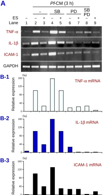
hu-Fig. 3. MAPK inhibitors differentially modulate the
Pf-CM-induced TNF-α, IL-1β and ICAM-1 mRNA expres-sions in THP-1 monocytes. THP-1 monocytes were pre-treated with 5 µg/mL ES products and 10 µg/mL of poly-myxin B for 6 h. The cells were then washed and the medium was replaced. The cells were then either treated with PD98059 (50 µM), SB20358, (10 µM) or a combi-nation of these inhibitors for 1 h prior to the stimulation with 250 µL Pf-CM for an additional 3 h incubation. Total RNA (1 µg/lane) was obtained and TNF-α, ICAM-1 and β-actin mRNA was assessed by semiquantitative RT-PCR. One representative of 3 experiments is shown.
Pf-CM (3 h) – SB PD SB PD – – + – + – + – + 1 2 3 4 5 6 7 8 9 ES Lane (%) (%) (%) TNF-α IL-1β ICAM-1 GAPDH TNF-α mRNA IL-1β mRNA ICAM-1 mRNA 160 120 80 40 0 160 120 80 40 0 160 120 80 40 0 Relativ e e x pression Relativ e e xpression Relativ e e xpression
A
B-1
B-2
B-3
man serum-treated monocytes in the absence of LPS or Pf-CM (lanes 1 and 6). These results indi-cate that Pf-CM has the ability of some stimulatory activity to induce the gene expression of pro-inflam-matory cytokines and ICAM-1, and this activity ex-hibited in Pf-CM-cultured THP-1-cells is a Plasmo-dium falciparum origin, without a trace of endo-toxin contamination.
Treatment of THP-1 cells with ES products prior to stimulation with Pf-CM prevent induc-tion of TNF-α, IL-1β and ICAM-1 gene expres-sion
In order to evaluate the impact of ES products on TNF-α, IL-1β and ICAM-1 gene expressions of THP-1 cells, these monocytes were left untreated or treated with 5 µg/mL ES products and polymyxin B prior to stimulation with 250 µL Pf-CM in the final medium volume of 3 mL. As shown in Fig. 2, ES products potently reduced TNF-α mRNA (by 76%), IL-1β (by 85%) and ICAM-1 mRNA expression (by 60%) in response to Pf-CM (lane 3). THP-1 cells treated with human serum showed a band pattern similar to the controls (lanes 1 and 5). The levels of constitutively expressed β-actin mRNA, however, did not change with the treatment of ES products. This result shows that ES products are a potent in-hibitor for TNF-α, IL-1β and ICAM-1 gene expres-sion in human monocytes in response to Pf-CM.
Role of ERK1/2 and p38 MAPK pathways in the induction of TNF-α, IL-1β and ICAM-1 gene expression in Pf-CM-stimulated THP-1 cells
Using well-known pharmacological inhibitors of the ERK1/2 and p38 MAPK pathways, PD98059 and SB203580, respectively (Dudley et al., 1995), we investigated the suppressive mechanisms of ES products on TNF-α, IL-1β and ICAM-1 gene ex-pressions in human monocytes stimulated with Pf-CM. THP-1 cells were pretreated with 5 µg/mL ES products and polymyxin B for 6 h, washed and then the medium was replaced. The monocytes were then treated with SB203580 and/or PD98059 for 1 h
before the addition of 250 µL Pf-CM in the final vol-ume of 3 mL for further 3 h incubation. As shown in Fig. 3, PD98059 treatment suppressed TNF-α, IL-1β and ICAM-1 mRNA expression by 83%, 75%
and 73% (lane 6), respectively; however, SB203580 treatment induced approximately 1.5-fold levels of the TNF-α, IL-1β mRNA and ICAM-1 mRNA ex-pression (lane 4), compared to the positive controls (lane 2). Combination of PD98059 and SB203580 profoundly reduced the 3 genes to near control lev-els (lane 8). These data indicate that the ERK1/2 MAPK pathway, but not p38 MAPK is important for induction of TNF-α, IL-1β and ICAM-1 mRNA expression in THP-1 cells stimulated with Pf-CM. We observed an additional suppressive effect by ES products on IL-1β, but not TNF-α, and ICAM-1 mRNA expression in the cells treated with PD98059 (lane 7).
Discussion
A complex parasite such as human Plasmodium is likely to generate a variety of substances that injure the hosts directly or cause immunopathology. It has been widely known that severe malaria is associat-ed with a failure of the host defense system to con-trol parasite replication, excessive secretion of pro-inflammatory cytokines and sequestration of para-sitized-erythrocytes in vital organs, such as the kid-ney, the lungs, the brain, etc. The excessive secre-tion of pro-inflammatory cytokines, such as TNF-α, IL-1β and IL-6 (Aikawa et al., 1990) has been as-sumed because of the stimulation of effector cells by certain parasite-derived toxins [substance(s)], re-leased by ruptured schizonts that cause paroxysms of fever. This substance(s) has been shown to con-tain glycosylphosphatidylinositol (Schofield and Hacket, 1993; Schofield et al., 1996; Tachado et al., 1996) and malarial pigments (Pichyangkul et al., 1994). Several studies have shown that glycosyl-phosphatidylinositol anchor, a membrane attach-ment mechanism for cell surface proteins widely used in eukaryotes, plays a crucial role in the pa-thology of malaria by inducing pro-inflammatory cytokines, including iNOS (Schofield et al., 1996; Tachado et al., 1996), TNF-α (Schofield et al., 1993) and ICAM-1 (Schofield et al., 1996). Pichyangkul et al. (1994) demonstrated that sequestered malaria pig-ment in the microvasculature also induced production
and induction of TNF-α and IL-1β gene expression of murine macrophages. In agreement with several previous studies (Abdalla and Wickramasinghe, 1985; Taverne et al., 1990a, 1990b), we demonstrated that Pf-CM induced TNF-α, IL-1β and ICAM-1 mRNA expression in THP-1 cells, which was not abolished by the treatment of 10 µg/mL polymyxin B (Fig. 1), suggesting that the stimulatory activity of Pf-CM was of parasite origin. We found that pretreatment of THP-1 monocytes with ES products prior to stimulation by Pf-CM reduced TNF-α, IL-1β and ICAM-1 gene expression (Fig. 2). We are confident that the results are not a consequence of toxicity since treatment of macrophages by ES products for over 24 h neither inhibited the growth of cell num-bers (unpublished data) nor affected the expression level of the house-keeping gene β-actin.
Further investigation through the MAPK path-way showed that pretreatment of THP-1 cells with PD98059 partially inhibited TNF-α, IL-1β and ICAM-1 mRNA expression, whereas SB203580, instead of inhibition, induced all gene expressions approximately 1.5 times compared to the positive controls (Fig. 3) and a combination of PD98059 and SB203580 profoundly reduced TNF-α, IL-1β and ICAM-1 mRNA expression to near control levels, indicating that the ERK1/2 pathway is important for optimal induction of TNF-α, IL-1β and ICAM-1 mRNA expression. We observed that ES products additionally suppress IL-1β mRNA expression in LPS-stimulated THP-1 cells treated with PD98059, suggesting that ES products may suppress IL-1β mRNA by inhibiting phosphorylation of the ERK1/2 pathway. The reason for this additional suppressive effect only observed for IL-1β mRNA expression is still unclear.
Paroxysmal fever is the hallmark of malaria. Several observations indicate that TNF-α is a criti-cal mediator of malarial fever: i) it is a potent endoge-nous pyrogen (Dinarello et al., 1986); ii) when P. falciparum is cultured with human monocytes, the rate of TNF production rises sharply at the time of schizont rupture (Kwiatkowski, 1990; Kwiatkowski et al., 1990); iii) in P. vivax infection, paroxysmal fever is associated with a sharp rise in circulating TNF levels (Karunaweera et al., 1992) and iv) anti-TNF
therapy inhibits fever in children with cerebral ma-laria (Kwiatkowski et al., 1993). However, TNF-α is not the only mediator of malaria fever. Other cytokines also have pyrogenic properties, including IL-1β (Rocket et al., 1994), IL-1α (Kwiatkowski et al., 1990) and IL-6 (Kern et al., 1989), which are produced mainly by the monocytes/macrophages series. In addition to the pyrogenic effect, TNF-α, has been shown to induce ICAM-1 gene expression of the endothelial cell, the major adherence receptor in the brain (Aikawa et al., 1990; Udomsangpetch et al., 1997). A study by Udomsangpetch et al. (2002) demonstrated that febrile temperatures in the range usually encountered during malaria induced cyto-adherence of the ring-stage of Plasmodium falci-parum-infected erythrocytes in vitro. This fever-induced cytoadherence was shown to be associated with increased expression of the Plasmodium falci-parum-erythrocyte membrane protein-1, a protein which is exported from the parasite and linked to the erythrocyte cytoskeleton during the 2nd half of the life cycle, on the surface of the infected red blood cell (Sharma, 1991). Up-regulating ICAM-1 expression of the endothelial cells allows parasites to bind and sequester in cerebral vessels, disrupting the function of the central nervous system (Adams et al., 2000), and in turn, leading to cerebral malar-ia. Based on the understanding of the pathogenesis of malaria, we suppose that suppression of TNF-α and IL-1β mRNA expression in THP-1 cells by ES products would, in turn, reduce the expression of ICAM-1 of endothelial cells by decreasing fever in-directly. In addition, ES products might also directly suppress ICAM-1 expression in monocytes.
In epidemic areas, helminthic infections com-monly coexist with malaria in an individual. The significance of such concurrent infection, however, is not yet clearly understood. However, results have revealed that worm infection is associated with protection from cerebral malaria (Nacher et al., 2000), acute renal failure and jaundice (Nacher et al., 2001). Thus, worms might be central to natu-ral anti-malarial immunity. Worms, which can in-duce a bias towards Th-2 cytokines and depress other cytokines could render the host more suscep-tible to liver-stage parasites and favor P. falciparum
reproduction but protect the host from severe com-plication of blood stage malaria. The beneficial or detrimental role of TNF-α, IL-1β and ICAM-1 dur-ing malaria attack could have important conse-quences, and modulation of these mediators during acute disease could lead to a novel approach in the treatment of malaria. Although co-infection of ma-laria and Spirometra erinaceieuropaei is unusual, the evidence presented here points to the possibility that ES products might be useful in reducing in-flammatory responses and the phenomenon of se-questration in severe malaria, especially in cerebral malaria.
Acknowledgments: We thank Professor Motomi Torii and Dr. Osamu Kaneko, the Department of Molecular Parasitology, School of Medicine Ehime University, Ja-pan, for kindly providing Plasmodium falciparum-cul-tured medium.
This work was supported by a Grant-in-Aid for Sci-entific Research (numbers 12670231, 09670258 and 13670246) from the Ministry of Education, Culture, Sports, Science and Technology, Japan.
References
1 Abdalla SH, Wickramasinghe SN. Monocytes activa-tion by supernatants from continuous cultures of
Plas-modium falciparum. Trans R Soc Trop Med Hyg 1985;
79:657–662.
2 Adams S, Turner GD, Nash GB, Micklem K, Newbold CI, Craig AG. Differential binding of clonal variants of
Plasmodium falciparum to allelic forms of intracellular
adhesion molecule 1 determined by flow adhesion assay. Infect Immun 2000;68:264–269.
3 Aikawa M, Iseki M, Barnwell JW, Taylor D, Oo MM, Howard RJ. The pathology of human cerebral malaria. Am J Trop Med Hyg 1990;43:30–37.
4 Artavanis TK, Riley EM. Innate immune response to malaria:rapid induction of IFN-γ from human NK cells by live Plasmodium falciparum-infected erythrocytes. J Immunol 2002;169:2956–2963.
5 Ballet JJ, Druilhe P, Querleux MA, Schmitt C, Agrapart M. Parasite-derived mitogenic activity for human T cells in Plasmodium falciparum continuous cultures. Infect Immun 1981;33:758–762.
6 Clark IA, Chaudri G, Cowden WB. Role of TNF in the illness and pathology of malaria. Trans R Soc Trop Med Hyg 1989;83:436–440.
7 Clark IA, Cowden WB, Butcher GA. TNF and inhibi-tion of growth of Plasmodium falciparum. Immunol Lett 1990;25:175–178.
Beutler B, Cerami A, et al. Tumor necrosis factor (cachectin) is an endogenous pyrogen and induces production of interleukin 1. J Exp Med 1986;163: 1433–1450.
9 Dirgahayu P, Fukumoto S, Miura K, Hirai K. Ex-cretory/secretory products from plerocercoids of
Spiro-metra erinaceieuropaei suppress the TNF-α gene ex-pression by reducing phosphorylation of ERK1/2 and p38 MAPK in macrophages. Int J Parasitol 2002; 32:1155–1162.
10 Dudley DT, Pang L, Decker SJ, Bridges AJ, Saltiel AR. A synthetic inhibitor of the mitogen-activted protein kinase cascade. Proc Natl Acad Sci U S A 1995;92: 7686–7689.
11 Fukumoto S, Hirai K, Tanihata T, Ohmori Y, Stuehr DJ, Hamilton TA. Excretory/secretory products from plerocercoids of Spirometra erinacei reduce iNOS and chemokines mRNA levels in peritoneal macrophages stimulated with cytokines and/or LPS. Parasite Immunol 1997;19:325–323.
12 Grau GE, Taylor TE, Molyneux ME, Wirima JJ, Vassalli P, Hommel M, et al. Tumor necrosis factor disease severity in children with falciparum malaria. N Engl J Med 1989;320:1586–1591.
13 Guevara PJA, Holder AA, McBride JS, Blackman MJ. Antibodies that inhibit malaria merozoite surface protein-1 processing and erythrocyte invasion are blocked by naturally acquired human antibodies. J Exp Med 1997;186:1689–1699.
14 Karunaweera ND, Grau GE, Gamage P, Carter R, Mendis KN. Dynamics of fever and serum levels of tumor necrosis factor are closely associated during clinical paroxysms in Plasmodium vivax malaria. Proc Natl Acad Sci USA 1992;89:3200–3203.
15 Kern P, Hemmer CJ, Van Damme J, Gruss H-J, Dietrich M. Elevated tumor necrosis factor alpha and interleukin-6 serum levels as markers for complicated
Plasmodium falciparum malaria. Am J Med 1989;57:
139–143.
16 Kumaratilake LM, Ferrante A, Jaeger T, Morris-Jones SD. The role of complement, antibody, and tumor ne-crosis factor alpha in the killing of Plasmodium
falci-parum by the monocytic cell line THP-1. Infec Immun
1997;65:5342–5345.
17 Kurtzhals JA, Adabayeri V, Goka BQ. Low plasma concentrations of interleukin 10 in severe malarial anemia compared with cerebral and uncomplicated malaria. Lancet 1998;351:1768–1772.
18 Kwiatkowski, D. Tumor necrosis factor, fever and fatality in falciparum malaria. Immunol Let 1990;25: 213–216.
19 Kwiatkowski D, Cannon JG, Manogue KR, Cerami A, Dinarello CA, Greenwood BM. Tumour necrosis factor production in falciparum malaria and its assocition with schozont rupture. Clin Exp Immunol 1989;77:361–366.
20 Kwiatkowski D, Hill AVS, Sambou I, Twumasi P, Castracane J, Manogue KR, et al. TNF concentration
in fatal cerebral, non-fatal cerebral, and uncomplicated
Plasmodium falciparum malaria. Lancet 1990;336:
1201–1204.
21 Kwiatkowski D, Molyneux ME, Stephens S, Curtis N, Klein N, Pointaire P, et al. Anti-TNF therapy inhibits fever in cerebral malaria. Q J Med. 1993:86:91–98. 22 Luty AFJ, Perkins DJ, Lell B, Schmidt-Ott R, Lehman
LG, Luckner D, et al. Low interleukin 12 activity in severe Plasmodium falciparum malaria. Infect Immun 2000;68:3909–3915.
23 McGuire W, Hill AV, Allsopp CE, Greenwood BM, Kwiatkowski D. Variation in the TNF-α promoter re-gion associated with susceptibility to cerebral malaria. Nature 1994;371:508–510.
24 Miura K, Fukumoto S, Dirgahayu P, Hirai K. Ex-cretory/secretory products from plerocercoids of
Spiro-metra erinaceieuropaei suppress gene expression and
production of tumor necrosis factor-α in murine macro-phages stimulated with lipopolysaccharide or lipothei-choic acid. Int J Parasitol 2001;31:38–47.
25 Nacher M, Gay F, Singhasivanon P, Krudsood S, Treeprasertsuk S, Mazier D, et al. Ascaris
lumbricoi-des infection is associated with protection from
cere-bral malaria. Parasite Immun. 2000;22:107–113. 26 Nacher M, Singhasivanon P, Silachamroon U,
Treeprasertsuk S, Vannaphan S, Traore B, et al. Hel-minth infections are associated with protection from malaria-related acute renal failure and jaundice in Thailand. Am J Trop Med Hyg;65:834–836.
27 Pichyangkul S, Saengkrai P, Webster HK.
Plasmodi-um falciparPlasmodi-um pigment induces monocytes to release
high levels of tumor necrosis factor-alpha and inter-leukin-1 beta. Am J Trop Med Hyg 1994;51:430–435 28 Reyes L, Davidson MK, Thomas LC, Davis, JK. Effect
of Mycoplasma fermentans incognitos on differentia-tion of THP-1 cells. Infect Immun 1999;67:3188– 3192.
29 Riggione F, Pulido M, Noya O. Plasmodium
falcipa-rum: surface modifications of infected erythrocytes
from clinical isolates. Evidence of antigenic diversity using Venezuelan human malarial sera. Parasitol Res 1996;82:490–494.
30 Rockett KA, Awburn MM, Rockett EJ, Clark IA. Tumor necrosis factor and interleukin-1 synergy in the context of malaria pathology. Am J Trop Med Hyg 1994;50:735–742.
31 Schofield L, Hackett F. Signal transduction in host cells by a glycosylphosphatidylinositol toxin of malaria parasites. J Exp Med 1993;177:145–153.
32 Schofield L, Novakovic S, Gerold P, Schwarz RT, McConville MJ, Tachado SD. Glycosylphosphatidy-linositol toxin of Plasmodium up-regulates intercellular adhesion 1, vascular cell adhesion molecule-1, and E-selectin expression in vascular endothelial cells and increases leukocyte and parasite cyto-adherence via tyrosine kinase-dependent signal trans-duction. J Immunol 1996;156:1886–1896.
in falciparum malaria. Int J Biochem 1991;23:775– 789.
34 Tachado SD, Gerold P, McConville MJ, Baldwin T, Quilici D, Scwarz RT, et al. Glycosylphosphatidy-linositol toxin of Plasmodium induces nitric oxide syn-thase expression in macrophages and vascular endo-thelial cells by a protein tyrosine kinase-dependent and protein kinase C-dependent signaling pathway. J Immunol 1996;156:1897–1907.
35 Taverne J, Bate CAW, Kwiatkowski D, Jakobsen PH, Playfair JHL. Two soluble antigens of Plasmodium
falciparum induce tumor necrosis factor alpha release
from macrophages. Infec Immun 1990a;58:2923– 2928.
36 Taverne J, Bate CAW, Sarkar DA, Meager A, Rook GAW, Playfair JHL. Human and murine macrophages produce TNF in response to soluble antigens of
Plas-modium falciparum. Parasite Immunol 1990b;12:33–
43.
37 Trager W, Jensen JB. Human malaria in continuous
culture. Science 1976;193:673–675.
38 Tsuchiya S, Yamane M, Yamaguchi Y, Konno T, Tada K. Establishment and characterization of a human acute monocytic cell line (THP-1). Int J Cancer 1980; 26:171–176.
39 Udomsangpetch R, Reinhardt PH, Schollaardt T, Elliott JF, Kubes P, Ho M. Promiscuity of clinical
Plasmodium falciparum isolates for multiple adhesion
molecules under flow conditions. J Immunol 1997; 158:4358–4364.
40 Udomsangpetch R, Pipitaporn B, Silamut K, Pinches R, Kyes S, Looareesuwan S, Newbold C, White NJ. Fe-brile temperature induced cytoadherence of ring-stage
Plasmodium falciparum-infected erythrocytes. Proc
Natl Acad Sci USA 2002;99, 11852–11829.
Received January 5, 2004; accepted January 28, 2004 Corresponding author: Kazumitsu Hirai, MD, PhD