Car di ovas c ul ar t oxi c ef f ec t s of t ar get ed
c anc er t her apy
著者
Taj i r i Kaz uko, Aonum
a Kaz ut aka, Seki ne I kuo
j our nal or
publ i c at i on t i t l e
J apanes e j our nal of c l i ni c al onc ol ogy
vol um
e
47
num
ber
9
page r ange
779- 785
year
2017- 09
権利
( C) The Aut hor 2017. Publ i s hed by O
xf or d
U
ni ver s i t y Pr es s .
Thi s i s a pr e- c opyedi t ed, aut hor - pr oduc ed PD
F
of an ar t i c l e ac c ept ed f or publ i c at i on i n
J apanes e J our nal of Cl i ni c al O
nc ol ogy
f ol l ow
i ng peer r evi ew
. The ver s i on of r ec or d
Vol um
e 47, I s s ue 9, 1 Sept em
ber 2017, Pages
779 785 i s avai l abl e onl i ne at :
ht t ps : / / doi . or g/ 10. 1093/ j j c o/ hyx071.
U
RL
ht t p: / / hdl . handl e. net / 2241/ 00151256
Review article
Cardiovascular toxic effects of targeted cancer therapy
Kazuko Tajiri1, Kazutaka Aonuma1, Ikuo Sekine2
1Department of Cardiology, Faculty of Medicine, University of Tsukuba, Tsukuba, Japan
2Department of Medical Oncology, Faculty of Medicine, University of Tsukuba, Tsukuba,
Japan
Corresponding author: Ikuo Sekine, MD, PhD
Department of Medical Oncology, Faculty of Medicine, University of
Tsukuba
1-1-1 Tennodai, Tsukuba, Ibaraki 305-8575, Japan
Phone & fax: +81-29-853-3014
E-mail: [email protected]
Abstract
Over the past decade, there has been a major shift in chemotherapy from non-specific
cytotoxic drugs to molecular targeted drug therapies. As more molecular targeted therapies
are developed, new types of cardiovascular toxicities induced by targeted therapies are a
growing problem. Cardiotoxicity induced by the human epidermal growth factor receptor-2
inhibitor trastuzumab manifests as decreased left ventricular ejection fraction. In contrast to
anthracycline treatment, most cardiac events occur during trastuzumab treatment, but are
reversed quickly when treatment is interrupted and cardiac intervention is established.
Vascular endothelial growth factor pathway inhibitors decrease vascular tone, leading to
hypertension. After drug initiation, the early detection and aggressive pharmacological
management of hypertension are necessary to avoid severe complications. Cardiovascular
safety is an emerging challenge in patients treated with newer generations of BCR-ABL
inhibitors. Although rare, dasatinib-induced pulmonary hypertension is potentially fatal.
Vascular events including cardiac and cerebral ischemic events and peripheral arterial
occlusive disease have emerged as a new type of toxicity in patients treated with ponatinib
and nilotinib. Thus, a wide variety of cardiovascular toxicities have been observed in patients
treated with targeted drugs and have become a critically important topic of discussion for the
practicing oncologist and cardiologists. Awareness of the potential side effects, recognition of
signs and symptoms, and the establishment of therapeutic strategies are all crucial to
Introduction
The introduction of molecular targeted therapies has revolutionized cancer therapy and
contributed to a steady decline in cancer deaths since the late 1990s. However, both cardiac
and vascular toxicities have been reported for these agents, some as expected on-target effects
and others as off-target toxicities (Table 1). This review focuses on the cardiovascular
toxicities associated with three categories of targeted therapy agents: 1) human epidermal
growth factor receptor-2 (HER2) inhibitors; 2) vascular endothelial growth factor (VEGF)
signaling pathway inhibitors; and 3) BCR-ABL kinase inhibitors.
1. HER2 inhibitors
Trastuzumab, the first clinically used humanized monoclonal antibody directed against the
erythroblastic leukemia viral oncogene homolog 2 (ErbB2, also known as HER2), has
revolutionized the treatment of metastatic HER2-positive breast cancer. Although early phase
II trials indicated high efficacy and a favorable safety profile, an unexpectedly high rate of
adverse cardiac events during the first phase III trial was identified; 27% of patients receiving
concomitant trastuzumab and anthracycline-containing chemotherapy developed cardiac
dysfunction compared with 8% of patients receiving anthracycline alone (1–3). The rates of
cardiac dysfunction in patients who received paclitaxel and trastuzumab versus paclitaxel
alone were 13% and 1%, respectively. The incidence of New York Heart Association
(NYHA) class III or IV heart failure was highest among patients receiving anthracycline,
cyclophosphamide, and trastuzumab, 16%, compared with 3% for patients receiving
anthracycline and cyclophosphamide alone. In subsequent trials, the incidence of cardiac
events was reduced through changes in chemotherapy regimens, stricter patient selection, and
close cardiac assessment (4). However, cardiotoxicity remains a significant problem in
increase through improved survival.
1.1 ErbB2 signaling in the heart
The importance of ErbB receptors and their ligand neureglin-1 (NRG-1) during development
is evident from analyses of genetically modified mice. The deletion of ErbB2 (5), ErbB4 (6),
and NRG-1(7) led to embryonic lethality caused by cardiac malformations (5). Conditional
mutations of ErbB2 in cardiomyocytes resulted in the development of spontaneous dilated
cardiomyopathy with left ventricular chamber dilation, wall thinning, and reduced
contractility (8,9). Additionally, cardiomyocytes isolated from these conditional mutants were
more susceptible to anthracycline toxicity (9).
NRG-1 is expressed by the endocardium and endothelium of the cardiac
microvasculature (10). ErbB2 functions as a non-ligand-binding, pre-activated co-receptor; in
the myocardium, it heterodimerizes with ErbB4 upon NRG-1-induced activation (11). The
binding of NRG1 to ErbB4 increases its kinase activity and leads to heterodimerization with
ErbB2 or homodimerization with ErbB4 and stimulation of the intracellular signal
transduction pathways, such as the phosphoinositide 3-kinase (PI3K)/Akt, Ras/extracellular
signal-regulated kinases (ERK), and proto-oncogene tyrosine-protein kinase (Src)/focal
adhesion kinase (FAK) pathway. NRG-1/ErbB signaling induces cardiomyocyte growth and
proliferation via PI3K/Akt and ERK1/2 signaling. It also protects cardiomyocytes from
apoptosis and stimulates nitric oxide (NO) production through PI3K/Akt signaling (Figure 1)
(11).
NRG-1/ErbB signaling also plays important roles in adaptation of the heart to injury
in adults as well as attenuating myofibrillar disarray and promoting cell survival (10,12). In
animal models of myocardial ischemia, doxorubicin cardiomyopathy, viral myocarditis, and
infusion of recombinant NRG-1 receptor-active peptide (13). Furthermore, NRG-1
administration to adult mice promotes myocardial regeneration by inducing mononucleated
cardiomyocytes to divide, improving cardiac function after myocardial infarction (14). Thus,
exogenous NRG-1 agents have been developed and are being evaluated in clinical trials. Early
clinical trials with the epidermal growth factor (EGF)-like domain of NRG in heart failure
have demonstrated safety and efficacy (15,16), and is currently tested in phase III clinical trial.
Recent work from D’Uva et al. clearly showed that augmentation of ErbB2 signaling
awakened a dormant regenerative window in juvenile and adult mouse cardiomyocytes (17).
ErbB2 was necessary for NRG-1-induced cardiomyocyte proliferation during the transient
postnatal regenerative window and became limiting as cardiomyocytes stopped dividing.
They showed that transient reactivation of ErbB2 signaling in adult mice stimulated
cardiomyocyte proliferation and allowed anatomical and functional regeneration of hearts
after myocardial infarction. Taken together, these data point toward a fundamental role of
ErbB2 signaling in cardiac development during embryogenesis and in cardiomyocyte survival,
especially in situations of stress by promoting the survival and regeneration pathways that
maintain cardiac function.
1.2 Management of anti-HER2 therapy-associated cardiotoxicity
Initially, the incidence of cardiotoxicity was reportedly high when trastuzumab was
administered concurrently with anthracyclines in a trial of metastatic breast cancer (2).
Applying trastuzumab sequentially after anthracyclines or using an anthracycline-free
chemotherapy regimen substantially reduced the clinical heart failure rate. Based on several
large-scale trials of adjuvant therapy in breast cancer, the rate of cardiac dysfunction was
7.1-18.6%, with severe overt heart failure (NYHA class III and IV) rates of 0.4-4.1% (18–20).
dysfunction rates were 3.2-9.4%, with 0.4-0.5% developing clinical heart failure (20–22).
These data indicate that concomitant or previous use of anthracyclines substantially increases
trastuzumab-associated cardiotoxicity. Long-term follow-up data showed that, in contrast to
anthracyclines, most cardiac events occurred during trastuzumab treatment and were reversed
quickly when treatment was interrupted and cardiac intervention was established (23–25).
However, in most trials, patients were relatively young and had normal or nearly normal
cardiac function without a significant cardiovascular history. A large cohort of breast cancer
patients at least 66 years old looked at the rate of cardiotoxicity in patients who received
trastuzumab and chemotherapy (anthracycline and/or taxane) compared with chemotherapy
alone (26). Among trastuzumab-treated patients, the rate of congestive heart failure was
29.4% compared with 18.9% in nontrastuzumab users (P < 0.001). Among trastuzumab users,
older age (age >80 years) was one of the factors that increased the risk of congestive heart
failure (26). Another large cohort study of elderly women aged 67–94 years of age showed
that adjusted 3-year heart failure or cardiomyopathy incidence rates were higher for patients
receiving trastuzumab (32.1%) and anthracycline plus trastuzumab (41.9%) compared with no
adjuvant therapy (18.1%, P < 0.001) (27). Thus, the incidence of cardiotoxicity in older
patients treated with trastuzumab is expected to be higher than in the overall population
evaluated in large clinical trials. Therefore, cardiac risk assessment, cancer recurrence risk,
and discussion between cardiologists and oncologists should take place prior to making
decisions about cancer treatment, and long-term continuous cardiac monitoring is especially
advised in this population.
Previous studies revealed several risk factors for anti-HER2 drug-induced
cardiotoxicity, including previous anthracycline exposure, hypertension, a low baseline left
ventricular ejection fraction (LVEF), and older age (28). One of the most relevant clinical
increased cancer recurrence (29). The clinical benefit from HER2 inhibitors needs to be
balanced against cardiotoxicity. For patients with advanced cancer, the balance between
trastuzumab benefit and heart failure risk may remain finely balanced, if trastuzumab was
effective. Careful consideration should be given before trastuzumab discontinuation. It
remains unclear whether an asymptomatic LVEF decline is predictive of clinical heart failure
among patients treated with HER2 inhibitors. Therefore, some patients with reduced LVEF
may have the opportunity to continue trastuzumab under optimal cardioprotective treatment.
More recently, a double-blinded, placebo-controlled trial showed that for patients with
early-stage HER2-positive breast cancer, prophylactic treatment with angiotensin-converting
enzyme inhibitors (ACE inhibitors) or β-blockers attenuated cardiac dysfunction associated
with trastuzumab therapy by reducing the decrease in LVEF; however, the treatment could
not prevent trastuzumab-related left ventricular remodeling, the primary outcome of this trial
(30). Larger studies with longer follow-up are required to reaffirm the protective effects of
ACE inhibitors and β-blockers on cardiac function and to determine the impact of such
interventions on cardiovascular outcomes.
2. VEGF signaling pathway inhibitors
2.1 VEGF inhibitor-induced hypertension
Incidence
Soon after the VEGF pathway inhibitors entered the clinical arena, it became evident that
hypertension was a serious unexpected cardiovascular toxicity (31). The VEGF signaling
inhibitors and their cardiovascular side effects are summarized in Table 1. Systemic arterial
hypertension induction or worsening is caused by all of these drugs and is the most common
cardiovascular side effect, with a reported 20-44% incidence of overall hypertension and a
hypertension is not a side effect of treatment, but rather a mechanism-dependent on-target
toxicity (33). This has led to the concept that hypertension may serve as a surrogate for the
effective anti-angiogenic response and could be a biomarker of better outcomes (34–36).
Potential mechanisms of VEGF inhibitor-induced hypertension
VEGF inhibitors induce an imbalance between vasodilation and vasoconstriction; as a
consequence, they increase the peripheral vascular resistance and blood pressure (31,37,38).
VEGF binding to VEGF receptors (VEGFRs) initiates a tyrosine kinase signaling cascade
that leads to increased proliferation, survival, permeability, and migration (Figure 2) (39).
VEGFR2 activation leads to PI3K recruitment followed by protein kinase B (PKB)/Akt
activation and endothelial NO synthase (eNOS) phosphorylation, resulting in increased NO
production. In a paracrine fashion, NO diffuses to vascular smooth muscle cells, where it
activates guanylyl cyclase with a consequent increase in cyclic guanosine monophosphate
(cGMP) production and vasodilation, thus playing an important role in maintaining vascular
tone (40). VEGF also leads to the production of another vasodilator, prostacyclin I2 (PGI2),
and decreases endothelin-1 (ET-1) level, a potent vasoconstrictor. Thus, VEGF inhibitors
decrease the vascular tone, leading to hypertension (31,37,38).
Another possible mechanism is a net reduction in tissue microvessel density and
capillary rarefaction (loss of parallel capillary circulation), resulting in increased afterload
and thereby contributing to the pathogenesis of hypertension (41). VEGF is an important
mediatorof endothelial cell proliferation and survival. Therefore, the inhibition of VEGF
signaling would cause vascular rarefaction and endothelial cell apoptosis (38).
Monitoring and treatment of hypertension
remain to be established. However, preexisting hypertension has been considered an
independent risk factor for hypertension after VEGF pathway inhibition. In addition, an age >
60 years and elevated body mass index emerged as independent risk factors (38,42,43).
The recommendations for clinical practice are; (1) a formal risk assessment for
existing cardiovascular disease and potential cardiovascular complications before VEGF
pathway inhibitor treatment; (2) active monitoring for blood pressure elevations
and cardiac toxicity with more frequent assessments during the first therapy cycle; and (3)
aggressive management of blood pressure elevations and early symptoms and signs of cardiac
toxicity to prevent clinically limiting complications (44,45). Because the development of
hypertension in response to VEGF pathway inhibition can occur within hours to days, close
monitoring of blood pressure after the initiation of a VEGF signaling inhibitor is mandatory
(44). In patients with preexisting hypertension, the blood pressure target for initiating VEGF
inhibitor treatment should be < 140/90 mmHg, or lower in cases of overt proteinuria (32,46).
After the initiation of VEGF inhibitors, the early detection and aggressive
pharmacological management of hypertension are necessary to avoid severe complications
(46). ACE inhibitors, angiotensin II receptor blockers, and β-blockers are reasonable as
first-line therapies for VEGF inhibitor-induced hypertension (32,44,46). As the
non-dihydropyridine calcium channel blockers (verapamil and diltiazem) inhibit cytochrome
P450 3A4 and result in increased plasma concentrations of many VEGF inhibitors, they
should preferably be avoided. If blood pressure is uncontrolled (systolic blood pressure ≥ 160
mmHg or diastolic blood pressure ≥ 100 mmHg), dose reduction and reinforcement of
antihypertensive treatment or discontinuation of VEGF inhibitors should be considered
(32,37,46).
Numerous studies have shown that arterial and venous thromboembolic events are increased
in cancer patients treated with VEGF inhibitors. Meta-analyses of patients taking VEGF
inhibitors revealed that the incidence of arterial thrombotic events was 1.4-3.3% with a
relative risk compared to control of 1.4-3.0 (47–49), while the incidence of venous thrombotic
events was 2.8-11.9% with a relative risk of 1.1-1.3 compared to control (50,51).
The vascular endothelium is involved in the regulation and maintenance of vascular
homeostasis and prevents abnormal blood clotting and bleeding. VEGF plays a considerable
role in the maintenance of vascular integrity by activating survival and anti-apoptotic
signaling (52,53). VEGF inhibition can interfere with the regenerative capacity of endothelial
cells and cause defects of the endothelial layer that expose the highly prothrombotic basement
membrane (32,54). Exposure to subendothelial von Willebrand factor and tissue factor initiate
platelet aggregation and the coagulation cascade (54). VEGF also increases the bioavailability
of prostacyclin and NO, both of which have antiplatelet activities and promote thrombosis
when inhibited (32).
3. BCR-ABL tyrosine kinase inhibitors
For most patients with chronic myeloid leukemia (CML), small-molecule tyrosine kinase
inhibitors (TKIs) have turned a fatal disease into a manageable chronic condition. Imatinib,
the first BCR-ABL TKI granted regulatory approval, inhibits ABL kinase as well as
proto-oncogene c-KIT and platelet-derived growth factor receptor (PDGFR). Newer
generations of BCR-ABL kinase inhibitors (dasatinib, nilotinib, bosutinib, and ponatinib)
have been developed to overcome imatinib resistance or intolerance (55). Cardiovascular
safety is an emerging challenge in patients treated with newer generations of BCR-ABL
inhibitors.
roles in the vasculature. Long-term observations from phase III studies have revealed a lower
incidence of peripheral arterial disease in patients treated with imatinib compared with
patients not treated with TKI or treated with nilotinib (56). A randomized double-blind
placebo-controlled trial reported that imatinib significantly improved exercise capacity,
hemodynamics, and right ventricular function in patients with pulmonary hypertension
(57,58).
3.1. Pulmonary hypertension
In contrast to imatinib, dasatinib is known to cause drug-induced pulmonary hypertension at
an estimated lowest incidence of 0.45% and a median delay between drug initiation and
pulmonary hypertension diagnosis of 34 months (range, 8-48 months) (59). At diagnosis,
most patients had severe clinical, functional and hemodynamic signs of impairment with
minimal acute vasodilator response, some of which required vasoactive drugs and intensive
care unit management (59). Clinical and functional improvements were usually observed after
dasatinib discontinuation; however, the majority of patients failed to demonstrate complete
hemodynamic recovery, and some died of sudden death or cardiac failure during follow-up
(59). The mechanism of dasatinib-associated pulmonary hypertension is not yet completely
understood. One possible mechanism behind dasatinib-induced pulmonary hypertension is
that dasatinib causes pulmonary vascular endothelial cell damage, endoplasmic reticulum
stress, and mitochondrial reactive oxygen species production, which leads to increased
susceptibility to the development of pulmonary hypertension (60). Interestingly, dasatinib is
associated with a higher incidence of pleural effusion, reportedly, 14-39% (61). The presence
of symptoms (i.e., chest pain, dyspnea, dry cough, syncope) not explained by pleural effusion
should prompt the suspicion of pulmonary hypertension. Although rare, it is potentially fatal.
hypertension, but pharmacologic treatment may be needed and the referral to a suitable
specialist is mandatory (61).
3.2. Vascular adverse events
Vascular events including cardiac and cerebral ischemic events and peripheral arterial
occlusive disease have become an emerging new type of toxicity in CML patients treated with
ponatinib and nilotinib (61,62). The rates of vascular adverse events in clinical trials varied
considerably because the trials were not designed to assess this point, and vascular risk factors
were not properly assessed before and during the treatment. After a 2-year observation time,
the percentage of CML patients developing vascular adverse events during nilotinib was
reportedly 1-29% (62). A prospective study involving 159 patients on imatinib or nilotinib
showed a higher incidence of abnormal ankle-brachial index (ABI) in patients on nilotinib
(relative risk, 10.3). The incidence of abnormal ABI in patients treated with first- and
second-line nilotinib was 26% and 36%, respectively, compared with 6.3% for first-line
imatinib (63). In a recent study using the the French Pharmacovigilance Database, 25 cases
with peripheral aortic obstructive disease were identified, and the mean time from initiation of
nilotinib to the event onset was 24 months (64). The frequency of arterial occlusive events in
patients treated with ponatinib in the Evaluation of Ponatinib versus Imatinib in Chronic
Myeloid Leukemia (EPIC) study was 7.1% compared with 2.0% for imatinib after a median
follow-up of 5.1 months (65). Notably, median time to onset of first arterial occlusive event
was 3.6 months for ponatinib-treated patients. In the phase 2 trial of ponatinib in refractory
CML, arterial occlusive events were observed in 27% of patients after a median follow-up of
38 months, in which the median time to onset was 11 months (66). Thus, in contrast to other
vascular toxic agents, e.g. tobacco, steroids, BCR-ABL TKIs seem to affect vascular
assessment repeatedly.
The mechanisms behind the vascular toxicity of nilotinib and ponatinib remain
unclear. Several clinical studies suggest that nilotinib is associated with hyperglycemia and
hypercholesterolemia (61), which are major risk factors for developing atherosclerosis.
Nilotinib may accelerate atherosclerosis, leading to ischemic vascular adverse events. Given
the high frequency of vascular adverse events associated with nilotinib and ponatinib, in the
first-line treatment of chronic-phase CML in patients at very high risk of cardiovascular
disease, imatinib or dasatinib seems to be the preferred option.
4. Future directions
Without question, targeted therapies have revolutionized the treatment of cancer across
multiple histologies. Therefore, the cardiac impact of targeted therapies is a critically
important topic of discussion, not only for the practicing oncologist as well as cardiologists
and researchers. Awareness of the potential side effects, recognition of the signs and
symptoms, and establishment of therapeutic strategies are all crucial for providing quality
patient care. Long-term follow-up is needed as the field continues to improve survival
outcomes with new and exciting therapies.
Conflict of Interest
The authors declare no conflicts of interest.
Funding
This work was supported by grants from JSPS KAKENHI Grant Numbers JP15K19364 and
Acknowledgements
References
1. Cobleigh MA, Vogel CL, Tripathy D, et al. Multinational study of the efficacy and
safety of humanized anti-HER2 monoclonal antibody in women who have
HER2-overexpressing metastatic breast cancer that has progressed after chemotherapy
for metastatic disease. J Clin Oncol 1999;17:2639–48.
2. Slamon DJ, Leyland-Jones B, Shak S, et al. Use of chemotherapy plus a monoclonal
antibody against HER2 for metastatic breast cancer that overexpresses HER2. N Engl J
Med 2001;344:783–92.
3. Seidman A, Hudis C, Pierri MK, et al. Cardiac dysfunction in the trastuzumab clinical
trials experience. J Clin Oncol 2002;20:1215–21.
4. Pondé NF, Lambertini M, de Azambuja E. Twenty years of anti-HER2
therapy-associated cardiotoxicity. ESMO Open 2016;1:e000073.
5. Lee KF, Simon H, Chen H, Bates B, Hung MC, Hauser C. Requirement for neuregulin
receptor erbB2 in neural and cardiac development. Nature 1995;378:394–8.
6. Gassmann M, Casagranda F, Orioli D, et al. Aberrant neural and cardiac development in
mice lacking the ErbB4 neuregulin receptor. Nature 1995;378:390–4.
7. Meyer D, Birchmeier C. Multiple essential functions of neuregulin in development.
Nature 1995;378:386–90.
8. Ozcelik C, Erdmann B, Pilz B, et al. Conditional mutation of the ErbB2 (HER2) receptor
in cardiomyocytes leads to dilated cardiomyopathy. Proc Natl Acad Sci U S A
2002;99:8880–5.
9. Crone SA, Zhao Y-Y, Fan L, et al. ErbB2 is essential in the prevention of dilated
cardiomyopathy. Nat Med 2002;8:459–65.
ventricular myocytes. J Biol Chem 1998;273:10261–9.
11. Vermeulen Z, Segers VFM, De Keulenaer GW. ErbB2 signaling at the crossing between
heart failure and cancer. Basic Res Cardiol 2016;111:60.
12. Sawyer DB, Zuppinger C, Miller TA, Eppenberger HM, Suter TM. Modulation of
anthracycline-induced myofibrillar disarray in rat ventricular myocytes by
neuregulin-1beta and anti-erbB2: potential mechanism for trastuzumab-induced
cardiotoxicity. Circulation 2002;105:1551–4.
13. Liu X, Gu X, Li Z, et al. Neuregulin-1/erbB-activation improves cardiac function and
survival in models of ischemic, dilated, and viral cardiomyopathy. J Am Coll Cardiol
2006;48:1438–47.
14. Bersell K, Arab S, Haring B, Kühn B. Neuregulin1/ErbB4 signaling induces
cardiomyocyte proliferation and repair of heart injury. Cell 2009;138:257–70.
15. Gao R, Zhang J, Cheng L, et al. A Phase II, randomized, double-blind, multicenter,
based on standard therapy, placebo-controlled study of the efficacy and safety of
recombinant human neuregulin-1 in patients with chronic heart failure. J Am Coll
Cardiol 2010;55:1907–14.
16. Jabbour A, Hayward CS, Keogh AM, et al. Parenteral administration of recombinant
human neuregulin-1 to patients with stable chronic heart failure produces favourable
acute and chronic haemodynamic responses. Eur J Heart Fail 2011;13:83–92.
17. D’Uva G, Aharonov A, Lauriola M, et al. ERBB2 triggers mammalian heart
regeneration by promoting cardiomyocyte dedifferentiation and proliferation. Nat Cell
Biol 2015;17:627–38.
18. Romond EH, Perez EA, Bryant J, et al. Trastuzumab plus adjuvant chemotherapy for
operable HER2-positive breast cancer. N Engl J Med 2005;353:1673–84.
chemotherapy in HER2-positive breast cancer. N Engl J Med 2005;353:1659–72.
20. Slamon D, Eiermann W, Robert N, et al. Adjuvant trastuzumab in HER2-positive breast
cancer. N Engl J Med 2011;365:1273–83.
21. Jones SE, Collea R, Paul D, et al. Adjuvant docetaxel and cyclophosphamide plus
trastuzumab in patients with HER2-amplified early stage breast cancer: a single-group,
open-label, phase 2 study. Lancet Oncol 2013;14:1121–8.
22. Tolaney SM, Barry WT, Dang CT, et al. Adjuvant paclitaxel and trastuzumab for
node-negative, HER2-positive breast cancer. N Engl J Med 2015;372:134–41.
23. Slamon D, Eiermann W, Robert N, et al. Adjuvant trastuzumab in HER2-positive breast
cancer. N Engl J Med 2011;365:1273–83.
24. de Azambuja E, Procter MJ, van Veldhuisen DJ, et al. Trastuzumab-associated cardiac
events at 8 years of median follow-up in the Herceptin Adjuvant trial (BIG 1-01). J Clin
Oncol 2014;32:2159–65.
25. Advani PP, Ballman K V, Dockter TJ, Colon-Otero G, Perez EA. Long-term cardiac
safety analysis of NCCTG N9831 (Alliance) adjuvant trastuzumab trial. J Clin Oncol
2016;34:581–7.
26. Chavez-MacGregor M, Zhang N, Buchholz TA, et al. Trastuzumab-Related
Cardiotoxicity Among Older Patients With Breast Cancer. J Clin Oncol
2013;31:4222–8.
27. Chen J, Long JB, Hurria A, Owusu C, Steingart RM, Gross CP. Incidence of Heart
Failure or Cardiomyopathy After Adjuvant Trastuzumab Therapy for Breast Cancer. J
Am Coll Cardiol 2012;60:2504–12.
28. Ezaz G, Long JB, Gross CP, Chen J. Risk prediction model for heart failure and
cardiomyopathy after adjuvant trastuzumab therapy for breast cancer. J Am Heart Assoc
29. Yu AF, Yadav NU, Lung BY, et al. Trastuzumab interruption and treatment-induced
cardiotoxicity in early HER2-positive breast cancer. Breast Cancer Res Treat
2015;149:489–95.
30. Pituskin E, Mackey JR, Koshman S, et al. Multidisciplinary Approach to Novel
Therapies in Cardio-Oncology Research (MANTICORE 101-Breast): A Randomized
Trial for the Prevention of Trastuzumab-Associated Cardiotoxicity. J Clin Oncol
2016;35:870-7.
31. Small HY, Montezano AC, Rios FJ, Savoia C, Touyz RM. Hypertension due to
antiangiogenic cancer therapy with vascular endothelial growth factor inhibitors:
understanding and managing a new syndrome. Can J Cardiol 2014;30:534–43.
32. Li W, Croce K, Steensma DP, McDermott DF, Ben-Yehuda O, Moslehi J. Vascular and
metabolic implications of novel targeted cancer therapies. J Am Coll Cardiol
2015;66:1160–78.
33. Simons M, Eichmann A. “On-target” cardiac effects of anticancer drugs. J Am Coll
Cardiol 2012;60:626–7.
34. Rini BI, Cohen DP, Lu DR, et al. Hypertension as a biomarker of efficacy in patients
with metastatic renal cell carcinoma treated with sunitinib. J Natl Cancer Inst
2011;103:763–73.
35. Cai J, Ma H, Huang F, et al. Correlation of bevacizumab-induced hypertension and
outcomes of metastatic colorectal cancer patients treated with bevacizumab: a systematic
review and meta-analysis. World J Surg Oncol 2013;11:306.
36. George S, Reichardt P, Lechner T, Li S, Cohen DP, Demetri GD. Hypertension as a
potential biomarker of efficacy in patients with gastrointestinal stromal tumor treated
with sunitinib. Ann Oncol 2012;23:3180–7.
system? Curr Oncol Rep 2016;18:33.
38. Kruzliak P, Novak J, Novak M. Vascular endothelial growth factor inhibitor-induced
hypertension: from pathophysiology to prevention and treatment based on long-acting
nitric oxide donors. Am J Hypertens 2014;27:3–13.
39. Hoeben A, Landuyt B, Highley MS, Wildiers H, Van Oosterom AT, De Bruijn EA.
Vascular endothelial growth factor and angiogenesis. Pharmacol Rev 2004;56:549–80.
40. Hood JD, Meininger CJ, Ziche M, Granger HJ. VEGF upregulates ecNOS message,
protein, and NO production in human endothelial cells. Am J Physiol
1998;274:H1054-8.
41. Inai T, Mancuso M, Hashizume H, et al. Inhibition of vascular endothelial growth factor
(VEGF) signaling in cancer causes loss of endothelial fenestrations, regression of tumor
vessels, and appearance of basement membrane ghosts. Am J Pathol 2004;165:35–52.
42. Hamnvik O-PR, Choueiri TK, Turchin A, et al. Clinical risk factors for the development
of hypertension in patients treated with inhibitors of the VEGF signaling pathway.
Cancer 2015;121:311–9.
43. Robinson ES, Khankin EV, Karumanchi SA, Humphreys BD. Hypertension induced by
vascular endothelial growth factor signaling pathway inhibition: mechanisms and
potential use as a biomarker. Semin Nephrol 2010;30:591–601.
44. Steingart RM, Bakris GL, Chen HX, et al. Management of cardiac toxicity in patients
receiving vascular endothelial growth factor signaling pathway inhibitors. Am Heart J
2012;163:156–63.
45. Maitland ML, Bakris GL, Black HR, et al. Initial assessment, surveillance, and
management of blood pressure in patients receiving vascular endothelial growth factor
signaling pathway inhibitors. J Natl Cancer Inst 2010;102:596–604.
and cardiovascular toxicity developed under the auspices of the ESC Committee for
Practice Guidelines The Task Force for cancer treatments and cardiovascular toxicity of
the European Society of Cardiology (ESC). Eur Heart J 2016;37:2768-801.
47. Ranpura V, Hapani S, Chuang J, Wu S. Risk of cardiac ischemia and arterial
thromboembolic events with the angiogenesis inhibitor bevacizumab in cancer patients:
A meta-analysis of randomized controlled trials. Acta Oncol 2010;49:287–97.
48. Choueiri TK, Schutz FAB, Je Y, Rosenberg JE, Bellmunt J. Risk of arterial
thromboembolic events with sunitinib and sorafenib: a systematic review and
meta-analysis of clinical trials. J Clin Oncol 2010;28:2280–5.
49. Qi W-X, Shen Z, Tang L-N, Yao Y. Risk of arterial thromboembolic events with
vascular endothelial growth factor receptor tyrosine kinase inhibitors: An up-to-date
meta-analysis. Crit Rev Oncol Hematol 2014;92:71–82.
50. Nalluri SR, Chu D, Keresztes R, Zhu X, Wu S. Risk of venous thromboembolism with
the angiogenesis inhibitor bevacizumab in cancer patients. JAMA 2008;300:2277.
51. Sonpavde G, Je Y, Schutz F, et al. Venous thromboembolic events with vascular
endothelial growth factor receptor tyrosine kinase inhibitors: A systematic review and
meta-analysis of randomized clinical trials. Crit Rev Oncol Hematol 2013;87:80–9.
52. Nachman RL, Rafii S. Platelets, petechiae, and preservation of the vascular wall. N Engl
J Med 2008;359:1261–70.
53. Byrne AM, Bouchier-Hayes DJ, Harmey JH. Angiogenic and cell survival functions of
vascular endothelial growth factor (VEGF). J Cell Mol Med 2005;9:777–94.
54. Elice F, Rodeghiero F, Falanga A, Rickles FR. Thrombosis associated with angiogenesis
inhibitors. Best Pract Res Clin Haematol 2009;22:115–28.
55. Moslehi JJ, Deininger M. Tyrosine kinase inhibitor-associated cardiovascular toxicity in
56. Giles FJ, Mauro MJ, Hong F, et al. Rates of peripheral arterial occlusive disease in
patients with chronic myeloid leukemia in the chronic phase treated with imatinib,
nilotinib, or non-tyrosine kinase therapy: a retrospective cohort analysis. Leukemia
2013;27:1310–5.
57. Hoeper MM, Barst RJ, Bourge RC, et al. Imatinib mesylate as add-on therapy for
pulmonary arterial hypertension: results of the randomized IMPRES study. Circulation
2013;127:1128–38.
58. Shah AM, Campbell P, Rocha GQ, et al. Effect of imatinib as add-on therapy on
echocardiographic measures of right ventricular function in patients with significant
pulmonary arterial hypertension. Eur Heart J 2015;36:623–32.
59. Montani D, Bergot E, Gunther S, et al. Pulmonary arterial hypertension in patients
treated by dasatinib. Circulation 2012;125:2128–37.
60. Guignabert C, Phan C, Seferian A, et al. Dasatinib induces lung vascular toxicity and
predisposes to pulmonary hypertension. J Clin Invest 2016;126:3207–18.
61. Steegmann JL, Baccarani M, Breccia M, et al. European LeukemiaNet recommendations
for the management and avoidance of adverse events of treatment in chronic myeloid
leukaemia. Leukemia 2016;30:1648–71.
62. Valent P, Hadzijusufovic E, Schernthaner G-H, Wolf D, Rea D, le Coutre P. Vascular
safety issues in CML patients treated with BCR/ABL1 kinase inhibitors. Blood
2015;125:901–6.
63. Kim TD, Rea D, Schwarz M, et al. Peripheral artery occlusive disease in chronic phase
chronic myeloid leukemia patients treated with nilotinib or imatinib. Leukemia
2013;27:1316–21.
64. Bondon-Guitton E, Combret S, Pérault-Pochat MC, et al. Cardiovascular risk profile of
2016;11:549-52.
65. Lipton JH, Chuah C, Guerci-Bresler A, et al. Ponatinib versus imatinib for newly
diagnosed chronic myeloid leukaemia: an international, randomised, open-label, phase 3
trial. Lancet Oncol 2016;17:612–21.
66. Cortes JE, Kim D-W, Pinilla-Ibarz J, et al. Long-Term Follow-up of Ponatinib Efficacy
Figure Legends
Figure 1. Schematic diagram of ErbB2 downstream signalling pathways and modulators
regulating cardiomyocyte dedifferentiation, proliferation, contraction, and hypertrophic
growth.
Neuregulin-1 (NRG-1) secreted from endothelial cells binds to ErbB4, induces
phosphorylation of ErbB2/ErbB4 heterodimers, and is expressed in cardiomyocytes. This
results in cell signaling through the Ras/ERK, Src/FAK, and PI3K/AKT pathways, which
leads to cardiomyocyte proliferation, hypertrophy, dedifferentiation, and
contraction. Trastuzumab and pertuzumab bind to the ErbB2, while lapatinib binds the
intracellular adenosine triphosphate binding domain of ErbB2, which results in cell signaling
inhibition.
Figure 2. Mechanisms of VEGF signaling pathway inhibitor-induced cardiovascular
toxicities.
VEGF binding to VEGF receptors initiates a tyrosine kinase signaling cascade that leads to
increased proliferation, survival, permeability, and migration. VEGF inhibitors induce an
imbalance between vasodilation and vasoconstriction by reducing NO and PGI2 and
increasing ET-1 and, as a consequence, increase peripheral vascular resistance and blood
Table
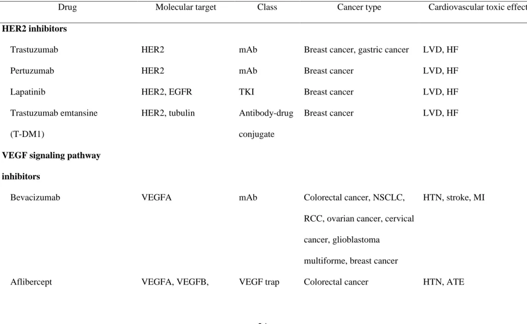
Table 1. Targeted cancer therapies and associated cardiovascular toxic effects
Drug Molecular target Class Cancer type Cardiovascular toxic effects
HER2 inhibitors
Trastuzumab HER2 mAb Breast cancer, gastric cancer LVD, HF
Pertuzumab HER2 mAb Breast cancer LVD, HF
Lapatinib HER2, EGFR TKI Breast cancer LVD, HF
Trastuzumab emtansine
(T-DM1)
HER2, tubulin Antibody-drug
conjugate
Breast cancer LVD, HF
VEGF signaling pathway
inhibitors
Bevacizumab VEGFA mAb Colorectal cancer, NSCLC,
RCC, ovarian cancer, cervical
cancer, glioblastoma
multiforme, breast cancer
HTN, stroke, MI
PIGF
Ramucirumab VEGFR2 mAb Colorectal cancer, gastric
cancer, NSCLC
HTN, ATE
Sunitinib VEGFRs, PDGFRs,
FLT3, CSF1R
TKI RCC, GIST, pancreatic
neuroendocrine tumors
HTN, QTc prolongation,
torsade de points, ATE, VTE,
HF
Sorafenib VEGFRs, PDGFRs,
FLT3, RAF1, BRAF
TKI RCC, hepatic cell carcinoma,
thyroid cancer
HTN, ATE, VTE, HF
Pazopanib VEGFR1, VEGFR3,
PDGFRs, c-KIT
TKI RCC, soft-tissue sarcoma HTN, QTc prolongation,
torsade de points, ATE, VTE
Axitinib VEGFRs, PDGFRs,
FLT3, CSF1R
TKI RCC HTN, ATE, LVD
Vandetanib VEGFR2, EGFR, RET TKI Medullary thyroid cancer HTN, QTc prolongation,
torsade de points, sudden
death
PDGFR, RET, RAF,
FGFR
BCR-ABL TKIs
Bosutinib BCR-ABL, Src family TKI CML Pericardial effusion,
pulmonary edema
Dasatinib BCR-ABL, PDGFR,
c-KIT, Src family
TKI CML, Ph+ALL Pulmonary artery
hypertension, pleural effusion
Nilotinib BCR-ABL, PDGFR,
c-KIT
TKI CML QTc prolongation, CAD, PAD
Ponatinib BCR-ABL, FGFR,
VEGFR, PDGFR, Src
family, c-KIT, RET,
FLT3
TKI CML, Ph+ALL HTN, CAD, PAD, stroke,
VTE, atrial fibrillation
ALL, acute lymphocytic leukemia; CAD, coronary artery disease; CML, chronic myeloid leukemia; CSF1R, colony stimulating factor-1
receptor; EGFR, epidermal growth factor receptor; FGFR, fibroblast growth factor receptor; GIST, gastrointestinal stromal tumor; HER2, human
myocardial infarction; NSCLC, non-small cell lung cancer; PAD, peripheral artery disease; PDGFR, platelet-derived growth factor receptor; Ph,
Philadelphia chromosome; RCC, renal cell carcinoma; TKI, tyrosine kinase inhibitor; VEGF, vascular endothelial growth factor; VEGFR, VEGF
NRG-1 Endothelial cell
ADAM17-19
proteolytic activation
Cardiomyocyte Trastuzumab
Pertszumab
Lapatinib
℗
ERK Ras Src
PI3K
FAK Akt
↓Hypertrophy ↓Proliferation
↓Dedifferentiation Heart Failure
Figure 1
Figure 2
VEGFR
Bevacizumab Aflibercept
Ramucirumab
TKI with anti-VEGF activity: sunitinib, sorafenib,
pazopanib, axitinib, vandetanib, regorafenib
↓Proliferation ↓Survival ↓Vasodilation ↓Vascular permeability
↓Migration
VEGF