Biological and Pharmaceutical Bulletin Regular Article
Evaluation of CYP2D6 Protein Expression and Activity in the Small Intestine to Determine its Metabolic Capability in the Japanese Population
Momoko KAWAKAMIa,c, Sachiko TAKENOSHITA-NAKAYAb, Yuko TAKEBA*c, Yuki NISHIMURAd, Masayuki ODAe, Minoru WATANABEf, Yuki OHTAc, Shinjiro KOBAYASHIg, Takehito OHTSUBOg, Shinichi KOBAYASHIb, Naoki UCHIDAa,b, Naoki MATSUMOTOc
aDepartment of Clinical Pharmacology, Showa University School of Medicine, bShowa University Clinical Research Institute for Clinical Pharmacology and Therapeutics, 6-11-11 Kita-karasuyama, Setagaya-ku, Tokyo 157-8577, Japan, cDepartment of Pharmacology, St. Marianna University School of Medicine, 2-16-1 Sugao, Miyamae-ku, Kawasaki-shi, Kanagawa 216-8511, Japan, dDepartment of Pharmacology, Showa University School of Medicine, 1-5-8 Hatanodai, Shinagawa-ku, Tokyo 142- 8555, Japan, eDepartment of Pharmacogenomics, St. Marianna University Graduate School of Medicine, fInstitute of Animal Experimentation, St. Marianna University Graduate School of
Medicine, and gDivision of Gastroenterological and General Surgery, St. Marianna University School of Medicine, 2-16-1 Sugao, Miyamae-ku, Kawasaki, Kanagawa 216-8511, Japan.
To whom correspondence should be addressed:
Yuko Takeba, DVM., PhD.,
Department of Pharmacology, St. Marianna University School of Medicine, 2-16-1 Sugao, Miyamae-ku, Kawasaki, Kanagawa 216-8511, Japan
Phone: +81-44-977-8111 Ext. 3531 Fax: +81-44-975-0509
e-mail: [email protected]
2 Summary
CYP2D6 plays an important role in the metabolism of many drugs such as opioids and
antidepressants. Polymorphisms of the CYP2D6 gene are widely observed in the Japanese population,
and can affect the first-pass metabolism of orally administered drugs. Several CYP enzymes have
been identified in the small intestine of Caucasians, but intestinal CYP enzymes have not been
reported in the Japanese population, except for CYP3A4 and CYP2C19. In this study, we evaluated
the CYP2D6 metabolic capacity by measurement of CYP2D6 mRNA and protein levels and activity in the small intestine of Japanese individuals. Normal jejunal tissues were obtained from 31 patients
who had undergone pancreaticoduodenectomy, and the CYP2D6*10 variant was identified in these
tissues. CYP2D6 mRNA and CYP2D6 protein levels were analyzed using real-time reverse
transcription-polymerase chain reaction and Western blotting, respectively. Bufuralol 1′-
hydroxylation, a marker of CYP2D6 activity, was analyzed using high-performance liquid
chromatography. Frequencies of the CYP2D6*1/*1, *1/*10, and *10/*10 genotypes in the jejunal
tissue were 29.0% (n = 9), 35.5% (n = 11), and 35.5% (n = 11), respectively. CYP2D6 protein and
activity levels did not differ significantly between the genotypes. A positive correlation was found
between CYP2D6 protein and activity levels. Furthermore, CYP2D6 protein levels and activity in the
small intestine were significantly lower than those in the liver. These findings suggest that the
metabolic capacity of CYP2D6 in the small intestine of the Japanese population has a relatively small
effect on drug metabolism.
3 Keywords;
Cytochrome P450 2D6; small intestine; Japanese; polymorphism; bufuralol
4 Introduction
Cytochrome P450 (CYP) enzymes are present in most tissues in the body, but are most abundant
in the liver. There, they play a pivotal role in the metabolism of endogenous substrates and of
exogenous compounds such as xenobiotics.1) Their presence in extrahepatic tissues, such as the
esophagus, stomach, and small intestine,2–4) also contributes to drug metabolism.5–7) Paine et al.
reported a significant contribution of CYP enzymes in the small intestine to the first-pass metabolism
of midazolam, specifically by intestinal CYP3A4.7) Expressions of CYP3A, CYP2C9, CYP2C19,
CYP2D6, CYP2J2, and CYP4F proteins have been demonstrated in the small intestine of
Caucasians.4,8) Of these, CYP2D6 plays a particularly important role in drug metabolism. Although
CYP2D6 accounts for only 2% of the total CYP protein in the liver, it is a very important isoform
because it metabolizes approximately 20–25% of commonly used drugs, including codeine, tramadol,
and tamoxifen.9) However, CYP2D6 expression and activity have not yet been characterized in the
small intestine of the Japanese population.
CYP2D6 gene polymorphisms are produced from over 109 allelic variants.10) The phenotypes
resulting from these polymorphisms are associated with abolished, decreased, and increased enzyme
activity, and the resulting phenotypes can be classified as a poor metabolizer (PM), intermediate
metabolizer (IM), extensive metabolizer (EM), or ultra-rapid metabolizer (UM), based on the level of
enzyme activity.11) Thus, CYP2D6 polymorphisms greatly affect the pharmacokinetics of CYP2D6
substrates,12,13) though it is unknown whether CYP2D6 present in the small intestine also has an
5 influence on the blood concentrations of its substrates.
CYP2D6*1 is the wild-type, or the so-called normal, allele and results in normal enzyme
activity. Nishida et al. have reported allele frequencies for *10, *5, *4, and *3 of 38.1%, 4.5%, 0.2%,
and 0%, respectively, in healthy Japanese individuals.14) The reported frequencies of CYP2D6 *5, *4,
and *3, which confer the PM phenotype and deficient CYP2D6 activity, are approximately 10% in
Caucasians and less than 1% in the Japanese population. However, the frequency of CYP2D6*10,
which confers the IM phenotype, is relatively high in Asians, and most Asian individuals have been
categorized as IMs. CYP2D6.10. exhibits relatively low catalytic activity toward dextromethorphan,
metoprolol, and nortriptyline.15–17)
Recently, we reported that the protein level and activity of CYP must be determined to estimate its
metabolic capacity because we could not predict the metabolic capacity based on the CYP mRNA level and polymorphism in the human small intestine. 18) A major purpose of this study was to evaluate the
CYP2D6 metabolic capacity in the small intestine of Japanese individuals. We then examined
CYP2D6 mRNA and protein levels and activity in small intestine tissue of Japanese surgical patients.
Additionally, we compared the protein and enzyme activity levels in the intestine with those observed
in the liver tissue. Finally, we determined whether CYP2D6*10 affects the metabolic capacity of
CYP2D6 in the small intestine.
6 Materials and Methods
Human Intestine and Liver Samples
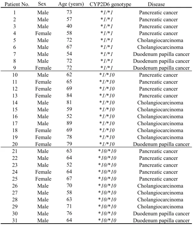
We obtained tissue specimens from the jejunal portion of the small intestine from 31 Japanese
patients (21 men, 10 women) undergoing pancreaticoduodenectomy for various cancers (Table 1). All
specimens were taken from normal (unaffected) tissue. The mucosa of the tissues was immediately
scraped off with a knife, frozen in liquid nitrogen, and preserved at −80°C until use. We also obtained
normal liver tissue from 13 Japanese patients (9 men and 4 women; age range 44 to 81 years, mean =
66.7 years) undergoing partial hepatectomy for hepatocellular carcinoma (n = 3), cholangiocarcinoma
(n = 5), metastatic liver cancer (n = 4), and hepatoma (n = 1). The tissue was taken at least 10 cm
from the tumorous part of the liver, examined pathologically, and confirmed to be free of tumor cells.
Our study was approved by the St. Marianna University School of Medicine Ethics Committee (approval number #1082).
Immunohistochemistry
Immunohistochemistry was used to detect CYP2D6 protein. Tissue samples were fixed in 10%
formalin, embedded in paraffin, and cut into 3-µm-thick sections. The paraffin-embedded sections
were deparaffinized in xylene and rehydrated in a graded series of ethanol washes. Endogenous
peroxidase activity in the tissue sections was inactivated with 0.1% hydrogen peroxide.
Immunoreactivity in sections was visualized with a Dako EnVision system (Carpinteria, CA, U.S.A.)
according to the manufacturer’s instructions. Tissues were then counterstained with hematoxylin.
7
Extraction of Genomic DNA and CYP2D6 Genotyping
Genomic DNA was extracted from the small intestine tissue using the illustra Tissue and Cells
genomicPrep Mini Spin Kit (GE Healthcare, Buckinghamshire, U.K.). Genotyping for the
CYP2D6*10 allele was performed by polymerase chain reaction (PCR).19) The CYP2D6*10 allele was identified as the 188C→T mutation using a 2-step PCR protocol, as described by Johansson et al.20)
The thermal cycling process was performed in a PTC-1152 thermal cycler (Bio-Rad, Tokyo, Japan)
under the following conditions: 40 cycles of denaturation at 94°C for 1 min, annealing at 58°C for 2
min, and extension at 72°C for 3 min for a double-stranded sequence; or 27 cycles of denaturation at
95°C for 7 min, annealing at 95°C for 30 s, and extension at 58°C for 4 min and 15 s for a single-
stranded sequence. CYP2D6*5 and CYP2D6*10 were the main focus of the investigation, based on
the estimated percentage of Japanese individuals that carry each CYP2D6 allele; all other alleles were
regarded as CYP2D6*1. CYP2D6*5 was detected by allele-specific primer PCR using a commercial
kit (SNP typing kit Cytochrome p450 CYP2D6*5, Toyobo, Osaka, Japan). Mutant alleles that cause
defective enzyme activity and are identified in very low frequencies in Japanese individuals (*2, *4,
*14, and *36)21–23) were not examined.
RNA Extraction and Real-Time Reverse Transcription PCR
Total RNA was extracted from the small intestine tissue using a commercial kit (RNeasy Mini,
Qiagen, Germantown, MD, U.S.A.). cDNA was synthesized from 1 µg of total RNA using a
RETROscript kit (Ambion Inc., Austin, TX, U.S.A.). Quantitative mRNA analysis was performed
8
with a 3502S Light Cycler (Roche Diagnostics, Basel, Switzerland). Briefly, the PCR was performed
in 20 μL of total reaction volume containing 2 μL cDNA, 10 pmol primers, 3 mM MgCl2, and 2 μL
SYBR Green with a Light Cycler Fast Start DNA Master SYBER Green Kit (Roche Diagnostics). The
primers used for CYP2D6 and CYP3A4 were obtained from the Light Cycler–Primer Set for human
cytochrome P450 2D6 and 3A4 (Search LC, Heidelberg, Germany). The primers for GAPDH were 5ʹ-
GACAACTTTGGTATCGTGGA-3ʹ (sense) and 5ʹ-TACCAGGAAATGAGCTTGAC-3ʹ
(antisense). The threshold cycles (Ct) were calculated using the Light Cycler software (Version. 5.32).
Standard curves were plotted as Ct-versus-log cDNA quantities, and the sample quantities were
determined from the standard curves. Expression was quantified by the multiplex comparative
threshold method, in which the gene expression data were normalized to the expression level of the
housekeeping gene for GAPDH in each sample.
Preparation of Microsomes
Microsomes were prepared as described previously.24) Briefly, the jejunal mucosae and liver were
homogenized with 3 volumes of 1.15% KCL, and microsomes were prepared by sequential
centrifugation of the homogenate at 9,000 × g for 20 min and of the resultant supernatant at 105,000 ×
g for 60 min at 4°C. The protein concentration was determined with a protein assay kit (Bio-Rad,
Hercules, CA, U.S.A.).
Western Blotting
The microsomes (100 µg protein) were resolved by sodium dodecyl sulfate-polyacrylamide gel
9
electrophoresis (10% acrylamide), and then transferred to Hybond-ECL (Roche Diagnostics)
membranes for 60 min. The membrane was blocked overnight at 4°C with 5% skimmed milk in Tris- buffered saline containing Tween 20 (NaCl 150 mM, Tris-HCl 100 mM, pH 7.5, Tween 0.5%). Anti-
CYP2D6 mouse monoclonal antibody (BD Biosciences, Franklin Lakes, NJ, U.S.A.) was applied to
the blots for 2 h at room temperature. Anti-polyclonal antibody (Wako, Osaka, Japan) was used as the
loading control. Reactive proteins were viewed using enhanced chemiluminescence (ImmunoStar,
Wako). Signal intensities of the detected bands were analyzed using a C-DiGit Chemiluminescent
Western Blot Scanner (LI-COR Biosciences, Lincoln, NE, U.S.A.). Protein expression was quantified
by densitometric analysis. The expression level of -tubulin was confirmed as a control. The inter-
individual and inter-tissue differences in -tubulin levels in the small intestine and liver samples were
less than three-fold. The data of the CYP2D6 protein level was shown as the density in 1mg protein.
When we measured the quantity of protein in some intestinal samples using recombinant CYP2D6
(BD Biosciences) as standard, we confirmed that there was correlation between the quantified protein
levels and band density (R=0.864).For comparison of protein levels in the liver and in the small
intestine, the sample loading was simultaneously performed individual samples and positive control in a gel. Each data was corrected using positive control.
Determination of CYP2D6 Activity by High-Performance Liquid Chromatography (HPLC)
To investigate CYP2D6 activity, 1ʹ-hydroxybufuralol was assayed as described previously, with
minor modifications.25) The reaction mixtures contained human jejunal microsomes (HJMs) (50 µg
10
protein) or liver microsomes (HLMs) (15 µg protein), 10 µM bufuralol, and an NADPH-generating
system in 100 mM potassium phosphate buffer (pH 7.4), and were incubated at 37°C for 20 min
(HJMs) or 15 min (HLMs). A Capcell Pak C18 SG120 HPLC column (4.6 × 250 mm2, Shiseido,
Tokyo) was used for detecting 1′-hydroxybufuralol. A portion of the reaction mixture was injected
into the column and separated with a mobile phase of 20 mM NaClO4 (pH 2.5) and acetonitrile
(70:30) at a rate of 1 mL/min. The HPLC instrumentation included an SCL-10A VP system controller,
a VC-10AD VP pump, a DGU-14-A degasser, an SIL-10AD VP auto-sampler (Shimadzu, Kyoto), a Hitachi L-7485 fluorescence detector (excitation 252 nm, emission 302 nm), and a Fujitsu computer
running Shimadzu CLASS-VP software.
Statistical Analyses
All values are expressed as mean ± SD. The Steel-Dwass test was used for multiple comparisons,
and the non-parametric Mann–Whitney test was used to analyze between-group differences in protein
levels and in protein activity. Correlation between CYP2D6 activity and protein expression levels was
tested by Pearson’s product–moment correlation coefficient. All statistical analyses were performed
with KyPlot 5.0 (Kyeus Lab, Tokyo, Japan), and p < 0.05 was considered significant.
Results
Localization of CYP2D6 in the Small Intestine
11
To determine whether CYP2D6 protein is present in the small intestine of Japanese individuals,
we immunostained jejunal sections for CYP2D6. CYP2D6 was detected in enterocytes of the jejunal
mucosal epithelium and lamina propria (Fig. 1).
Effect of CYP2D6*10 Genotype on CYP2D6 mRNA Levels in the Small Intestine
To certify the quality of the intestinal samples, we analyzed CYP2D6 mRNA expression in the
same samples as those used for CYP3A4 mRNA analysis. CYP3A4 mRNA levels (353.3 ± 341.8 ×
106) were 13-fold higher than CYP2D6 mRNA levels (26.8 ± 25.4 × 10⁶) (Fig. 2A).
The frequencies of the CYP2D6*1/*1, *1/*10, and *10/*10 genotypes in the small intestine
samples were 29.0% (n = 9), 35.5% (n = 11), and 35.5% (n = 11), respectively. The CYP2D6*5 allele
was not be detected in any sample. To clarify the effect of these CYP2D6 genotypes on CYP2D6
transcription, we analyzed CYP2D6 mRNA levels. The mean CYP2D6 mRNA level in the
CYP2D6*10 homozygotic tissue (45.6 ± 19.9 × 10⁶) was significantly (p < 0.01) higher than that in
the CYP2D6*1 homozygotic tissue (5.8 ± 3.7 × 10⁶) or the CYP2D6*1/10 heterozygotic tissue (19.6 ±
12.0 × 10⁶), although individual differences in CYP2D6 mRNA levels were observed within each
genotype (Fig. 2B).
CYP2D6 Protein Levels and Activity in the Small Intestine
Next, we evaluated the effect of CYP2D6 polymorphisms on CYP2D6 protein levels and activity.
When we measured the quantity of protein in some intestinal samples using recombinant CYP2D6
12
(BD Biosciences) as standard, we confirmed that there was correlation between the quantified protein
levels and band density (R = 0.864).
The correlation (R= 0.625, p = 0.011) was found between CYP2D6 protein levels and enzymatic
activity (Fig. 3A). CYP2D6 protein levels did not differ significantly between the genotypes (Fig.
3B).
The mean CYP2D6 activity associated with CYP2D6*1/*1 (2.6 ± 1.4 pmol/mg protein/min)
tended to be higher than that associated with CYP2D6*1/*10 (0.8 ± 0.4 pmol/mg protein/min, p =
0.106) and with CYP2D6*10/*10 (1.3 ± 0.4 pmol/mg protein/min, p = 0.348) (Fig. 3C).
CYP2D6 Protein levels and Activity in Small Intestine vs. Liver
Results for western blotting for CYP2D6 in the small intestine and liver of Japanese are shown in Fig 4A. CYP2D6 protein levels were significantly lower in the small intestine (5.5 ± 2.7× 10⁴
counts/mg protein) than in the liver (16.2 ± 3.2 × 10⁴ counts/mg protein) (Fig.4B). Specific CYP2D6
genotypes were not determined in this experiment because of the limited volume of the liver sample.
CYP2D6 activity in the small intestine (1.6 ± 1.2 pmol/mg protein/min) was much lower than that
in the liver (67.9 ± 43.6 pmol/mg protein/min) (Fig. 4C).
Discussion
Human small intestine epithelial cells are the site of initial biotransformation of orally ingested
xenobiotics. Recent studies have shown that metabolism by small intestine CYP proteins can affect
13
the toxicity and therapeutic efficacy of xenobiotics.26) In this study, we evaluated the metabolic
capacity of CYP2D6 in the small intestine of Japanese patients based on CYP2D6 protein level and
activity, and investigated the effect of genetic polymorphisms on CYP2D6 metabolic capacity.
Previous studies have shown that CYP2D6 is expressed in the human small intestine.27,28) Using
immunohistochemistry, we found that CYP2D6 protein was localized in the enterocytes and lamina
propria of the small intestine of Japanese individuals. Although CYP2D6 mRNA levels were lower
than the CYP3A4 mRNA levels in the samples, we did find that CYP2D6 mRNA was expressed in
the small intestine of Japanese individuals. Previous studies have reported that CYP3A4 mRNA levels
were quantified in human intestine29) 30). The present results of CYP3A4 mRNA levels cannot be simply compared with already reported results because of differences in intestinal sampling and ethnicity.
However, mRNA extraction and real time-PCR analysis was performed with almost the same procedure.
We consider that CYP3A4 mRNA stability in the small intestine is preserved. We verified that CYP3A4
mRNA levels were expressed on average at high levels. Further, these findings are consistent with those
of previous studies.4, 27)
CYP2D6 genetic polymorphisms have been shown to be the major cause of variation in the
metabolism of tamoxifen, and the polymorphisms can thus lead to adverse effects or lack of
therapeutic efficacy.31) CYP2D6 genotypes include CYP2D6*1 (wild-type), *2, *4, *5, and *10. Of the
possible CYP2D6 alleles, CYP2D6*10 occurs at a frequency of 34.6–71.0% in Japanese individuals,
but only 1–2% in Caucasians10,11,15,19) Because the IM phenotype produced by CYP2D6*10 exhibits
14
lower catalytic activity than that of the phenotype produced by CYP2D6*1,32,33) CYP2D6*10 is an
important allele in the Japanese population, as it confers interindividual differences in the
pharmacokinetics of CYP2D6 substrates. The allele frequency CYP2D6*10 was 53.2%. The allele
frequency for CYP2D6*10 we observed was similar to that reported previously (34.6% -71.0%).
10,11,15,19) Additionally, the mean CYP2D6 mRNA levels in patients with the CYP2D6*10/*10 genotype
were significantly higher than those in patients with CYP2D6*1/*1 and CYP2D6*1/*10 genotypes.
Consistent with this, CYP2D6 *10 is believed to be the allele that encodes CYP2D6 protein with
lowered stability, and the higher mRNA levels in patients with CYP2D6*10/*10 may be due to a
tandem-type CYP2D6*36-*10/*36-*10 allele, which produces four copies of CYP2D6 mRNA.34)
Further study is needed to evaluate whether the other CYP2D6 alleles, including the tandem-type *36-
*10 might be included in the examined Japanese subjects.
In contrast to the mRNA results, there was no statistical difference in CYP2D6 protein or activity
levels between the genotypes, though the protein and activity levels in patients with the CYP2D6*10
allele tended to be lower than those in the other groups. These findings indicate that the CYP2D6*10
allele has only a small influence on CYP2D6 activity levels in the small intestine. Additionally,
CYP2D6 activity levels in the small intestine of the subjects were significantly lower than those in the
liver. Therefore, CYP2D6 in the small intestine of Japanese individuals does not appear to contribute
as much to drug metabolism as does CYP2D6 in the liver. We showed that CYP2D6 protein levels in
the liver ware two-hold higher than that in the small intestine, whereas the activity in the liver was 42-
15
fold higher than that in the small intestine. For example, the presence of enteric bacteria is known to
alter the mRNA or protein expression and activity of hepatic drug metabolizing enzymes. 35) The
difference in the enzyme activity in the small intestine and liver may be related to induction or
inhibition factors included in each tissue microsome. 36) This will have to be investigated in detail
because the cause has not been clarified yet in human tissues.
We found no correlation between CYP2D6 mRNA levels and CYP2D6 protein levels.
This seemingly paradoxical finding is attributable to mechanisms that are involved in post-
translational processing steps of CYP2D6 in the small intestine. For example, CYP3A4 protein
expression is partly regulated by post-translational machinery.37) Additionally, glycoprotein 78 (gp78)
and C-terminal heat shock protein 70 (Hsp70)-interacting protein (CHIP) E3 ligases cooperatively
degrade CYP3A4 protein via its ubiquitination in human liver.38) This mechanism is believed to be
independent of the transcriptional steps; therefore, the discrepancy between CYP2D6 mRNA and
CYP2D6 protein levels might be the result of inter-individual differences in post-translational
regulation. Furthermore, it remains unclear whether CYP2D6 more rapidly degraded in the small
intestine than in the liver. Further investigation is necessary to clarify this issue.
A limitation of this study is that we did not examine other CYP2D6 alleles besides the CYP2D6*10
allele in the small intestine samples we obtained. Additionally, we used jejunal tissues for the study,
but did not examine all parts of the small intestine. Further studies are needed to fully characterize
CYP2D6 genetics in the small intestine of the Japanese population.
16
In conclusion, we found that CYP2D6*10 polymorphism did not influence the metabolic
capacity of CYP2D6 in the small intestine. Our findings suggest that the metabolic capacity of
CYP2D6 in the small intestine of Japanese individuals has a relatively small effect on the metabolism
of orally administered drugs in comparison to that of CYP2D6 in the liver.
Conflict of Interest
The authors declare no conflict of interest.
17 References
1) Nebert DW, Russell DW. Clinical importance of the cytochromes P450. Lancet, 360, 1155–
1162 (2002).
2) Lechevrel M, Casson AG, Wolf CR, Hardie LJ, Flinterman MB, Montesano R, Wild CP.
Characterization of cytochrome P450 expression in human oesophageal mucosa.
Carcinogenesis, 20, 243–248 (1999).
3) Yokose T, Doy M, Kakiki M, Horie T, Matsuzaki Y, Mukai K. Expression of cytochrome P450 3A4 in foveolar epithelium with intestinal metaplasia of the human stomach. Jpn. J.
Cancer Res., 89, 1028–1032 (1998).
4) Paine MF, Hart HL, Ludington SS, Haining RL, Rettie AE, Zeldin DC. The human intestinal cytochrome P450 "pie". Drug Metab. Dispos., 34, 880–886 (2006).
5) Pinto LF, Moraes E, Albano RM, Silva MC, Godoy W, Glisovic T, Lang MA. Rat
oesophageal cytochrome P450 (CYP) monooxygenase system: comparison to the liver and
relevance in N-nitrosodiethylamine carcinogenesis. Carcinogenesis, 22, 1877–1883 (2001).
6) Vences-Mejía A, Caballero-Ortega H, Dorado-González V, Gamboa-Domínguez A, Gómez- Ruiz C, Camacho-Carranza R, Espinosa-Aguirre JJ. Cytochrome P450 expression in rat
gastric epithelium with intestinal metaplasia induced by high dietary NaCl levels. Environ.
Toxicol. Pharmacol., 20, 57–64 (2005).
18
7) Paine MF, Shen DD, Kunze KL, Perkins JD, Marsh CL, McVicar JP, Barr DM, Gillies BS, Thummel KE. First-pass metabolism of midazolam by the human intestine. Clin. Pharmacol.
Ther., 60, 14–24 (1996).
8) Wang MZ, Wu JQ, Bridges AS, Zeldin DC, Kornbluth S, Tidwell RR, Hall JE, Paine MF.
Human enteric microsomal CYP4F enzymes O-demethylate the antiparasitic prodrug pafuramidine. Drug Metab. Dispos., 35, 2067–2075 (2007).
9) Samer CF, Lorenzini KI, Rollason V, Daali Y, Desmeules JA. Applications of CYP450 testing in the clinical setting. Mol. Diagn. Ther., 17, 165–184 (2013).
10) The Human Cytochrome P450 (CYP) Allele Nomenclature Database:
〈www.cypalleles.ki.se/〉, cited 1 February, 2016.
11) Ingelman-Sundberg M, Sim SC, Gomez A, Rodriguez-Antona C. Influence of cytochrome P450 polymorphisms on drug therapies: pharmacogenetic, pharmacoepigenetic and clinical
aspects. Phramacol. Ther., 116, 496–526 (2007).
12) Ingelman-Sundberg M. Genetic polymorphisms of cytochrome P450 2D6 (CYP2D6): clinical consequences, evolutionary aspects and functional diversity. Pharmacogenomics J., 5, 6–13
(2005).
13) Kirchheiner J, Nickchen K, Bauer M, Wong ML, Licinio J, Roots I, Brockmöller J.
Pharmacogenetics of antidepressants and antipsychotics: the contribution of allelic variations
to the phenotype of drug response. Mol. Psychiatry., 9, 442–473 (2004).
19
14) Nishida Y, Fukuda T, Yamamoto I, Azuma J. CYP2D6 genotypes in a Japanese population:
low frequencies of CYP2D6 gene duplication but high frequency of CYP2D6*10.
Pharmacogenetics, 10, 567–570 (2000).
15) Kubota T, Yamaura Y, Ohkawa N, Hara H, Chiba K. Frequencies of CYP2D6 mutant alleles in a normal Japanese population and metabolic activity of dextromethorphan O-demethylation
in different CYP2D6 genotypes. Br. J. Clin. Pharmacol., 50, 31–34 (2000).
16) Taguchi M, Nozawa T, Kameyama T, Inoue H, Takesono C, Mizukami A, Hashimoto Y.
Effect of CYP2D6*10 on pharmacokinetic variability of routinely administered metoprolol in
middle-aged and elderly Japanese patients. Eur. J. Clin. Pharmacol., 59, 385–388 (2003).
17) Yue QY, Zhong ZH, Tybring G, Dalén P, Dahl ML, Bertilsson L, Sjöqvist F. Pharmacokinetics of nortriptyline and its 10-hydroxy metabolite in Chinese subjects of different CYP2D6
genotypes. Clin. Pharmacol. Ther., 64, 384–90 (1998).
18) Hayashi M, Matsumoto N, Takenoshita-Nakaya S, Takeba Y, Watanabe M, Kumai T, Takagi M, Tanaka M, Otsubo T, Kobayashi S. Individual metabolic capacity evaluation of
cytochrome P450 2C19 by protein and activity in the small intestinal mucosa of Japanese
pancreatoduodenectomy patients. Biol. Pharm. Bull., 34, 71–76 (2011).
19) Kimura S, Umeno M, Skoda RC, Meyer UA, Gonzalez FJ. The human debrisoquine 4-
hydroxylase (CYP2D) locus: sequence and identification of the polymorphic CYP2D6 gene, a
related gene, and a pseudogene. Am. J. Hum. Genet., 45, 889–904 (1989).
20
20) Johansson I, Oscarson M, Yue QY, Bertilsson L, Sjöqvist F, Ingelman-Sundberg M. Genetic analysis of the Chinese cytochrome P4502D locus: characterization of variant CYP2D6 genes
present in subjects with diminished capacity for debrisoquine hydroxylation. Mol.
Pharmacol., 46, 452–459 (1994).
21) Nakamura K, Goto F, Ray WA, McAllister CB, Jacqz E, Wilkinson GR, Branch RA.
Interethnic differences in genetic polymorphism of debrisoquin and mephenytoin
hydroxylation between Japanese and Caucasian populations. Clin. Pharmacol. Ther., 38, 402–
408 (1985).
22) Ishizaki T, Eichelbaum M, Horai Y, Hashimoto K, Chiba K, Dengler HJ. Evidence for
polymorphic oxidation of sparteine in Japanese subjects. Br. J. Clin. Pharmacol., 23, 482–485
(1987).
23) Horai Y, Nakano M, Ishizaki T, Ishikawa K, Zhou HH, Zhou Bl, Liao CL, Zhang LM.
Metoprolol and mephenytoin oxidation polymorphisms in Far Eastern Oriental subjects:
Japanese versus mainland Chinese. Clin. Pharmacol. Ther., 46, 198–207 (1989).
24) Watanabe M, Tateishi T, Tanaka M, Kumai T, Kobayashi S. Role of CYP3A in bromperidol metabolism in rat in vitro and in vivo. Xenobiotica, 29, 839–846 (1999).
25) Nishimura Y, Kurata N, Sakurai E, Yasuhara H. Inhibitory effect of antituberculosis
drugs on human cytochrome P450-mediated activities. J Pharmacol. Sci., 96, 293–300
(2004).
21
26) Thelen K, Dressman JB. Cytochrome P450-mediated metabolism in the human gut wall. J.
Pharm. Pharmacol., 61, 541–558 (2009).
27) Zhang QY, Dunbar D, Ostrowska A, Zeisloft S, Yang J, Kaminsky LS. Characterization of human small intestinal cytochromes P-450. Drug Metab Dispos., 27, 804-809 (1999).
28) Maximov PY, McDaniel RE, Fernandes DJ, Korostyshevskiy VR, Bhatta P, Mürdter TE,
Flockhart DA, Jordan VC. Simulation with cells in vitro of tamoxifen treatment in
premenopausal breast cancer patients with different CYP2D6 genotypes. Br J Pharmacol.,
171, 5624–5635 (2014).
29) Nakamura T, Sakaeda T, Ohmoto N, Tamura T, Aoyama N, Shirakawa T, Kamigaki T, Nakamura T, Kim KI, Kim SR, Kuroda Y, Matsuo M, Kasuga M, Okumura K.Real-time
quantitative polymerase chain reaction for MDR1, MRP1, MRP2, and CYP3A-mRNA levels
in Caco-2 cell lines, human duodenal enterocytes, normal colorectal tissues, and colorectal
adenocarcinomas. Drug. Metab. Dispos., 30, 4-6 (2002).
30) Thörn M, Finnström N, Lundgren S, Rane A, Lööf L. Cytochromes P450 and MDR1 mRNA
expression along the human gastrointestinal tract. Br. J. Clin. Pharmacol,. 60, 54–60 (2005).
31) Sakuyama K, Sasaki T, Ujiie S, Obata K, Mizugaki M, Ishikawa M, Hiratsuka M. Functional characterization of 17 CYP2D6 allelic variants (CYP2D6.2, 10, 14A-B, 18, 27, 36, 39, 47-51,
53-55, and 57). Drug Metab. Dispos., 36, 2460–2467 (2008).
32) Honda M, Nozawa T, Igarashi N, Inoue H, Arakawa R, Ogura Y, Okabe H, Taguchi M,
22
Hashimoto Y. Effect of CYP2D6*10 on the pharmacokinetics of R- and S-carvedilol in
healthy Japanese volunteers. Biol Pharm Bull., 28, 1476–1479 (2005).
33) Senda C, Yamaura Y, Kobayashi K, Fujii H, Minami H, Sasaki Y, Igarashi T, Chiba K.
Influence of the CYP2D6*10 allele on the metabolism of mexiletine by human liver
microsomes. Br J Clin Pharmacol., 52, 100–103 (2001).
34) Gaedigk A, Bradford LD, Alander SW, Leeder JS. CYP2D6*36 gene arrangements within the cyp2d6 locus: association of CYP2D6*36 with poor metabolizer status. Drug Metab. Dispos.,
34, 563–569 (2006).
35) Toda T, Ohi K, Kudo T, Yoshida T, Ikarashi N, Ito K, Sugiyama K. Ciprofloxacin suppresses Cyp3a in mouse liver by reducing lithocholic acid-producing intestinal flora. Drug Metab
Pharmacokinet., 24, 201-208 (2009).
36) Shimizudani T, Nagaoka K, Hanioka N, Yamano S, Narimatsu S. Comparative study of the oxidation of propranolol enantiomers in hepatic and small intestinal microsomes from
cynomolgus and marmoset monkeys. Chem Biol Interact., 183, 67–78 (2010).
37) Goodwin B, Hodgson E, D'Costa DJ, Robertson GR, Liddle C. Transcriptional regulation of the human CYP3A4 gene by the constitutive androstane receptor. Mol Pharmacol., 62, 359-
365 (2002).
38) Kim SM, Acharya P, Engel JC, Correia MA. Liver cytochrome P450 3A ubiquitination in
vivo by gp78/autocrine motility factor receptor and C terminus of Hsp70-interacting protein
23
(CHIP) E3 ubiquitin ligases: physiological and pharmacological relevance. J Biol. Chem.,
285, 35866–35877 (2010).
24 Figure Legends
Fig. 1. Immunohistochemical localization of CYP2D6 protein in jejunal tissue of Japanese patients.
(A) Anti-rabbit IgG polyclonal antibody was used as a negative control. (B) and (C) Immunostaining
for CYP2D6 in jejunal tissue. The brown stain indicates CYP2D6 expression. Scale bars represent
100 μm (A and B) and 20 μm (C). CE, crypt epithelium; EC, enterocyte; GC, goblet cells; LP, lamina
propria.
Fig. 2. CYP3A4 and CYP2D6 mRNA levels in the small intestine of the Japanese patients. (A)
Individual variation in CYP3A4 and CYP2D6 mRNA levels. (B) Influence of the CYP2D6 genotypes
on CYP2D6 mRNA levels in the small intestine of the Japanese patients. CYP2D6 mRNA levels in
the jejunal tissues are quantified in relation to each CYP2D6 genotype (*1/*1, *1/*10, *10/*10). Data
are shown as mean ± SD; p < 0.01 versus *10/*10.
Fig. 3. CYP2D6 protein levels and it’s activity in the small intestine. (A) Correlation between
CYP2D6 protein levels and bufuralol 1′-hydroxylation activity in the small intestine of the Japanese
patients. Measurement of bufuralol 1′-hydroxylation activity as a marker of CYP2D6 activity
(described in Materials and Methods). The effects of CYP2D6 protein levels (B) and bufuralol 1′-
hydroxylation activity (C) in the small intestine in relation to the different genotypes. Data are shown
as mean ± SD (n = 4–6).
25
Fig. 4. CYP2D6 protein levels and bufuralol 1′ hydroxylation activity in the liver and small intestine
of Japanese patients. (A) Western blotting for CYP2D6 in the small intestine and liver of Japanese
patients. Representative images are shown. α-tubulin was used as a loading control. (B) CYP2D6
protein levels in the small intestine and liver. Data are shown as mean ± SD (n = 13–14). p < 0.01
versus the small intestine, Mann-Whitney test. (C)Bufuralol 1′-hydroxylation activity in the liver
and small intestine of the Japanese patients. CYP2D6 activity in the liver was significantly higher
than that in the small intestine. Data are shown as mean ± SD (n = 13–16), p < 0.001, Mann–Whitney
test. CYP2D6 protein and activity levels in the liver were higher than those in the small intestine.
26
Fig. 1
27
Fig. 2
28
Fig. 3
29
Fig. 4
30
Table 1. Characteristics of the 31 Patients From Whom Normal Jejunal Samples Were Obtained