Title
Effect of Osteogenic Differentiation Medium on Proliferation and Differentiation of Human
Mesenchymal Stem Cells in Three‑dimensional Culture with Radial Flow Bioreactor
Author(s) 西村, 逸郎 Journal , (): ‑
URL http://hdl.handle.net/10130/3631 Right
Original Article
Effect of Osteogenic Differentiation Medium on Proliferation and Differentiation of Human Mesenchymal Stem Cells in Three-dimensional Culture with Radial Flow Bioreactor
Itsurou Nishimura
Department Crown and Bridge Prosthodontics
Abstract
Human mesenchymal stem cells (hMSCs) are pluripotent cells, and have been expanded and
differentiated into several kinds of mesodermal tissue in vitro. In order to promote bone repair, the
enhancement of proliferation and differentiation of hMSCs towards osteoblasts in vitro is
recommended prior to therapeutic delivery. However, for clinical application, it is still unclear which
method is more advance for tissue engineering to transplant undifferentiated or to some extent
differentiated stem cell. Therefore, the present study aimed to investigate how osteogenic
differentiation medium (ODM) affect the hMSCs cultured in 3D scaffold by a radial-flow bioreactor
(RFB) besides cell growth medium (GM). To produce precultured sheets, the hMSCs were first
seeded onto type 1 collagen sheets and incubated for 12 h, after which they were placed in the RFB
for fabrication of scaffolds. The culture medium was circulated at 3 mL/min and the cells
dynamically cultured for 1 week at 37°C. Static cultivation in a culture dish was also carried out.
Cell proliferations were evaluated by histological analysis and DNA-based cell count. Alkaline
phosphatase (ALP) activity, immunocytochemical analysis with BMP-2 and osteopontin on the
hMSCs in the collagen scaffold were performed. In 14 days ODM culture, significant increase in cell
number and higher density of cell distribution in the scaffold were observed both static and dynamic
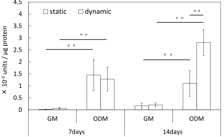
cultivation compared to GM culture. Significant increase of ALP activity in 14 days ODM is
recognized in dynamic cultivation compared to static cultivation. Cells that BMP-2 was expressed
were frequently observed for 14 days in dynamic culture compared with other conditions, and
expression of osteopontin was confirmed in dynamic cultivation for both 7 days and 14 days. Results
in this study revealed that both the proliferation and bone differentiation of hMSCs in 3D culture by
RFB were accelerated by the culture of osteogenic differentiation medium, suggesting an
advantageous future clinical application of RFB cell culture and cell transplantation for tissue
engineering.
Introduction
Large bone defects caused by trauma, inflammation, tumors, or congenital abnormalities
are often treated with autologous or allogenic bone grafts. The implantation of autologous bone
grafts are the most popular due to their high performance in terms of osteogenesis, the only
drawback being there limited availability due to donor site morbidity. Allogenic bone grafts are less
attractive due to the risk of immunogenicity, donor-to-host transmission of disease (e.g. HIV), graft
failure as a consequence of the reduced osteoinductivity of allograft bone.1
Recently, cell based tissue engineering has drawn much interest as an alternative to these
approaches, offering the potential for creation of bioartificial tissues and even organs. Human
mesenchymal stem cells (hMSCs) are pluripotent cells, can be readily isolated from adult donors
with a little damage and are inducible osteoprogenitor cells, making them the cell of choice in bone
tissue engineering and regeneration.2,3 However, the amount of hMSCs harvested from donor tissue
is limited to directly apply to clinical treatment. Static culturing of MSCs on porous scaffolds and
maturation is the simplest method of developing cell-scaffold complex for in vivo implantation.4
Several studies have reported low seeding efficiencies and non-uniform cell distributions within
scaffolds, owing, in part, to the manual- and operator-dependent nature of the process.5-7 Moreover,
lack of influence from shear stress and mechanical loading, in static culture efficient osteoinduction
seems difficult to achieve. Accordingly, specialized dynamic culture systems, called bioreactors,
have been used in bone tissue engineering. Such a dynamic three-dimensional (3D) culture system
may represent a more physiological environment than a dish and that fluid flow is an important
component for seeding and culturing BMSCs in 3D environments.8-11 This increased interest in tissue
engineering has led to the development of various types of equipment for the construction of
bioreactors, including spinner flasks, rotating wall vessels, and direct perfusion bioreactors, have
been extensively investigated in bone tissue engineering.12,13
The radial-flow bioreactor (RFB) has shown the ability to maintain an even cell culture
environment by radial provision of the medium, allowing comparatively larger tissues to be
constructed.14-17 To allow even distribution of oxygen, culture medium is pumped to the center of the
chamber from the periphery under low shear stress.
In order to promote bone repair as one approach, the enhancement of proliferation and
differentiation of hMSCs towards osteoblasts in vitro is recommended prior to therapeutic delivery.4
In the previous study, it is reported that the preosteoblast-like cells and hMSCs were expand
uniformly over a 3D scaffold under dynamic cultivation using an RFB, and the hMSCs was not
changed in cellular characteristics compare to static cultivation in DMEM without bone
differentiation medium.14,18 However, for clinical application, it is still unclear which method is more
advantageously for tissue engineering to transplant undifferentiated or to some extent differentiated
stem cell.
Therefore, the present study aimed to investigate how osteogenic differentiation medium
(ODM) affect the hMSCs cultured in 3D scaffold by a radial-flow bioreactor (RFB).
Materials and Methods
Figure.1 shows a summary of the study protocol.
Culture of human MSCs
hMSCs derived from human bone marrow (PT-2501; Lonza Walkersville,) and donated by
a 19-year-old man were passaged 5 times for use in this study. Dulbecco’s modified Essential
medium (DMEM; Gibco) supplemented with 10% fetal bovine serum (Sigma-Aldrich) and 100 unit/
mL penicillin–streptomycin (Gibco) was used as growth media (GM) for static and dynamic
cultivation as well as preculture. On the other hand, GM supplemented with 50nM dexamethasone,
0.2mM ascorbic acid and 10mM b-glycerophosphate was used as an osteogenic differentiation
medium (ODM) for static and dynamic cultivation.
Cells suspension containing 5.0×10⁵ cells was seeded into 75cm2 flasks, and 20 mL fresh
culture medium added to each flask. Cultures were maintained at 37°C in a humidified atmosphere
with 5% CO2. The culture medium was changed every 3 days. At 1-week cultivation, before reaching
confluence, the cells were harvested by trypsin treatment and seeded onto type-1 collagen sheets
(Gunze) (pore size, 70-110 µm; porosity, 80%-95%; diameter, 12 mm; thickness, 3 mm).
Preculture
Method of cell seeding is based on Katayama’s study.18 Briefly, type 1 collagen sheets
were placed in a 12-well plate and cell suspension (80µL) containing 1.0×10⁵ cells was seeded onto
them. The sheets were then incubated in a humidified atmosphere at 37°C with 5% CO2 for 6 h. Next,
they were turned over, and a further 80µL cell suspension containing 1.0×10⁵ cells was added before
a further 6 h incubation (Finally, total cell seeding density is 2.0 ×10⁵ cells per sheet).
Dynamic cultivation
Figure.2 shows the RFB (Able) and RFB cell culture system used. To form a scaffold, 3
precultured sheets were placed in the RFB in layers after incubation for 12 h (6 h + 6 h). Temperature
(37°C), pH (7.4), and dissolved oxygen (DO, 6.86 ppm) in the medium reservoir were controlled and
monitored. The medium volume was maintained at 100 mL. After commencement of culture, the
medium was changed every day from the third day onward. The medium flow rate was set at 3
mL/min. Culture was carried out for 7 days and 14days in each of the GM and ODM as shown in
Figure 1
Static cultivation
The preculture protocol for static cultivation was the same as that for dynamic cultivation.
An individual precultured sheet was placed in each well of a 12-well plate. The culture medium was
maintained at 2mL. Culture was carried out in a humidified atmosphere at 37°C and 5% CO2 for 7
days and 14 days in each of the GM and ODM with no control of DO or pH values. The culture
medium was changed every 3 days. Table.1.shows cultivation condition.
Histological analysis
Figure 3 shows cross section of layered 3 collagen sheets used for each analysis.
Histological analysis was carried out at 7 days or 14 days of culture. Scaffolds that
harvested after culture were fixed with 10% neutral-buffered formalin and dehydrated through a
series of ethanol. After embedding in paraffin, 4-µm-thick sections were prepared from both types of
specimen and stained with hematoxylin-eosin (H-E staining). Finally, they were observed with an
universal photomicroscope (Axiophot 2, Carl Zeiss )
DNA-based cell count
At 7 days or 14 days of culture, scaffolds with 3 cultured sheets from the RFB were
selected for a DNA-based cell count. They were divided into upper, middle, and lower areas from top
to bottom (Fig.3). Single collagen sheets were also selected from the static cultivation, because
precultured collagen sheet was not laminated in static cultivation. This method of cell counting was
selected based on an earlier study by this group.18
Total DNA was quantified with the ND-1000 (NanoDrop Technologies). The cell number
was then calculated using a working curve based on the cell number and total DNA. The mean
DNA-based cell count of the 3 areas under dynamic cultivation was compared with that under static
cultivation.
Alkaline phosphatase (ALP) activity
Scaffolds ware harvested from RFB ,and placed in a 12-well plate. The scaffolds were
rinsed with cold phosphate-buffered saline (PBS), cut into small fragments, and sonicated for 30s
after application of 200 µL Triton-X. The lysates obtained were centrifuged at 15,000rpm for 15min,
and the supernatant was used as sample. ALP activity was assayed using LabAssay ALP (Wako).
Sample absorbance was measured in a 96-well plate at 405nm. Amount of total protein in the sample
was then examined with the Pierce BCA Protein Assay Kit (Thermo Fisher Scientific, Inc.). Finally,
ALP activity was expressed as units/µg protein.
Immunocytochemical analysis
Immunocytochemical analysis was carried out at 7 days or 14 days of culture in ODM.
Proteins in the collagen sheet were visualized with antibodies of BMP-2 and osteopontin. The
sections were washed in 10 nmol / L with pH 7.4 phosphate-buffered saline (PBS), and endogenous
peroxidase activity was blocked by incubating section with 0.3 % H2O2 in methanol for 30 min. The
sections were then reacted with the primary antibodies, BMP-2 (diluted 1:250) (Abcam ab14933)
and osteopontin (diluted 1:140) (Abcam ab 63856), overnight below 4°C. The sections were washed
in PBS and then incubated with the secondary antibody, peroxidase-labeled anti-mouse IgG
polyclonal antibody (Histofine Simple Stain MAX-PO, MULTI, Nichirei), for 30 min and washed
with PBS. Subsequently, the sections were stained with 3,3‘- diaminobenzidine (DAB substrate kit,
Nichirei), washed in sterilized water, and counterstained with hematoxylin. The sections were then
dehydrated according to established procedure and the sections were observed using a universal
photomicroscope (Axiophot 2).
Statistical analysis
The DNA-based cell count and ALP activity was statistically analyzed using a one-way
analysis of variance, followed by the multiple-comparison with fisher’s LSD.
Results
Histological analysis
Figure 4 and 5 shows optical micrographs of hematoxylin-eosin staining. The cells were
distributed densely in the scaffold both static and dynamic culture by ODM at 7 days of culture.
(Fig.4). A higher cell density was observed in ODM under dynamic culture and no difference was
observed between static and dynamic in GM. For 14 days culture, cells are distributed more densely
under both static and dynamic culture by ODM. (Fig.5) Compare to static culture, a higher cell
density was observed in both GM and ODM under the dynamic culture.
DNA-based cell count
The DNA-based cell count of each area is shown in Figure 6 and 7. Figure 6 shows the
comparison of cell numbers in each area (upper, middle, lower) under dynamic cultivation. No
significant difference in cell numbers was observed among the 3 areas.
The number of cells increased for 14 days compared to 7 days on both static and dynamic
culture in the ODM. Comparison of cell numbers between GM and ODM under static and dynamic
cultivation at 7 days and 14 days of cultivation is shown in Figure 7. On dynamic cultivation, mean
of 3 areas was chosen for comparison with that under static cultivation because of no significant
difference in cell numbers among the 3 areas. At 7 days of cultivation, no significant difference in
cell numbers between GM and ODM was found under either static or dynamic culture. On the other
hand, a significant difference in cell numbers between GM and ODM was observed under both static
and dynamic culture (approximately 1.7-1.9 times) in ODM for 14days (** p <0.01). Significant
increases in cell numbers under dynamic cultivation were noted in both GM and ODM for 7 and 14
days compare to static culture (approximately 1.4-1.8 times) (*p< 0.05, **p < 0.01).
ALP activity
Comparison of ALP activity is shown in Fig. 8. ALP activity was hardly observed on GM under both
static and dynamic culture. In ODM, ALP activity was increased under both dynamic and static
culture after 7 days as well as 14 days. Only a significant difference in ALP activity was observed
between static and dynamic culture after 14 days (approximately 2.5 times) (** p <0.01) even though
no significant difference was observed after 7 days.
Immunocytochemical analysis
The results of immunocytochemical analysis in ODM were shown in Figure 9 and 10.
BMP-2 was confirmed by color development in the cell both static and dynamic cultures at 7 days
and 14 days. (Fig.9) Especially, cells that BMP-2 was expressed were frequently observed for 14
days in dynamic culture compared with other conditions. Expression of osteopontin was observed in
dynamic cultivation for both 7 days and 14 days, but not in static cultivation. (Fig.10)
Discussion
This study aimed to investigate the effects of osteogenic differentiation medium on hMSCs
seeded in 3D scaffold under a perfusion culture by RFB. The results showed that the
three-dimensional culture of hMSCs in RFB with osteogenic differentiation medium accelerated both
cell proliferation and osteogenic differentiation.
As reported by Yoshinari et al., there is a possibility that the culture medium cannot
penetrate the scaffold equally due to calcification under long term 3D culture of hMSCs in
osteogenic differentiation medium using RFB.19 To avoid the influence, we set the culture period as 7
days and 14 days.
In this study, by culturing osteogenic differentiation medium, cell proliferation was
promoted significantly both in static and dynamic cultivation for 14 days while not for 7 days culture.
A possible explanation for the result is that the ODM induce hMSCs to differentiate to
preosteoblast-like cells, which can expand more quickly than totally undifferentiated hMSCs
although it is still unclear that in which differentiation stage the proliferation speed will increase, and
when the proliferation ability will lose.14,18
Compared to static culture, dynamic culture demonstrated significantly increased cell
numbers and higher cell distribution density in scaffold regardless culture time and culture medium.
It is possibly due to a more efficient delivery of nutrients and exchange of gas, along with the
elimination of metabolic waste under dynamic cultivation.20, 21 Accordingly, the cell death associated
with usual 3D culture is partially prevented by the dynamic cultivation.22
In this study, ALP activities as well as expression of BMP-2 and osteopontin were
analyzed to investigate how ODM affects osteogenic differentiation of hMSCs in 3D culture by RFB.
ALP activity and BMP-2 are known as early markers of osteoblastic differentiation.22, 23 By culturing
osteogenic differentiation medium, increase of ALP activity was observed in all experiment
conditions and there is a significant difference between static and dynamic culture for 14 days. ALP
activity is upregulated and the bone differentiation speed is promoted in dynamic cultivation
compared to static cultivation. Dynamic culture of hMSCs by RFB is believed to facilitate
osteogenic differentiation due to shear stress caused by medium perfusion and enhanced delivery of
ODM.25-27 Effective circulation of ODM provides necessary differentiation medium to hSMCs in
scaffolds uniformly and the shear stress are also important for cell differentiation.
BMP-2 was confirmed by color development in all condition, and especially in 14 days
dynamic culture, higher cell density and stronger color development were observed. This phenomena
are also in agreement with the prior observation that dynamic culture improves BMP-2 expression of
hMSCs in 3D scaffolds.28 Osteopontin is reported as secreted by osteoblasts at an early stage of bone
development and promotes cell attachment necessary for mineralization of the matrix.29 In this study,
expression of osteopontin was only confirmed in dynamic cultivation for both 7 days and 14 days,
but not in static cultivation. This finding is consistent with previous reports that osteopontin can be
readily induced by the fluid flow in the dynamic culture30, and the osteoblasts are sensitive even to
limited mechanical influences.10
Flow shear stress increases with an increase in the perfusion speed, which stimulates
proliferation of cells and the formation of the extracellular matrix, including collagen, under dynamic
cultivation.31 However, the benefit of flow shear stress to the proliferation of hMSCs may depend on
the flow rate and the type of bioreactor, cell, scaffold, or medium.32,33 Accordingly, further study is
necessary to clarify the appropriate perfusion speed for the combination of materials.
I n conclusion, the results in this study revealed that by culturing osteogenic differentiation
medium, both the proliferation and bone differentiation of hMSCs were accelerated in 3D culture
with dynamic cultivation using RFB. Thus, the method using the preosteoblast-like cells may reduce
the recognition of a foreign substance as well as the hindrance of medium penetration in the scaffold
caused by the calcification, indicating that the cultivation used in this study is believed to be better
for bone tissue engineering compared to using the conventional cultured bone that is calcified
scaffold in vitro. The possibility and effectivity to use ODM for 3D culture of hMSCs by RFB is
confirmed, which suggested an advantageous future clinical application of RFB cell culture and cell
transplantation for tissue engineering.
Acknowledgements
This research was supported by a Grant-in-Aid for Scientific Research (Challenging Exploratory
Research: 24390446) from the Japan Society for the Promotion of Science.
References
1. James L.M. Ferrara., Yanik, G. Acute graft versus host disease: pathophysiology, risk factors and prevention strategies. Clin Adv Hematol Oncol. 3, 415, 2005
2. Pittenger, M.F., Mackay, A.M., Beck, S.C., Jaiswal, R.K., Douglas, R., Mosca, J.D., Moorman, M.A., Simonetti, D.W., Craig, S., Marshak, D.R. Multilineage potential of adult human mesenchymal stem cells. Science. 284, 143, 1999
3. Prockop, D.J. Marrow stromal cells as stem cells for nonhematopoietic tissues. Science. 276, 71, 1997
4. Ceccarelli, G., Bloise, N., Vercellino, M., Battaglia, R., Morgante, L., De Angelis, MG., Imbriani, M., Visai, L. In vitro osteogenesis of human stem cells by using a three-dimensional perfusion bioreactor culture system: a review. Recent Pat Drug Deliv Formul. 7, 29, 2013
5. Holy, C.E., Shoichet, M.S., Davies, J.E. Engineering three-dimensional bone tissue in vitro using biodegradable scaffolds: investigating initial cell-seeding density and culture period. J Biomed Mater Res. 51,376. 2000
6. Xiao, Y.L., Riesle, J., Van Blitterswijk, C.A. Static and dynamic fibroblast seeding and cultivation in porous PEO/PBT scaffolds. J Mater Sci Mater Med. 10, 773, 1999
7. Xie, Y., Yang, S.T., Kniss, D.A. Three-dimensional cell-scaffold constructs promote efficient gene transfection: implications for cell-based gene therapy. Tissue Eng. 7,585,2001
8. Abbott, A. Cell culture: biology's new dimension. Nature. 424, 870, 2003
9. Jacks, T., Weinberg, R.A. Taking the study of cancer cell survival to a new dimension. Cell. 111, 923, 2001
10. Bancroft, G.N., Sikavitsas, V.I., van den Dolder, J., Sheffield, T.L., Ambrose, C.G., Jansen, J.A., Mikos, A.G. Fluid flow increases mineralized matrix deposition in 3D perfusion culture of marrow stromal osteoblasts in a dose-dependent manner. Proc Natl Acad Sci U S A. 99, 12600, 2002
11. Wendt, D., Marsano, A., Jakob, M., Heberer, M., Martin, I. Oscillating perfusion of cell suspensions through three-dimensional scaffolds enhances cell seeding efficiency and uniformity.
Biotechnol Bioeng. 84, 205, 2003
12. McCoy, R.J., O'Brien, F.J. Influence of shear stress in perfusion bioreactor cultures for the development of three-dimensional bone tissue constructs: a review. Tissue Eng Part B Rev. 16, 587 ,2010
13. Yeatts, A.B., Fisher, J.P. Bone tissue engineering bioreactors: dynamic culture and the influence of shear stress. Bone. 48, 171, 2011
14. Arano, T., Sato, T., Matsuzaka, K., Ikada, Y., Yoshinari, M. Osteoblastic cell proliferation with uniform distribution in a large scaffold using radial-flow bioreactor Tissue Eng Part C Methods.
16, 1387, 2010
15. Kataoka, K., Nagao, Y., Nukui, T., Akiyama, I., Tsuru, K., Hayakawa, S., Osaka, A., Huh, NH.
An organic-inorganic hybrid scaffold for the culture of HepG2 cells in a bioreactor. Biomaterials.
26, 2509, 2005
16. Hongo, T., Kajikawa, M., Ishida, S., Ozawa, S., Ohno, Y., Sawada, J., Umezawa, A., Ishikawa, Y., Kobayashi, T., Honda, H. Three-dimensional high-density culture of HepG2 cells in a 5-ml radial-flow bioreactor for construction of artificial liver. J Biosci Bioeng. 99, 237, 2005
17. Hongo, T., Kajikawa, M., Ishida, S., Ozawa, S., Ohno, Y., Sawada, J., Ishikawa, Y., Honda, H.
Gene expression property of high-density three-dimensional tissue of HepG2 cells formed in radial-flow bioreactor. J Biosci Bioeng. 101, 243 ,2006
18. Katayama, A., Arano, T., Sato, T., Ikada, Y., Yoshinari, M. Radial-flow bioreactor enables uniform proliferation of human mesenchymal stem cells throughout a three-dimensional scaffold.
Tissue Eng Part C Methods. 19, 109, 2013
19. Yoshinari, M., Seshima, H., Oda, Y., Inoue, T., and Matsuzaka, K. Three dimensional culture of osteoblastic cells using radial flow bioreactor. Regen Med 5(Suppl), 215, 2006.
20. Bjerre, L., Bünger, C.E., Kassem, M., and Mygind, T. Flow perfusion culture of human mesenchymal stem cells on silicate-substituted tricalcium phosphate scaffolds. Biomaterials 29, 2616, 2008. �
21. Orr, E.D., and Burg, J.L.K. Design of a modular bioreactor to incorporate both perfusion flow and hydrostatic compression for tissue engineering applications. Ann Biomed Eng 36, 1228, 2008. �
22. Volkmer, E., Drosse, I., Otto, S., Stangelmayer, A., Stengele, M., Kallukalam, B.C., Mutschler, W., Schieker, M. Hypoxia in static and dynamic 3D culture systems for tissue engineering of bone. Tissue Eng Part A. 14, 1331, 2008
23. Long, M.W. Osteogenesis and bone-marrow-derived cells. Blood Cells, Molecules, and Diseases 27, 677, 2001
24. Zhu, J.X., Sasano, Y., Takahashi, I., Mizoguchi, I., Kagayama, M. Temporal and spatial gene expression of major bone extracellular matrix molecules during embryonic mandibular osteogenesis in rats. Histochem J. 33, 25, 2001
25. Reich, K.M., McAllister, T.N., Gudi, S., Frangos, J.A. Activation of G proteins mediates flow-induced prostaglandin E2 production in osteoblasts. Endocrinology. 138,1014,1997
26. Nauman, E.A., Satcher, R.L., Keaveny, T.M., Halloran, B.P., Bikle, D.D. Osteoblasts respond to pulsatile fluid flow with short-term increases in PGE(2) but no change in mineralization. J Appl Physiol (1985). 90,1849, 2001
27. Bakker, A.D., Soejima, K., Klein-Nulend, J., Burger, E.H. The production of nitric oxide and prostaglandin E(2) by primary bone cells is shear stress dependent. J Biomech. 34, 671, 2001 28. Stiehler, M., Bünger, C., Baatrup, A., Lind, M., Kassem, M,. Mygind, T. Effect of dynamic 3-D
culture on proliferation, distribution, and osteogenic differentiation of human mesenchymal stem cells. J Biomed Mater Res A. 89, 96, 2009
29. Butler, W.T. The nature and significance of osteopontin. Connect Tissue Res. 23, 123, 1989 30. Braccini, A., Wendt, D., Jaquiery, C., Jakob, M., Heberer, M., Kenins, L., Wodnar-Filipowicz,
A., Quarto, R., Martin, I. Three-dimensional perfusion culture of human bone marrow cells and generation of osteoinductive grafts. Stem Cells. 23,1066, 2005
31. Nirmalanandhan, V.S., Shearn, J.T., Juncosa-Melvin, N., Rao, M., Gooch, C., Jain, A., Bradica, G., and Butler, D.L. Improving linear stiffness of the cell-seeded collagen sponge constructs by varying the components of the mechanical stimulus. Tissue Eng Part A 14, 1883, 2008.
32. Li, D., Tang, T., Lu, J., and Dai, K. Effects of flow shear stress and mass transport on the construction of a large-scale tis- sue-engineered bone in a perfusion bioreactor. Tissue Eng Part A 15, 2773, 2009
33. Porter, B., Zauel, R., Stockman, H., Guldberg, R., and Fyhrie, D. 3-D computational modeling of media flow through scaffolds in perfusion bioreactor. J Biomech 38, 543, 2005.
Figure legends
TABLE 1. Cultivation condition
FIG. 1. A flowchart of the present study.
FIG.2. Radial-flow bioreactor (RFB) system used in this study. (A) Schematic of total system.
Medium was circulated between RFB and medium reservoir using a circulation pump. During
experiment, dissolved oxygen (DO), pH, and temperature in medium were monitored and controlled.
Volume of chamber medium was maintained at 100mL, and fresh medium added continuously. (B)
Schematic of RFB. Medium in RFB flows from periphery to the center of reactor chamber.
FIG.3. Cross section of layered 3 collagen sheets used for each analysis. Scaffolds in RFB were
divided horizontally and perpendicularly into 9 areas consisting of 3 sheets (from top to bottom:
upper, middle, and lower) × 3 areas (inside, middle, and outside). Histological analysis and
immunocytochemical analysis were performed using the middle area of the middle sheet (shaded
area). DNA based cell count and ALP activity were evaluated using 3 sheets (upper, middle, and
lower).
FIG.4. Typical optical micrographs of specimens stained with hematoxylin–eosin. (7days)
(A) GM static cultivation.(B) GM dynamic cultivation. (C) ODM static cultivation. (D) ODM
dynamic cultivation. (×200; scale bar:100µm)
FIG.5. Typical optical micrographs of specimens stained with hematoxylin–eosin.(14days)
(A) GM static cultivation.(B) GM dynamic cultivation. (C) ODM static cultivation. (D) ODM
dynamic cultivation. (×200; scale bar:100µm)
FIG.6. Comparison of cell numbers (DNA-based cell count) in each area under dynamic cultivation.
No significant difference was observed among the 3 areas. Data are expressed as mean ± SD over 5
cultures.
FIG.7. Comparison of cell numbers between GM and ODM under static and dynamic cultivation.
Under dynamic cultivation, mean of 3 areas was chosen for comparison with that under static
cultivation. Data are expressed as mean ± SD over 5 cultures.
FIG.8. Comparison of ALP activity (units/µg protein) between GM and ODM under static and
dynamic cultivation. Under dynamic cultivation, mean of 3 areas was chosen for comparison with
that under static cultivation. Data are expressed as mean ± SD over 5 cultures.
FIG.9. Typical optical micrographs of specimens stained with BMP-2 antibodies in ODM. (A) 7
days static cultivation.(B) 7 days dynamic cultivation. (C) 14 days static cultivation. (D) 14 days
dynamic cultivation. (×320; scale bar:50µm)
FIG.10. Typical optical micrographs of specimens stained with osteopontin antibodies in ODM.
(A) 7 days static cultivation.(B) 7 days dynamic cultivation. (C) 14days static cultivation. (D) 14
days dynamic cultivation. (×320; scale bar:50µm)
TABLE 1. Cultivation condition
static dynamic
Cell number 2.0 × 10
5cells / scaffold
Temperature 37 ℃
CO
25%
pH 7.4
DO 6.86
Medium flow late 3ml / min
Medium volume 2ml / well 100ml / day
Medium change Every 3 days Changed daily after 3days
scaffold 1 sheet / well 3 sheets / reactor
Culture of hMSCs
Preculture
Static cultivation (well plate)
Dynamic cultivation ( RFB )
GM ODM
7 days or 14 days cultivation
Histological analysis DNA‐based cell count ALP‐activity
Immunocytochemical analysis
16mm
5mm
Air O
2CO
2DO
pH Temperature
Controller
PC
Medium flow
A B
RFB RFB RFB Medium reservoir Waste Fresh media bottle media bottle
FIG.2. Radial‐flow bioreactor (RFB) system used in this study. (A) Schematic of total system. Medium was
circulated between RFB and medium reservoir using a circulation pump. During experiment, dissolved
oxygen (DO), pH, and temperature in medium were monitored and controlled. Volume of chamber
medium was maintained at 100mL, and fresh medium added continuously. (B) Schematic of RFB.
upper
lower
Cen ter we ll
Upper
middle
lower
FIG.3. Cross section of layered 3 collagen sheets used for each analysis. Scaffolds in RFB
were divided horizontally and perpendicularly into 9 areas consisting of 3 sheets (from top
to bottom: upper, middle, and lower) × 3 areas (inside, middle, and outside). Histological
analysis and immunocytochemical analysis were performed using the middle area of the
C
static dynamic
GM
ODM
A B
D
FIG. 4.Typical optical micrographs of specimens stained with hematoxylin–eosin.(7days)
(A) GM static cultivation.(B) GM dynamic cultivation. (C) ODM static cultivation. (D) ODM
dynamic cultivation. ( × 200; scale bar:100µm)
static dynamic
GM
ODM
A B
C D
FIG.5. Typical optical micrographs of specimens stained with hematoxylin–eosin.(14days)
(A) GM static cultivation.(B) GM dynamic cultivation. (C) ODM static cultivation. (D) ODM
a
a a
a A
B
B
× 10
5cells / shee t 16 14 12 10 8 6 4 2
0
GM ODM GM ODM
7days 14days
upper middle lower
FIG.6. Comparison of cell numbers (DNA‐based cell count) in each area under
dynamic cultivation. No significant difference was observed among the 3 areas.
× 10
5cells / shee t 16 14 12 10 8 6 4 2 0
static dynamic
* * *
* *
*
*
*
*
*
*
GM ODM GM ODM
* *P<0.01
*
P<0.05
7days 14days
FIG.7. Comparison of cell numbers between GM and ODM under static and dynamic
cultivation. Under dynamic cultivation, mean of 3 areas was chosen for comparison with
× 10
‐3units / μ g pr ot ein
4.5 4 3.5 3 2.5 2 1.5 1 0.5 0
GM ODM GM ODM
7days 14days
static dynamic
* **
*
*
*
*
*
*
*
* *