Study on the Immune Responses in the Chicken with Eimeria Parasite Infection or Lactic Acid Bacteria Administration: Toward the Application of Lactobacillus acidophilus L-55 as an Anticoccidial Agent
2020, September Ho Thi Dung
Graduate School of Environmental and Life Science (Doctor’s Course)
OKAYAMA UNIVERSITY
I ABSTRACT
Avian coccidiosis is the most pathogenic disease of poultry and is caused by intracellular apicomplexan parasites that belong to several different species of Eimeria. Eimeria tenella (E.
tenella) is one of the most frequent species found in the chicken. Zhang (2013) has reported that the annual global cost of coccidiosis is about $ 2.4 billion mainly for drug related cost and clinical or subclinical treatment. The main problem in avian coccidiosis is the appearance of the drug- resistant parasite. Furthermore, safe and secure disease control does not rely on antibiotics is also required from the perspective of food security. Nowadays, probiotic bacteria are considered as potential alternatives solution to anticoccidial because it possesses characteristics against enteric disease such as balancing gut bacteria, reduce inflammation, preventing and treating diarrhea and boosting the immune system. In fact, probiotic bacteria such as lactic acid bacteria (LAB) have been used in livestock production for their benefits on bird performance, immunity and protection against enteric diseases. However, little is known about their use to control parasitic diseases, particularly avian coccidiosis. Therefore, understanding the immune responses to E. tenella could provide useful information for the development of agents to control the disease. The aims of this study were to (1) clarify the local cellular immune response in the site of E. tenella infection and (2) study on potential of Lactobacillus acidophilus L-55 (LaL-55) as an immune enhancer in chicken.
The aim of first experiment was to clarify the local immune response in chick cecum with E.
tenella infection. The results of the immune related gene expression showed that T helper (Th) cell marker CD4 was upregulated in the infected ceca, but not cytotoxic T cell marker CD8. The gene expressions of Th1 cytokines (Interleukin (IL)-18 and Interferon (IFN)-γ) were increased significantly, but not Th2 cytokines (IL-4, IL-13 and IL-10). The expression of granzyme A and perforin which thought to play a relevant role in cell-mediated cytotoxicity was increased after E.
tenella infection compared with the uninfected control group. These molecules are constitutively expressed in NK cells and cytotoxic T cells. We have found that the expression of IL-12 which stimulating T cells and NK cells to secrete IFN-γ was upregulated after E. tenella infection. Our data indicated that NK cells were induced by IL-12 and play an important role in the immune response to E. tenella infection by producing IFN-γ which to be involved in differentiation of T helper cells as well as exhibiting cytotoxic function. Taken together, these data suggested that E.
tenella infection induces cell-mediated immune response in the cecum, mainly Th1 and NK cells.
II
The aim of the second experiment was to evaluate the local cellular response in chick with E. tenella which was depleted mononuclear phagocytes by carrageenan. The macrophage-depleted chicks showed the reduction of the disease severity, significantly lower number of fecal oocysts, and parasite burden score in cecum compared with infected control group. The result of macrophages depletion chicks also indicated significantly lower histological score and the CD4 expression level than those of the infected control group. Cytokine responses in the ceca of macrophages depletion chicks appeared to be a mixed Th1/Th2 response, and perforin expression was higher in the macrophage depletion chicks than in the infected control group. The results of gene expression suggested that CD3−CD4−CD8− cells are induced by macrophage depletion and possibly regulate the local immune response in E. tenella infection which may contribute to the reduction of disease severity. Overall, our study indicated that the main immune cell type was induced after E. tenella infection is Th1 and NK cells and these responses might be regulated by macrophages during early E. tenella infection.
I have conducted the 3rd experiment to determine the potential of LaL-55 as an effective immune enhancer in chicken. L. acidophilus has been proven to elevate immunological activity by stimulating both the innate and adaptive immune responses. In vitro studies have indicated that L.
acidophilus is a strong inducer of Th1 cytokines, including IL-12 and IFN-γ. L. acidophilus is also involved with enhancing cytotoxic immune cell activity such as NK cells. Therefore, L. acidophilus, specifically LaL-55, was selected for this study to determine whether it may help protect chickens against diseases by enhancement of immune function. The results showed that oral administration of LaL-55 augmented the elevation in the total numbers of leukocytes and lymphocytes in the chicks with the Newcastle disease virus vaccine (NDV). NK cell activity was augmented by LaL- 55 administration at 2 weeks after the secondary NDV vaccine inoculation. IFN-γ-mRNA expression was significantly elevated in splenocytes from chicks administered with LaL-55.
Overall, our results suggested that LaL-55 augmented the cellular immune response and vaccine effect.
In conclusion, E. tenella infection induces cell-mediated immune response in the cecum, mainly Th1 and NK cells. LaL-55 could enhance cellular immune response, especially NK cell activity. Then our data may suggest that LaL-55 is safe and effective as an immuno-enhancer and can be applied as an antiparasitic formulation in chickens.
III
ACKNOWLEDGEMENT
I would first like to thank my supervisor, Assoc. Prof. Toshimitsu HATABU, without his training and support at every step, this paper would have never been accomplished. I will never forget that he always encourages and supports me when I do the experiments and writing the paper.
One again, I would like to thank him very much for his training and understanding.
I would also like to acknowledge my two PhD co-supervisors, Prof. Noboru SAITO and Prof. Takehito TSUJI for their kind help, and I am gratefully indebted to them for their very valuable comments which helped to improve my research and widen my knowledge.
I would like to show gratitude to all members of Animal Physiology Laboratory at Okayama University. They have helped and take care of me a lot during the time I study here. I have learned many things from them, not only experimental skill but also life skill. The mental encouragement from them helps me passed the home sick and familiar with Japan. Time to be with them would be one unforgettable memory in my life.
I would like to thank to Prof. Yasuhiro KONDO, Prof. Naotoshi TSUJI, Dr. Makoto MATSUBAYASHI, Dr. Yosuke SUNADA, Prof. Koji KIMURA and Assist. Prof. Yuki YAMAMOTO for their kindly support materials and equipment for my experiment.
I would like to acknowledge Okayama-Hue International Master’s Program which bring me the chance to get scholarship for study in Okayama, Japan.
A special acknowledgement would like to express to my boss and colleagues in Hue university of Agriculture and Forestry for their support during my study time in Japan.
I would like to thank the Ministry of Education, Culture, Sports, Science and Technology of Japan (MEXT) for their financial support during my doctoral course.
Finally, I must express my very profound gratitude to my family and to my Vietnamese friends for providing me with unfailing support and continuous encouragement throughout the time of study and through the process of researching and writing this thesis. This accomplishment would not have been possible without them. Thank you.
IV
TABLE OF CONTENTS
ACKNOWLEDGEMENT ... III TABLE OF CONTENTS ... IIV LIST OF TABLES ... VII LIST OF FIGURES ... VII LIST OF ABBREVIATIONS ... IIX
CHAPTER 1. GENERAL INTRODUCTION ... 1
1.1. Coccidiosis in poultry production ... 2
1.2. Eimeria tenella ... 3
1.2.1. Life cycle ... 3
1.2.2. Pathogenesis ... 4
1.3. Lactic acid bacteria ... 5
1.3.1. Main characteristic of lactic acid bacteria ... 5
1.3.2. Health benefits effect of Lactobacillus acidophilus ... 6
1.4. Overview of the immune system ... 8
1.4.1. Innate immunity ... 8
1.4.2. Adaptive immunity ... 10
1.4.3. Mucosal immune cells ... 14
1.5. Immune response to Eimeria ... 16
1.5.1. Innate responses to Eimeria ... 16
1.5.2. Adaptive immune response to Eimeria ... 18
1.6. Immune system response to LAB ... 20
1.6.1. Systemic immune system response ... 20
1.6.2. Mucosal immune system response ... 23
1.7. Objective ... 26
CHAPTER 2. LOCAL IMMUNE RESPONSES IN E.TENELLA INFECTED CHICKEN WITH OR WITHOUT MONONUCLEAR PHAGOCYTES ... 27
V
2.1. Introduction ... 28
2.2. Materials and methods ... 29
2.2.1. Chicks ... 29
2.2.2. Parasite ... 29
2.2.3. Experimental design ... 29
2.2.4. Fecal collection and oocyst quantification ... 30
2.2.5. Histopathologycal observation ... 30
2.2.6. Percentage of parasite in tissue ... 31
2.2.7. Gene expression analysis by RT-qPCR ... 31
2.2.8. Statistical analysis ... 33
2.3. Results ... 33
2.3.1. Gene expression analysis in cecum of E. tenella infected chicken with mononuclear phagocytes ... 33
2.3.2. Local immune response in E. tenella infected chicken without mononuclear phagocytes ... 36
2.4. Discussion ... 41
2.5. Conclusion ... 44
CHAPTER 3. THE EFFECT OF LACTOBACILLUS ACIDOPHILUS STRAIN L-55 ON CELLULAR IMMUNE RESPONSE AFTER NEWCASTLE DISEASE VIRUS-BASED LIVE ATTENUATED VACCINE INOCULATION ... 45
3.1. Introduction ... 46
3.2. Materials and Methods ... 47
3.2.1. Animals ... 47
3.2.2. Administration of L. acidophilus L-55 and NDV vaccine inoculation ... 47
3.2.3. Peripheral blood cell counts ... 48
3.2.4. Cytolytic activity of mononuclear cells ... 49
3.2.5. Semi-quantitative RT-PCR ... 49
VI
3.3. Results ... 51
3.3.1. Peripheral blood cell count ... 51
3.3.2. Pro-inflammatory gene expression... 52
3.3.3. The cytolytic activity of NK cell ... 53
3.4. Discussion ... 54
3.5. Conclusion ... 56
CHAPTER 4. GENERAL CONCLUSION ... 57
REFERENCES ... 60
VII
LIST OF TABLES
Table 1-1. Main characteristics of Eimeria species ... 2
Table 1-2. Chicken and mammalian Th subsets... ... 13
Table 2-1. Histomorphological evaluation of intestinal inflammation ... 31
Table 2-2. Percentage of parasite in tissue ... 31
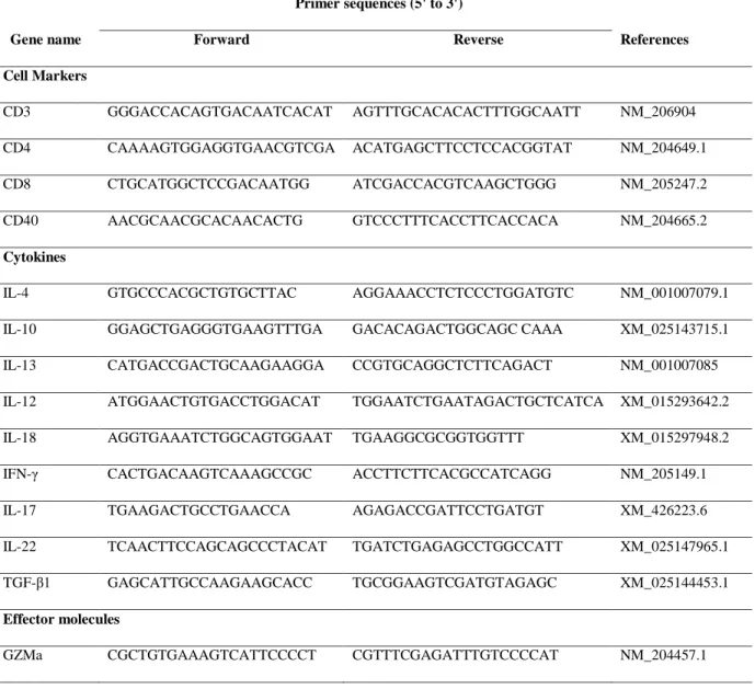
Table 2-3. The primer sets for Immune cell markers, cytokines, and Immune effector molecules ... 32
Table 3-1. Primer sets for semi-quantitative RT-PCR ... 50
LIST OF FIGURES Figure 1-1. E. tenella life cycle ... 3
Figure 1-2. Chicken T cells as classified by TCR, CD4 and CD8 expression and their putative functions. ... 12
Figure 1-3. Systemic immune response induced by probiotic bacteria after interaction with the immune cells of the Peyer's patches.. ... 22
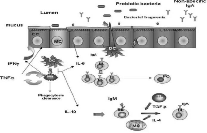
Figure 1-4. The local immune response in the gut induced by the interaction between probiotic bacteria and the epithelial and immune cells associated with the lamina propria of the small intestine.. ... 25
Figure 2-1. mRNA expression levels of immune cell markers in ceca of chicken with E. tenella.. ... 34
Figure 2-2. mRNA expression levels of cytokines in the ceca of chickens with E. tenella. ... 35
Figure 2-3. mRNA expression levels of immune effector molecules in ceca of chickens with E. tenella.. ... 36
Figure 2-4. Fecal oocyst shedding was monitored daily from 5 to 10 days post infection (dpi).. 37
Figure 2-5. Histopathology of HE-stained cecum sections. ... 38
Figure 2-6. mRNA expression levels of immune cell markers in ceca of chicken with E. tenella.. ... 39
VIII
Figure 2-7. mRNA expression levels of cytokines in the ceca of chickens with E. tenella.. ... 40 Figure 2-8. mRNA expression levels of immune effector molecules in ceca of chickens with E.
tenella.. ... 41
Figure 3-1. The course of leukocyte (a) or lymphocyte (b) counts in the peripheral blood of chicks that received the NDV vaccine and oral LaL-55. ... 52 Figure 3-2. Effect of LaL-55 administration on mRNA levels of cytokines in splenic
mononuclear cells. ... 53 Figure 3-3. Effect of LaL-55 administration on cytolytic activity of mononuclear cells ... 54
IX
LIST OF ABBREVIATIONS Eimeria tenella: E. tenella
LAB: Lactic acid bacteria
L. acidophilus: Lactobacillus acidophilus
BW: Body weight BWG: Body weight gain FCR: Feed conversion ratio C. diff: Clostridium difficile L. reuteri: Lactobacillus reuteri L. salivarius: Lactobacillus salivarius L. plantarum: Lactobacillus plantarum L. casei: Lactobacillus casei
L. rhamnosus: Lactobacillus rhamnosus L. bulgaricus: Lactobacillus bulgaricus
Th1: T helper type 1 Th2: T helper type 2 IL-12: Interleukin-12 IL-10: Interleukin-10 IL-4: Interleukin-4 IFN-γ: Interferon-γ IFN-α: Interferon IFN-α IFN-β: Interferon IFN-β NK: Natural killer
LaL-55: L. acidophilus strain L-55 DC: Dendritic cell
X IgA: Immunoglobulin A
IgG: Immunoglobulin G IgM: Immunoglobulin M M cell: Mast cell
TGF-β: Transforming Growth Factor β NDV: Newcastle disease virus
APC: Antigen-presenting cells TL: T lymphocytes
BL: B lymphocytes
SRBC: Sheep Red Blood Cell KLH: Keyhole limpet hemocyanin FAE: Follicle associated epithelium TNF-α: Tumor necrosis factor alpha EC: Epithelial cell
MIP-3α: Macrophage inflammatory protein-3 alpha CMI: Cell-mediated immunity
MHC: Major histocompatibility complex hr: Hour
L: Litter min: Minute
IL-1β: Interleukin-1β
MIP: Macrophage inflammatory protein HET: Heterophil extracellular trap LPS: Lipopolysaccharide
TLRs: Toll-like receptors
XI TCRs: T cell receptors
CTLs: Cytotoxic T lymphocytes PBLs: Peripheral blood leukocytes
STATs: Signal transducer and activator transcription factors Fox: Forkhead box
GC: Germinal center
ROR: RAR-related orphan receptor Tbet: T-box expressed in T cells Treg: T regulatory cell
STAT: Signal transducer and activator transcription factor ROS: Reactive oxygen species
RNS: Reactive nitrogen species LPL: Lamina Propria Lymphocytes IEL: Intraepithelial lymphocytes CRA: 51Chromium release assay
IELs: Intestinal intraepithelial lymphocytes dpi: Day post infection
1
CHAPTER 1. GENERAL INTRODUCTION
2 1.1. Coccidiosis in poultry production
Coccidiosis caused by several species of Eimeria that belong to the subkingdom Protozoa of the phylum Apicomplexa (Conway & McKenzie, 2008). Over 5000 species of Apicomplexa parasites have been reported consist of Cryptosporidium, Toxoplasma, Sarcocystis, and Eimeria (Wasmuth et al., 2009). More than 1000 species of Eimeria are known, and most of them parasitize the intestinal epithelium cells of vertebrates including horses, domestic dogs, cats, rabbits, cattle, sheep, pigs, turkeys, and chickens. In chicken, seven Eimeria spp. are reported, these are Eimeria acervulina, E. brunetti, E. maxima, E. mitis, E. necatrix, E. praecox, E. tenella, E. hagani and E.
mivati (Joyner & Long, 1974).
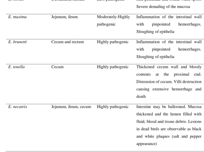
The prevalence of clinical and sub-clinical coccidiosis is approximately 5% and 20%, respectively. The annual cost incurred by the global poultry industry from the Eimeria infections has been estimated at approximately $2.4 billion, of which 76% is caused by clinical or sub-clinical coccidiosis and 24% by drug-related costs (Shirley & Lillehoj, 2012). Eimeria parasites invade and develop inside the intestinal epithelium cell of chickens. These parasites caused severe symptoms against chicken such as the reduction of feed intake, weight loss, bloody feces excretion, and die in case of heavy infection (Zaman et al., 2015). Each Eimeria species has a specific site in the gastrointestinal tract and display different pathogenic during infection. The summary of the main characteristics of Eimeria species is shown in Table 1-1 (Quiroz-Castañeda & Dantán-González, 2015).
Table 1-1. Main characteristics of Eimeria species
Species Infected site Pathogenicity Lesion
E. praecox Duodenum, jejunum Least pathogenic Watery intestinal contents. Mucus and mucoid casts
E. hanagi Duodenum, jejunum
and ileum
Least pathogenic Petechiae and white opacities in the upper small intestine. Intestinal content may be creamy or watery E. acervulina Duodenum, ileum Less pathogenic Limited enteritis causing fluid loss.
Malabsorption of nutrients.
E. mitis Ileum Less pathogenic Limited enteritis causing fluid loss.
Malabsorption of nutrients
3
E. mivati Duodenum, rectum Less pathogenic Red petechiae and round white spots.
Severe denuding of the mucosa
E. maxima Jejunum, ileum Moderately-Highly
pathogenic
Inflammation of the intestinal wall with pinpointed hemorrhages.
Sloughing of epithelia
E. brunetti Cecum and rectum Highly pathogenic Inflammation of the intestinal wall with pinpointed hemorrhages.
Sloughing of epithelia
E. tenella Cecum Highly pathogenic Thickened cecum wall and bloody
contents at the proximal end.
Distension of cecum. Villi destruction causing extensive hemorrhage and death
E. necatrix Jejunum, ileum, cecum Highly pathogenic Intestine may be ballooned. Mucosa thickened and the lumen filled with fluid, blood and tissue debris. Lesions in dead birds are observable as black and white plaques (salt and pepper appearance)
1.2. Eimeria tenella 1.2.1. Life cycle
E. tenella is the most pathogenic parasite caused avian coccidiosis in the world (Quiroz- Castañeda, 2018). E. tenella is a protozoan parasite causing haemorrhage in the caecum of the chicken was known at the end of the 19th century but it was not clear until the investigations of Tyzzer since 1929 (Chapman
& Shirley, 2003). The specific site of E. tenella infection is cecum of the chicken, particularly in the medial and distal regions of the cecum (Matsubayashi et al., 2012).
Figure 1-1. E. tenella life cycle
4
The life cycle of E. tenella is shown in Fig. 1-1. E. tenella life cycle has two phases of development including exogenous phase in the environment during which excreted oocysts (immature) undergo sporulation (sporogony) to become the maturation (acquired virulence), and endogenous phases in the cecum during which there are 3 times of asexual reproduction (schizogony) followed by sexual differentiation, fertilization and shedding of unsporulated oocyst within the feces.
Sporulated oocyst contains 4 sporocysts, including 2 sporozoites in each one. When sporulated oocysts are taken by chicken, sporozoites are released from the oocyst by enzymatic digestion and migrate to cecum where they invade villus enterocytes. Sporozoites are first seen within immune cells such as intestinal intraepithelial lymphocytes (IELs), primarily CD8+ cells and macrophages shortly after invasion, followed by developing inside epithelial cells by forming parasitophorous vacuole (Jiang et al., 2012). Sporozoites then undergo merogony (3 schizont generations) as asexual reproduction resulting release many merozoites into the gut. The first mature schizonts which contain merozoites appeared at 60hr after infection. The young second- generation schizonts grow larger in the lamina propria, and the number increased from 84hr. Large groups of seconds–generation schizonts are located within the mucosa and cause ulceration of mucosa surface. The third generation occurs and located in the superficial areas of the villous around crypts. The mature third-generation schizonts were found to be smaller than those of the first and second stages (approximately 10 μm in diameter). The merozoites from third-generation schizonts subsequently develop into the sexual forms, as micro and macro-gametocytes from 108h.
Macrogametocytes have a single nucleus at the central of the cytoplasm with a prominent wall- forming body, and were more frequently seen than multi-nucleated microgametocytes at the early sexual stage. Macrogametocytes and microgametocytes then fuse to form zygotes which were characterized by a central nucleus and the formation of an oocyst wall from 120h (Matsubayashi et al., 2012). Oocyst size increased in length and breadth with time but the shape index (length/breadth) remained constant (Fish, 1931). Mature oocysts were found from 132h are eventually released in the feces (Matsubayashi et al., 2012).
1.2.2. Pathogenesis
The main symptoms of E. tenella infection are reduction in weight gain and feed intake, bloody excrement and damaged cecum. Symptom and lesion of infected chicken depends on
5
developmental stage, host, Eimeria species and infected dose. Normally, as the life cycle of E.
tenella, bloody excrement in feces was observed from day 4 post infection and last for about 3 days. Oocyst shedding in feces is observed from 5 day post infection (dpi) and reached peak at 6 dpi. Then the number of oocyst in the feces gradually reduces (De Gussem, 2006). Pathological changes are observed in the cecum during E. tenella life cycle. From 96h post infection, many second-generation schizonts are observed. During this period, some areas of the superficial regions such as crypts, lamina propria, and submucosa were disrupted, and haemorrhage had occurred (Matsubayashi et al., 2012).
1.3. Lactic acid bacteria
1.3.1. Main characteristic of lactic acid bacteria
Lactic acid bacteria (LAB), including members of the genera Lactobacillus and Bifidobacterium, are normal inhabitants of the gastrointestinal tract that are classified as probiotic bacteria. LAB are capable of converting carbohydrate substrate acids (mainly lactic acid) and producing a wide range of metabolites. LAB are not a homogenous group. They share a low G + C content (Amann et al., 1995), are Gram-positive, non-spore forming, facultative anaerobic, rod shaped (bacillus), or spherical (coccus) microorganisms (Garvie, 1984), with genome sizes that can range from 1.8 to 3.2 Mb (Douillard & de Vos, 2014; Salvetti et al., 2013). Acid tolerance ability provides LAB the ability to outcompete other bacteria in a natural fermentation, as they can withstand the increased acidity from organic acid production, a property that makes them important in the final phases of many food fermentation processes, when other microorganisms are inhibited by the low pH. These bacteria are involved in the fermentation of a wide range of substrates, i.e., milk, fruit, vegetables, cereals, meat, and fish. They appear in the gut of many mammals and in the external environment. The fact that they can survive in so many different substrates is a clear indication of the diversity of their metabolism. Presently, the classification of LAB inserts them into six families, Aerococcaceae, Carnobacteriaceae, Enterococcaceae, Leuconostocaceae, Lactobacillacea, and Streptococcaceae. Due to their interesting beneficial properties, LAB are widely used as starter cultures, as probiotics, and as microbial cell factories (Ruiz Rodríguez et al., 2019).
Lactobacillus is the largest genus within the group of LAB. It contains 168 species, some of which are used in the manufacture of fermented dairy, sourdough, meat, vegetable foods, or used
6
as probiotics. The genus Lactobacillus belongs to the phylum Firmicutes, class Bacilli, order Lactobacillales, and family Lactobacillaceae. Lactobacilli are Gram-positive, catalase-negative, non-spore-forming, rod-shaped bacteria that produce lactic acid as the major end product of fermentation. Lactobacillus species can be divided into three groups: Obligately homofermentative including: L. acidophilus, L. delbrueckii, L. helveticus, L. salivarius. Facultatively heterofermentative including: L. casei, L. curvatus, L. plantarum, L. sakei and Obligately heterofermentative including: L. brevis, L. buchneri, L. fermentum, L. reuteri. Since the phylogenetic relatedness of lactobacilli was investigated by sequencing the 16S rRNA of all species successfully described up to now and this permitted the allotment of the lactobacilli to the following groups: Lactobacillus buchneri group (bu), Lactobacillus casei group (ca), Lactobacillus delbrueckii group (de), Lactobacillus plantarum group (pl), Lactobacillus reuteri group (re), Lactobacillus sakei group (sa), and Lactobacillus salivarius group (sl). In contrast, Lactobacillus brevis and Lactobacillus perolens as well as the related species Lactobacillus bifermentans and Lactobacillus coryneformis are uniquely positioned among the lactobacilli. (Ibrahim, 2016).
1.3.2. Health benefits effect of Lactobacillus acidophilus
Lactobacillus Acidophilus (L. acidophilus ) is a member of the Lactobacillus genus of bacteria, and it plays an important role in health improvement and are often used as probiotics (Walter, 2008). The World Health Organization defines probiotics as "live micro-organisms which, when administered in adequate amounts, confer a health benefit on the host" (Hill et al., 2014). L.
acidophilus has been extensively studied as a probiotic, and evidence has shown that it may provide a number of health benefits (Hütt et al., 2006).
Improves growth performance
The addition of either pure Lactobacillus cultures or mixtures of lactobacilli and other bacteria to broiler diets has been reported to improve the growth performance in chicken in many reports (Olnood et al., 2015). Broilers supplemented with L. acidophilus weighed 10.1% more than birds in control group at 42 days of age (Vantsawa et al., 2017). Dietary addition of L.
acidophilus could improve the intestinal health and reduce the mortality of broilers suffering from necrotic enteritis (Li et al., 2018). Kalavathy et al. (2003) found an improvement in body weight
7
gain (BWG) and feed conversion ratio (FCR) of broilers fed a mixture of different Lactobacillus strains from 1 to 42 days of age (Olnood et al., 2015). A consistent improvement in BWG of chickens fed a culture of Lactobacillus has also been reported (Awad et al., 2009). Feeding broiler chickens up to 6 weeks of age with a diet containing a single strain of Lactobacillus acidophilus or a mixture of lactobacilli significantly improved BWG and FCR (Jin et al., 1998). Cao et al. (2013) also found that supplementation of the broiler diets with a single strain of Lactobacillus (Enterococcus faecium) significantly improved the BW and BWG compared to the control (Cao et al., 2013).
Prevent and reduce diarrhea
A number of studies have shown that probiotics like L. acidophilus may help prevent and reduce diarrhea that's associated with various diseases. Evidence on the ability of L. acidophilus to treat acute diarrhea in children is mixed. Some studies have shown a beneficial effect, while others have shown no effect. Addition of L. acidophilus to oral rehydration therapy was effective in the treatment of children with acute diarrhea by decreasing the duration of diarrhea (Simakachorn et al., 2000). Evaluating the evidence by types of acute diarrhea suggests that probiotics significantly reduced antibiotic-associated diarrhoea by 52%, reduced the risk of travellers' diarrhea by 8%, and that of acute diarrhea of diverse causes by 34%. Probiotics reduced the associated risk of acute diarrhea among children by 57%, and by 26% among adults (Sazawal et al., 2006). Live L.
acidophilus reduced the incidence of radiation-induced diarrhea and the need for anti-diarrheal medication and had a significant benefits on stool consistency (Chitapanarux et al., 2010).
Similarly, it may help reduce diarrhea associated with antibiotics and a common infection called Clostridium difficile (Gao et al., 2010).
Boosting the immune system
L. acidophilus can boost the immune system and thus help reduce the risk of viral infections.
There are some studies suggested that probiotics may prevent and improve symptoms of the common cold in human (Kang et al., 2013; King et al., 2014). Another study carefully examined the effects of L. acidophilus on the gut. It found that taking it as a probiotic increased the expression of genes in the intestines that are involved in immune response (Leyer et al., 2009; Van Baarlen et al., 2011). L. acidophilus has been proven to elevate immunological activity by stimulating both
8
the innate and adaptive immune responses (Konstantinov et al., 2008; Liu et al., 2010a; Zhang et al., 2008). In vitro studies have indicated that L. acidophilus is a strong inducer of Th1 cytokines, including IL-12 and IFN-γ (Gackowska et al., 2006; Zeuthen et al., 2006). Also, L. acidophilus is involved in the enhancement of the activity of NK cells (Sunada et al., 2007). Clinical trials involving oral administration of L. acidophilus strain L-55 (LaL-55) show that this strain effectively suppresses experimental allergic rhinitis (Sunada et al., 2007) and experimental atopic dermatitis in mice (Sunada et al., 2008). Oral treatment of chicken with Lactobacilli including L.
acidophilus, L. reuteri, and L. salivarius modulated systemic antibody- and cell-mediated immune responses (Brisbin et al., 2011). These results suggested that L. acidophilus may support a healthy immune system.
1.4. Overview of the immune system
There are two major pillars of the immune system including; the innate and the adaptive immunity. Both systems work closely together and take on different tasks. The innate immune system is the first line of defense against pathogens, including physical barriers (skin, mucous membranes, pH), innate cells (NK cells, DCs, macrophages) antimicrobial peptides (i.e. β- defensins, cathelicidins, the complement system) and the inflammatory response. The adaptive response is pathogen-specific and occurred following the innate response. Adaptive immunity is consists of cell-mediated (T cell) and humoral (antibody) immunity. The immune system detects a pathogen through pathogen recognition receptors (PRRs) which mainly present on the antigen presenting cells (APC) such as macrophages, DCs, epithelial cells and NK cells. APC digest the invading pathogens and present the peptides to naïve T cells through major histocompatibility molecules (MHC) class II protein, depending on the cytokines produced, encourage their development into different T helper subtypes, thus driving cell–mediated and humoral adaptive responses (Chaplin, 2010).
1.4.1. Innate immunity
Chicken innate cells include macrophages, DCs, NK cells, thrombocytes, mast cells and heterophils (Petrone et al., 2002). Macrophages are one of the most important innate cells in the immune system (Ortiz et al., 2015). Macrophages are also considered as an effectors of the adaptive immune response. Macrophages take part in inflammation and induce T cell responses
9
through the production of pro-inflammatory cytokines such as interleukin (IL)-1β, IL-6 and chemokines (macrophage inflammatory protein (MIP)1 and MIP2). Macrophages also plays the role in immune regulation through the production of anti-inflammatory cytokines such as IL-10 (Klasing, 1998). Besides, macrophages also involved in the recognition of antibody and complement opsonized pathogens of humoral immunity. In mammals, macrophages have been described to polarized towards an M1 or M2 phenotype (Mills et al., 2000), although their presence has not yet been established in the chicken. M1 macrophages are considered important to inhibition of the pathogen, produce pro-inflammatory cytokines and are driven by IFN-γ. M2 macrophages are important to resolution of inflammation, tissue repair, exhibit enhanced phagocytosis and production of anti-inflammatory mediators such as IL-10 (Mills et al., 2000).
Along with macrophages, DCs are considered professional APCs and work as an important bridge between innate and adaptive immunity. Immature DCs reside in tissues, when they detect pathogens, they migrate to lymph nodes which containing naïve T cells to present pathogenic antigens via MHC class II molecules (Wu et al., 2010).
NK cells are large lymphocytes containing dense granules (Göbel et al., 1994) and are found in the intestinal epithelium and peripheral blood (Gö Bel et al., 2001). These cell release IFN-γ and has cytotoxic activity to kill pathogen (Merlino & Marsh, 2002). It has been shown that mammalian NK cells can respond to MHC-I cell (Ljunggren & Kärre, 1990). In addition, mammals also have NKT cells, cells that share both NK and T cell properties (Godfrey et al., 2010) although the presence of NKT cells has not yet been confirmed in the chicken.
White blood cells of the avian blood smear including granulocytes (heterophils, eosinophils, basophils) and non-granulocytes (monocytes and lymphocytes). Heterophils are the most abundant granulocyte in peripheral blood in most avian species. These cells tend to be round with a lobed nucleus containing dense, purple–staining chromatin. The cytoplasm is typically colorless with eosinophilic rod– shaped granules. Heterophils migrate towards sites of inflammation where they release pro-inflammatory cytokines and chemokines (Kogut et al., 1994). Heterophils are highly phagocytic and are able to undergo degranulation and oxidative burst (Kogut. et al., 1994). Chicken heterophils are also capable of heterophil extracellular trap (HET) formation in response to pathogens, whereby intracellular contents are released, trapping and killing pathogens (Chuammitri et al., 2009). Eosinophils are approximately the same size and shape as heterophils making them difficult to distinguish by size. The nucleus is lobed and typically stains darker than that of heterophils. The granules are large, round, and eosinophilic. Although functional eosinophils have
10
not yet been formally identified in the chicken, polymorphonuclear granulocytes (PMNs) with granules that readily stain with eosin have been identified (Maxwell, 1987). Eotaxins, a chemoattractant for mammalian eosinophils, and the eotaxin receptor have not been identified in the chicken genome. Mammalian eosinophils are granulocytes which circulate within the blood and function against multicellular parasites, such as helminths, by releasing chemical mediators such as histamine, ROS and pro-inflammatory cytokines and, as are mast cells and basophils, implicated in allergy (reviewed by (Rosenberg et al., 2013). Basophils are rarely seen in avian peripheral blood. Basophils have large, blue–staining granules separating them from other avian blood cells. Chicken basophils are found circulating in the blood and are similar to mammalian basophils in that they are phagocytic (Dhodapkar et al, 1982). Mammalian basophils are similar to mammalian eosinophils in that they are able to produce histamine and bind IgE-opsonised pathogens (Stone et al., 2010).
1.4.2. Adaptive immunity
The adaptive immune system, also called acquired immunity, uses specific antigens to strategically mount an immune response. Unlike the innate immune system, which attacks only based on the identification of general threats, the adaptive immunity is activated by exposure to pathogens, and uses an immunological memory to learn about the threat and enhance the immune response accordingly. The adaptive immune response is much slower to respond to threats and infections than the innate immune response. APCs are vital for effective adaptive immune response, as the functioning of both cytotoxic and helper T cells is dependent on APCs. Antigen presentation allows for specificity of adaptive immunity and can contribute to immune responses against both intracellular and extracellular pathogens. T cells must be activated before they can divide and perform their function. This is achieved by interacting with a professional APCs which present an antigen recognized by their T cell receptor. The APCs involved in activating T cells is usually a dendritic cell. T cells cannot recognize and therefore cannot respond to free or soluble antigens. They can only recognize and respond to antigen that has been processed and presented by cells via carrier molecules like MHC molecules. Helper T cells can recognize exogenous antigen presented on MHC class II; cytotoxic T cells can recognize endogenous antigen presented on MHC class I. Most cells in the body can present antigen to CD8+ cytotoxic T cells via MHC class I.
11 Cell-mediated immunity
T cell subsets
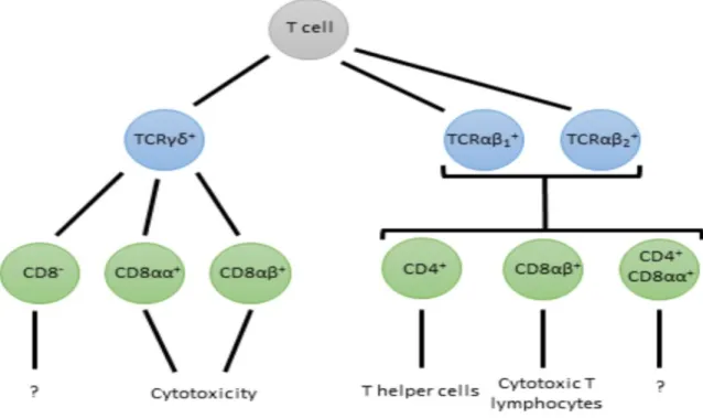
Fig. 1-2 provides an overview of chicken T cell subsets as defined by their TCR, CD8 and CD4 expression. Mammalian T cells express one of two types of TCR; TCRαβ and TCRγδ. The TCRαβ type is present on CD4+ and CD8+ T cells and recognizes antigen in the context of MHC molecules, a requirement to mounting adaptive immune responses. Chickens also have the TCRαβ however, in contrast to mammals, chickens have two types of β chain giving rise to two types of TCRαβ; TCRαβ1 and TCRαβ2 which differ in function and tissue distribution. High proportions of TCRαβ1 + cells are observed in the intestinal epithelium and lamina propria, whereas TCRαβ2+
cells are largely absent from the intestine, however both are present in the spleen (Chen et al., 1994). Cells expressing TCRγδ are much less common than those expressing TCRαβ in the lamina propria of the gut, however high numbers of TCRγδ+ cells are present in the chicken gut epithelium (Bucy et al., 1988a). The chicken TCRγδ appears able to recognize antigens without the requirement of antigen processing and presentation, and can exhibit cytotoxic activity similar to that of CTLs (Chen et al., 1994; Schild et al., 1994). The frequency of T cells expressing TCRγδ in chicken peripheral blood leukocytes (PBLs) can reach up to 20%, much higher than that of humans and mice (Cooper et al., 1991).
12
Figure 1-2. Chicken T cells as classified by TCR, CD4 and CD8 expression and their putative functions. Chicken T cells express one of three TCRs, and can be further subdivided by their expression of CD4, CD8αα homodimers and CD8αβ heterodimers.
TCRγδ+ T cells which co-express CD8αα or CD8αβ have higher levels of IFN-γ, FasL and lymphotactin expression than their CD8- counterparts indicating an activated phenotype which may be involved in mediating cytotoxic activity and polarisation of a Th1 response through IFN-γ production. TCRαβ1+ and TCRαβ2+ cells co-expressing the CD8αβ heterodimer are considered cytotoxic T lymphocytes (Pieper et al., 2008) whereas those co-expressing CD4 are thought to be T helper cells, although their function has not been confirmed in the chicken.
T helper cells
In mammals, depending on which cytokines are present, presentation of antigens via MHC class II to naïve CD4+ T cells results in the activation of different signaling molecules, known as signal transducer and activator transcription factors (STATs), which upregulate the master transcription factors for different T helper subsets. These transcription factors drive the production of cytokines which polarise the naïve T cell towards a particular Th phenotype. Different Th subsets
13
are defined by the cytokines that they produce, that drive their differentiation, and the transcription factors that they express (Table 1-2).
Table 1-2. Chicken and mammalian Th subsets. TGF-β4 is named TGF-β1 in mammals. CTL;
cytotoxic T lymphocyte. Fox; forkhead box. GC; germinal center. ROR; RAR-related orphan receptor. Tbet; T-box expressed in T cells. Treg; T regulatory cell. STAT; signal transducer and activator transcription factor.
Th subset
Driving cytokines
Signature cytokines
Signature transcription factors
Effector functions
Th1 IFN-γ, IL-12 IFN-γ Tbet, STAT1 Phagocyte and CTL mediated defence against intracellular microbes
Th2 IL-4 IL-4, IL-5,IL-13 GATA3,
STAT6
Stimulate antibody response to helminths
Tregs IL-2, TGF-β4 TGF-β4, IL-10 FoxP3 Suppress immune responses Th17 TGF-β4, IL-6,
IL-1β
IL-17A, IL-17F, IL-21, IL-22, IL-23
RORγt, STAT3
Recruit neutrophils/heterophils, defence against extracellular bacteria and fungi
Th9 TGF-β4, IL-4 IL-9 PU.1, STAT6 Pro-inflammatory, involved in autoimmunity and allergy
Tfh IL-21 IL-21 BCL6 GC formation, B cell selection and
differentiation
The Th1/Th2 paradigm was first proposed by Mosmann and Coffman (1989), and describes the polarisation of adaptive responses towards a Th1 or Th2 phenotype (Mosmann & Coffman, 1989). This paradigm appears to be conserved in the chicken; responses to intracellular pathogens such as Marek’s disease virus (Volpini et al., 1995; Xing & Schat, 2000) and infectious bursal disease virus (Liu et al., 2010) are governed by IFN-γ/Th1 cells and responses to helminths such as Histomonas meleagridis are governed by IL-4/IL-13/Th2 cells (Powell et al., 2009). Polarization of naïve T cells towards a Th1 phenotype is characterized by the production of IFN-γ (Mosmann
& Coffman, 1989). The IFN-γ produced mediates the effector functions of the Th1 response
14
including enhancing phagocytosis, release of Reactive oxygen species (ROS) and Reactive Nitrogen species (RNS), cytokine production and antigen presentation in macrophages, enhancing NK and CTL cytotoxicity and IFN-γ production (He et al., 2011; Merlino & Marsh, 2002) and, in mammals, is shown to enhance IL-12R expression on T cells to further polarize the Th1 response (Mullen et al., 2001). Polarization of naïve T cells towards a Th2 phenotype is characterized by the production of IL-4 and IL-13 (Mosmann & Coffman, 1989). Th2 responses mediate immunity through B cell activation, proliferation and differentiation into plasma cells, antibody class- switching, antibody production and polarization of macrophages towards a regulatory and wound- healing phenotype through their production of IL-10 and TGF-β. Chicken TGF-β4 is the equivalent of mammalian TGF-β1 (Halper et al., 2004). In mammals, IL-5 is also a hallmark cytokine of Th2 responses and activates eosinophils and basophils (Desreumaux et al., 1992). The absence of IL-5 during Th2 responses in the chicken may explain the lack of eosinophil and basophil activity observed. IL-5 is present in the chicken genome as a pseudogene (Avery et al., 2004). Th17 cells were first discovered in mammals by (Harrington et al., 2005) and are characterized by their production of IL-17A, IL-17F, IL-21 and IL-22 (Korn et al., 2009). Th17 cells exhibit microbicidal activity, are key regulators of inflammation and provide immunity at epithelial and mucosal barriers. All mammalian Th17-driving and effector cytokines (IL-1β, IL-6, TGF-β4, members of the IL-17 family, IL-21, IL-22 and IL-23) have also been identified in the chicken.
1.4.3. Mucosal immune cells
Immune cell populations in the gut include macrophages, heterophils, DCs, NK cells, B cells and T cells and these can be further subdivided by their location, either to the lamina propria (LPLs) or epithelium (IELs). In the chicken, the IEL population consists of NK and T cells expressing either TCRγδ or TCRαβ1. A large fraction of IELs express the CD8αα homodimer, whereas circulating CD8+ cells express the CD8αβ heterodimer. Relatively few IELs express CD4, similarly to mammals (Hoggenmueller & Wakenell, 1993). Many chicken T cell IELs express TCRγδ (Cooper et al., 1991). B cells are largely absent from the chicken intestinal epithelium, although a subset of epithelial NK cells expressing the B cell marker, chB6, are present (Vervelde
& Jeurissen, 1993). Macrophages and heterophils are more or less absent from the chicken intestinal epithelium (Hoggenmueller & Wakenell, 1993), however infection can lead to translocation of heterophils across the epithelium (Kogut, 2002). The location of NK and T cells in the epithelial layer means that they are some of the first cells to establish contact with invading
15
pathogens and are mediators of innate immunity. NK and T cells are able to recognize pathogens via Pattern recognition receptor (PRRs) on their surface (Chai & Lillehoj, 1988). TCRγδ+ cells have been shown to be capable of cytotoxic activity, cytokine production, including IFN-γ, and regulatory functions (Chen et al., 1994; Quere et al., 1990). The lamina propria is populated with a more diverse array of cell types than the epithelium including T cells, B cells, macrophages, DCs, NK cells and heterophils. In the chicken, the vast majority of LPLs are T cells, B cells, macrophages and DCs with some NK cells. In chickens, T cells in the lamina propria are mainly CD4+, CD8α+, TCRαβ1+ cells although some TCRγδ+ cells are present (Bucy et al., 1988b). Of the TCRαβ1 + population, the majority of cells co-express CD4 as opposed to CD8. Of the TCRγδ+ LPL population, some are CD8+ but the population of CD4+TCRαβ1 + T cells is much larger and TCRαβ2+ cells are largely absent from the intestine. Adaptive immunity in the gut is characterized by increased numbers of CD4+, CD8+and TCRαβ1 + T cells and chB6+ B cells in the lamina propria. NK cells in the lamina propria are mostly TCR-chB6-, but do possess an intracellular CD3 complex and surface CD8αα, are similar to mammalian NK cells in morphology and display cytotoxicity against the LSCC-RP9 tumor cell line, an NK cell target (Göbel et al., 1994). The B cells in the lamina propria are mainly IgA-secreting and account for the large concentrations of IgA observed in the intestinal mucosa in comparison with IgM and IgY (Lebacq-Verheyden et al., 1972). Chicken IgA is transported through the epithelial layer through interaction with the polymeric Ig receptor (M. Elaine Rose et al., 1981). Macrophages (expressing the chicken mannose receptor 1L-β and colony stimulating factor 1R) also constitute a large population of cells in the lamina propria and many heterophils are also present (Mast et al., 1998; Wells et al., 1998). During infection, macrophages are able to recognize pathogens and mediate direct pathogen killing through phagocytosis and production of ROS and RNS, help mediate inflammatory responses through cytokine production, and act as a bridge between the innate and adaptive response, through antigen presentation and production of T priming cytokines. Heterophils infiltrate the gut early on during infection and contribute to pathogen destruction and recruitment of other cells through expression of proinflammatory cytokines (Kogut, 2002). Heterophils and macrophages are also producers of anti-microbial peptides such as β-defensins and cathelicidins, contributing to innate mucosal immunity (Evans et al., 1994; Zhao et al., 2001).
16 1.5. Immune response to Eimeria
Infection of chickens with Eimeria induces both innate and adaptive responses, including increased heterophil activity, NK cell activity, T cell activation and antibody production and the outcome is dependent on a host of factors including the spp. of Eimeria, the genetics of the host and the severity of the infection.
1.5.1. Innate responses to Eimeria
Eimeria infection results in an inflammatory immune response characterized by increased production of pro-inflammatory mediators and infiltration and activation of innate cells such as macrophages and heterophils, capable of phagocytosis and release of ROS and RNS to control the pathogen (Lillehoj & Li, 2004; Petrone et al., 2002b). It is likely that APCs, including macrophages and DCs, fulfill multiple roles during Eimeria infection including phagocytosis, production of pro- inflammatory, regulatory and Th-promoting cytokines, production of ROS and RNS (such as H2O2
and NO) and antigen presentation for the development of cell-mediated and humoral responses.
During first exposure to E. tenella sporozoites, significantly more sporozoites are located next to or within macrophages compared to that of immune chickens indicating that macrophages are important in the first line of defense against Eimeria (Lillehoj & Li, 2004). Alternatively, it has been suggested that macrophages are involved in the transport of sporozoites to the crypt regions as indicated in early studies where large numbers of E. tenella and E. acervulina sporozoites were observed within macrophage-like cells (Challey & Buns, 1959; Doran, 1966). Increased macrophage numbers have also been identified in the gut following E. acervulina primary infection and E. maxima secondary challenge in chickens (Cornelissen et al., 2009). Therefore, it follows that macrophages are important cells in the response to Eimeria. While little is known about the direct responses of macrophages to Eimeria infection in vivo, the response of various chicken macrophage cell lines to various Eimeria antigens has been examined. Chow et al. (2011) investigated the response of a cell line, characterized by Rath et al. (2003) as having macrophage- like properties, to various recombinant surface antigens expressed on E. tenella sporozoites. The cell line, named HTC cells, increased nitrite production and IL1-β, IFN-γ, IL10 and NOS2 messenger RNA (mRNA) production in response to E. tenella surface antigens. In the same cell line in response to E. acervulina sporozoites, increased IL-18 expression was also observed and
17
differential Th1 promoting responses were observed in response to E. tenella (IFN-γ was upregulated) (Dalloul et al., 2007).
To date, the role of DCs in Eimeria infection has not been widely explored due to the lack of clearly defined markers to distinguish chicken DCs from macrophages, although some success in reducing oocyst shedding and increasing weight gains has been observed in using so called DC derived exosomes loaded with E. tenella, E. maxima or E. acervulina antigens to immunize chickens prior to challenge (Del Cacho et al., 2012). Heterophils form one of the first lines of defense against invading pathogens that display high levels of phagocytosis and can undergo processes such as oxidative burst and degranulation. Increased heterophils infiltrate the mucosa following E. tenella infection (Petrone et al., 2002) and heterophils (as did macrophages) isolated from peripheral blood showed increased transcripts of TLR4 and TLR15 when stimulated with both live and heat-killed E. tenella oocysts (Zhou et al., 2013).
NK cells, present in the intestinal epithelium, are one of the first innate cells to come into contact with Eimeria sporozoites therefore it is likely they are an important first line of defense against Eimeria. Hong et al. (2006) demonstrated that chicken NK lysin, an anti-microbial and anti- tumor peptide expressed by NK cells and T cells, demonstrates cytolytic activity when incubated with E. acervulina and E. maxima sporozoites in vitro. Following E. acervulina infection, NK lysin expression was upregulated from 7 dpi onwards, at 4 dpi with E. maxima infection and 3 dpi and then 6 dpi onwards with E. tenella infection in chicken IELs (Hong et al., 2006). Another study found that IEL NK cell activity decreased initially following E. maxima and E. acervulina infection in chickens but increased at 8 dpi and following homologous challenge (Lillehoj, 1989). In contrast to these findings, E. vermiformis challenge experiments in mice revealed no significant role for NK cell activity during infection. Mice with defective NK cell cytotoxic activity were not more susceptible than control mice to E. vermiformis infection and similar levels of parasite replication were observed in both groups of mice (Rose et al., 1995). Additionally, Smith et al., (1994) found that NK cell activity correlated with enhanced control of infection with E. vermiformis, however treatment of mice with an anti-aslio GM (anti-NK cell) antibody did not reduce control of the parasite (Smith et al., 1994). Eimeria antigens, including profilin, enhance human NK cell activity and IFN-γ production and are being explored as potential adjuvants for treatment of infectious disease and cancer (Aylsworth et al., 2013).
18 1.5.2. Adaptive immune response to Eimeria
T cells are an important aspect of primary and secondary Eimeria infection and in the development of protective immunity (Rose & Long, 1970). It appears that CD4+ T cells coordinate and induce protective immunity and CD8+ T cells execute effector functions to control parasite development during primary and secondary infection. Both CD4+ and CD8+ T cells increase following Eimeria infection. During E. tenella infection in chickens, the population of both CD4+
and CD8+ T lymphocytes increased the lamina propria in both naïve and immune chickens (Vervelde et al., 1996). Following E. maxima infection, CD4+ T cells infiltrated the lamina propria and CD8+ cells increased both the lamina propria and the epithelium of the chicken jejunum (Rothwell et al., 1995). Swinkels et al. (2006) also reported an increased percentage of CD8α+
cells in the duodenum of day old chicks following E. acervulina infection, but birds of this age failed to increase IFN-γ production following infection. Increased numbers of CD4+ T cells were observed in the jejunum, caecum and duodenum following single infection with E. maxima, E.
tenella and E. acervulina respectively, however increased numbers of CD8+ T cells were observed in the duodenum of E. acervulina–infected birds only. However, co-infection with all three of these Eimeria spp. resulted in increased numbers of CD8+ T cells in both the duodenum and caecum (Cornelissen et al., 2009). Increased numbers of CD8+ were also observed following primary E.
tenella infection, but these increases were less pronounced following secondary and tertiary infection (Wattrang et al., 2016). Various studies have also examined the effects of CD4 and CD8 depletion on immunity to Eimeria. Antibody-mediated depletion of CD4 during E. tenella infection enhanced oocyst shedding, but not during E. acervulina infection and that CD4-depletion did not affect the development of protective immunity. The same study showed that in CD8- or TCRαβ- depleted chickens, reduced oocyst shedding occurs following E. tenella or E. acervulina primary infection but increased following secondary infection. During E. acervulina infection in chickens, intestinal CD8+ T cells did not increase after primary infection, but increased with secondary infection, indicating a role in protective immunity (Trout & Lillehoj, 1996). These studies indicate that CD4+ T cells are required for immunity during primary infection, whereas CD8+ T cell effector functions are required for immunity to Eimeria following secondary exposure to the parasite.
TCRγδ+ cells are present in the intestinal epithelium and lamina propria although their importance during Eimeria infection is not well understood. In response to E. acervulina an increase in TCRγδ+ cells is observed in the duodenum (Swinkels et al., 2006). In murine studies,
19
TCRαβ-deficient mice had higher oocyst outputs than TCRαβ/γδ- deficient mice following E.
vermiformis infection and adoptive transfer of TCRγδ+cells from the mesenteric lymph nodes (MLN) of both naïve and previously infected mice resulted in decreased oocyst shedding (Smith
& Hayday, 2000), indicating that TCRγδ+ cells are important to protection against Eimeria but do not help impart specific protective immunity. Another study found that, in the absence of TCRγδ+cells, exacerbated immunopathology was observed during E. vermiformis infection, indicating that TCRγδ+ cells may have a regulatory role during Eimeria infection. However in contrast, another study in mice challenged with E. vermiformis, depletion of TCRγδ+ IELs had no effect on fecal oocyst count in the mice or on immunity to secondary challenge (Rose et al., 1996).
During Eimeria infection, increased IFN-γ is produced from early time points thus contributing to the innate inflammatory response and orchestrating the Th1 response. Increased IFN-γ is produced at the site of infection in chickens infected with E. acervulina, E. maxima and E. tenella (Byrnes et al., 1993; Rothwell et al., 2000; Martin Shirley et al., 2007). Furthermore, pre-treatment of chickens with recombinant IFN-γ resulted in improved weight gains compared to those not treated with IFN-γ following infection with E. acervulina (Lowenthal et al., 1997). In chickens also pre-treated with recombinant IFN-γ, the ability of E. tenella to replicate within the host was greatly reduced and as a result, chickens given IFN-γ reduced oocyst output following infection (Choi et al., 1999). Breed et al. (1997) suggested that at 8 dpi with E. tenella, a subset of antigen-specific CD4+ T cells circulate within the blood, primed to proliferate and produce IFN-γ in response to secondary exposure of E. tenella sporozoites (Breed et al., 1997). A similar population appears to exist in the spleen which migrate towards sites of infection following secondary E. tenella challenge (Rothwell et al., 2000). During E. falciformis and E. pragensis infection in mice, IFN-γ deficient mice display severe pathology compared with those with intact IFN-γ production and have reduced weight gain at both primary and secondary infection (Pogonka et al., 2010; Rose et al., 1992). A Th2 immune response to Eimeria has not yet been observed.
However, the Th2 response drives antibody-mediated immunity and a small number of roles for humoral immunity have been observed in response to Eimeria infection (Davis et al., 1979;
Zigterman et al., 1993). Immunity to other protozoan parasites including Toxoplasma (Dupont et al., 2012), Leishmania (Castellano et al., 2009), Cryptosporidium (Ehigiator et al., 2007) and Plasmodium (Radošević et al., 2010) is also mediated by Th1 responses. However, Th2-related cytokines appear to be important for regulating Th1 responses and cell mediated immunity. IL-10 reduces macrophage IL-12 production, thereby reducing IFN-γ production by NK and Th1 cells
20
(D’Andrea et al., 1992). In addition, IL-10 synergizes with IL-4 and TGF-β to reduce the development of Th1 responses and therefore reducing subsequent immunopathology (Oswald et al., 1992). During Cryptosporidium parvum infection, antibody-mediated depletion of IL-12 results in reduced IFN-γ production and increased disease severity (Urban et al., 1996). However, during Cryptosporidium infection in mice, increased IL-4 producing T cells are observed during the resolution of infection, indicating they are important in establishing humoral immunity and preventing prolonged infection (Aguire et al., 1998).
1.6. Immune system response to LAB 1.6.1. Systemic immune system response
The systemic immune system including the innate and adaptive immune responses which affecting the whole body. The innate immune response comprises factors that exist prior to the advent of infection, and are capable of exclusion or rapid response to microbes. The primary components of innate immunity of poultry are physical and chemical barriers, such as feathers and skin, epithelia and production of mucus; phagocytic cells, including macrophages and natural killer cells; complement proteins and mediators of inflammation; and cytokines. Overall, the innate immune response to virus infection is an immediate reaction designed to control and inhibit virus growth and spread and aid in developing pathogen-specific protection through the adaptive immune response.
Recently, beneficial therapeutic effects of LAB as a probiotic on the immune response have been extensively studied. Treatments with various members of the Lactobacillus species have been shown to stimulate multiple aspects of the immune response. The majority of evidence from in vitro systems, animal models and humans suggested that probiotics can enhance both specific and nonspecific immune responses. These effects are believed to be mediated through activating macrophages, increasing levels of cytokines, increasing NK cell activity and/or increasing levels of immunoglobulins (Elbanna et al., 2018). The mechanisms of systemic immune response induced by probiotic bacteria are reviewed by Maldonado Galdeano et al., 2007 (Fig. 1-3). Systemic antibody- and cell-mediated immune responses can be modulated by oral treatment with lactobacilli but that these bacteria may vary in their ability to modulate the immune response (Brisbin et al., 2011). Both Lactobacillus strains, L. salivarius and L. plantarumcan promote naive Th1 cell differentiation and participate in immunomodulatory responses (Elbanna et al., 2018). The
21
cell-mediated immune response was evaluated at 21 days post-immunization by measurement of IFN-γ production in splenocytes stimulated with inactivated vaccine antigens. L. salivarius-treated birds had significantly more serum antibody to sheep red blood cell (SRBC) and keyhole limpet hemocyanin (KLH) than birds that were not treated with probiotics. L. salivarius-treated birds also had decreased cell-mediated immune responses to recall antigen stimulation. L. reuteri treatment did not significantly affect the systemic immune response, while L. acidophilus treatment increased the antibody response to KLH (Brisbin et al., 2011). L. acidophilus is more effective at inducing T-helper-1 cytokines in chicken spleen and cecal tonsil cells. In addition, Haghighi et al also reported that L. acidophilus and Bifidobacterium bifidum enhance the systemic antibody response to some antigens in chickens (Haghighi et al., 2005).
22
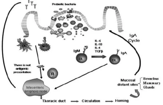
Figure 1-3. Systemic immune response induced by probiotic bacteria after interaction with the immune cells of the Peyer's patches. In the Peyer's patches, the probiotic bacteria or their fragments are internalized by M cells or in a paracellular way through follicle-associated epithelial cells of the Peyer's patches. After that, the bacteria or their particles interact with the macrophages and DC, which are activated to produce cytokines. As consequence of the bacterial stimulation to the immune cells in this inductor site of the immune response, cytokine production is enhanced, as well as the switch from IgM to IgA B cells. IL-10, IL-6, IL-4, and Transforming Growth Factor β (TGF-β) from immune cells could also promote this T- independent switch. Probiotic stimulation can induce the IgA cycle, increasing the number of IgA+ cells in mucosal sites distant to the intestine. The IgA+ cells migrate to the mesenteric lymphoid node and then via the thoracic duct to the circulation, arriving in the bronchus and mammary glands. The cytokines released by probiotic stimulation in Peyer's patches are the biological messengers of the complex network of signals that activate the systemic immune response. DC, dendritic cells; MQ, macrophages cells; APC, antigen-presenting cells; TL, T lymphocytes; BL, B lymphocytes. (Maldonado Galdeano et al., 2007).
23 1.6.2. Mucosal immune system response
Mucosal immunity is formed by mucosa-associated lymphoid tissue, which functions independently of the systemic immune system. Several beneficial effects of probiotics on the host intestinal mucosal defenses system have been identified. These include blocking pathogenic bacterial effects by producing bacteriocidal substances and competing with pathogens and toxins for adherence to the intestinal epithelium. For intestinal epithelial homeostasis, probiotics promote intestinal epithelial cell survival, enhance barrier function, and stimulate protective responses from intestinal epithelial cells (Yan & Polk, 2011).
Mucosal immunity has its own innate and adaptive components. The immune response is initiated by innate immunity following exposure to foreign substances or tissue injury. Innate immunity exerts protective roles in host homeostasis in part by priming adaptive immune responses against persisting insults and inducing inflammation. However, the unbalanced immune response leads to severe inflammation and uncontrolled tissue damage and disease. Sensing of the intestinal microbiota by the host mucosal immune system plays significant roles in maintaining intestinal homeostasis and inducing systemic protective responses. At the gut mucosal level, the innate immune response not only provides the first line of defense against pathogenic microorganisms but also provides the biological signals that instruct the adaptive immune system to elicit a response.
Noncommensal and probiotic bacteria are also able to induce a gut mucosal immune response (Tlaskalová-Hogenová et al., 2004).
LAB are present in the intestine of most animals. The beneficial role played by these microorganisms in humans and other animals, including the effect on the immune system, has been extensively reported. The activation of the systemic and secretory immune response by LAB requires many complex interactions among the different constituents of the intestinal ecosystem (microflora, epithelial cells and immune cells). Through different mechanisms, they send signals to activate immune cells (Tlaskalová-Hogenová et al., 2004). The study about the influence of the oral administration of different geneous of LAB including L. casei, L. acidophilus, L. rhamnosus, L. delbrueckii subsp. bulgaricus, L. plantarum, Lactococcus lactis and Streptococcus thermophiles on immune response demonstrated that all the bacteria assayed were able to increase the number of IgA producing cells associated with the lamina propria of small intestine. The increase in IgA+
producing cells was not always correlated with an increase in the CD4+ T cell number, indicating